

The DOI matches its Crossref record; 30 of the 30 references it cites were found in a registry or an index.
Pediatric neuroimaging using magnetic resonance imaging during non-sedated sleep
Open accessAbstract
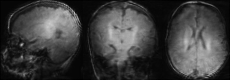
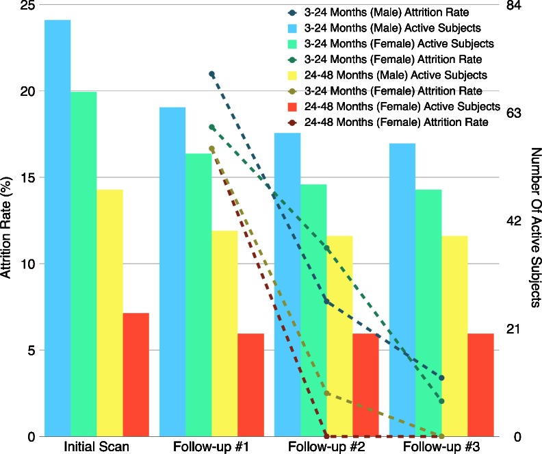
BACKGROUND: Etiological studies of many neurological and psychiatric disorders are increasingly turning toward longitudinal investigations of infant brain development in order to discern predisposing structural and/or functional differences prior to the onset of overt clinical symptoms. While MRI provides a noninvasive window into the developing brain, MRI of infants and toddlers is challenging due to the modality's extreme motion sensitivity and children's difficulty in remaining still during image acquisition. OBJECTIVE: Here, we outline a broad research protocol for successful MRI of children under 4 years of age during natural, non-sedated sleep. MATERIALS AND METHODS: All children were imaged during natural, non-sedated sleep. Active and passive measures to reduce acoustic noise were implemented to reduce the likelihood of the children waking up during acquisition. Foam cushions and vacuum immobilizers were used to limit intra-scan motion artifacts. RESULTS: More than 380 MRI datasets have been successfully acquired from 220 children younger than 4 years of age within the past 39 months. Implemented measures permitted children to remain asleep for the duration of the scan and allowed the data to be acquired with an overall 97% success rate. CONCLUSION: The proposed method greatly advances current pediatric imaging techniques and may be readily implemented in other research and clinical settings to facilitate and further improve pediatric neuroimaging.
Overview
The claims and statistics of this paper haven’t been checked yet.
Run the check — free, no account needed. It reads the open-access text and checks the sources it cites.
Integrity signals
Registry
Cites
Statistics
Figures
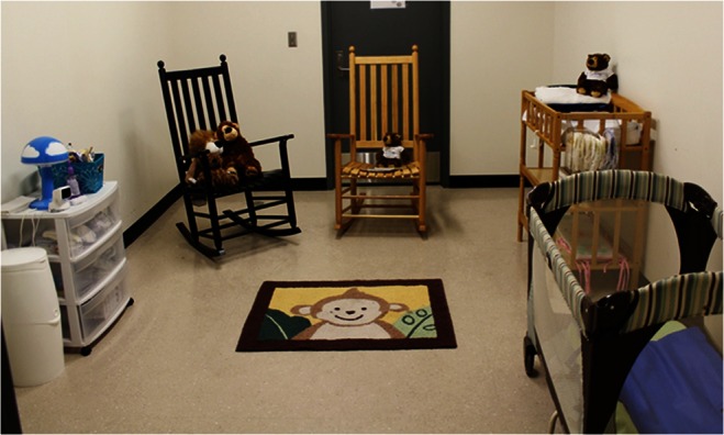
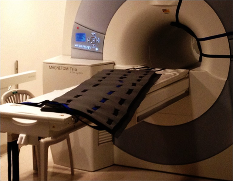
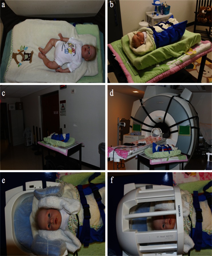
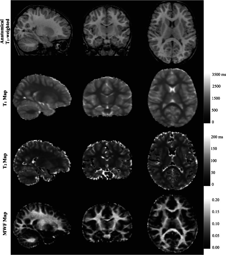
Verification
Claims the paper makes
The claims and statistics of this paper haven’t been checked yet.
Run the check — free, no account needed. It reads the open-access text and checks the sources it cites.
Claims other papers make about this one
Each is checked against this paper’s own text: does it say what the citing paper says it does?
In the citing paper
“this strategy of template creation mirrors our prior work that has been shown to successfully create structural and quantitative MRI templates of the pediatric brain (Deoni et al., 2012, Dean et al., 2014).”
From Quantifying cortical development in typically developing toddlers and young children, 1–6 years of age (2017) · cites this paper as Dean et al., 2014
Self-check
Percentages, sums and reported differences re-computed from the paper’s own numbers.
The self-check needs the paper’s full text.
Citations
Cites
Structural magnetic resonance imaging of the adolescent brain
GIEDD, J. N. (2004) · Annals of the New York Academy of Sciences · doi:10.1196/annals.1308.009 · Cited by 1,610
Sexual dimorphism of brain developmental trajectories during childhood and adolescence
Lenroot, R. K., Gogtay, N., Greenstein, D. K., Wells, E. M., Wallace, G. L., Clasen, L. S., Blumenthal, J. D., Lerch, J., Zijdenbos, A. P., Evans, A. C., et al. (2007) · NeuroImage · doi:10.1016/j.neuroimage.2007.03.053 · Cited by 1,309
Longitudinal Development of Human Brain Wiring Continues from Childhood into Adulthood
Lebel, C., & Beaulieu, C. (2011) · Journal of Neuroscience · doi:10.1523/jneurosci.5302-10.2011 · Cited by 1,193
Quantitative magnetic resonance imaging of human brain development: ages 4–18
Giedd, J. N., Snell, J. W., Lange, N., Rajapakse, J. C., Casey, B. J., Kozuch, P. L., Vaituzis, A. C., Vauss, Y. C., Hamburger, S. D., Kaysen, D., et al. (1996) · Cerebral Cortex · doi:10.1093/cercor/6.4.551 · Cited by 1,024
Structural MRI of pediatric brain development: what have we learned and where are we going?
Giedd, J. N., & Rapoport, J. L. (2010) · Neuron · doi:10.1016/j.neuron.2010.08.040 · Cited by 836
Resting-state networks in the infant brain
Fransson, P., Skiöld, B., Horsch, S., Nordell, A., Blennow, M., Lagercrantz, H., & Åden, U. (2007) · Proceedings of the National Academy of Sciences · doi:10.1073/pnas.0704380104 · Cited by 696
Wolff, J. J., Gu, H., Gerig, G., Elison, J. T., Styner, M., Gouttard, S., Botteron, K. N., Dager, S. R., Dawson, G., Estes, A. M., et al. (2012) · American Journal of Psychiatry · doi:10.1176/appi.ajp.2011.11091447 · Cited by 671
The NIH MRI study of normal brain development
Evans, A. C. (2006) · NeuroImage · doi:10.1016/j.neuroimage.2005.09.068 · Cited by 526
DiMaggio, C. J., Sun, L. S., & Li, G. (2011) · Anesthesia & Analgesia · doi:10.1213/ane.0b013e3182147f42 · Cited by 515
Gleaning multicomponent T 1 and T 2 information from steady‐state imaging data
Deoni, S., Rutt, B. K., Arun, T., Pierpaoli, C. M., & Jones, D. K. (2008) · Magnetic Resonance in Medicine · doi:10.1002/mrm.21704 · Cited by 473
Self-citation
Cited by
Sorted by citations
Items 101–138 of 138
Aksenov, D. P. (2021) · Frontiers in Neurology · doi:10.3389/fneur.2021.664706 · Cited by 5
Infant peripherally inserted central catheter insertion without general anesthesia
Laing, S., Burgoyne, L. L., Muncaster, M., Taranath, A., & Taverner, F. J. (2020) · Pediatric Anesthesia · doi:10.1111/pan.13950 · Cited by 4
Mairhörmann, B., Castelblanco, A., Häfner, F., Pfahler, V., Haist, L., Waibel, D., Flemmer, A. W., Ehrhardt, H., Stoecklein, S., Dietrich, O., et al. (2021) · medRxiv · doi:10.1101/2021.08.06.21261648 · Cited by 4
DiPiero, M. A., Rodrigues, P. G., Justman, M., Roche, S., Bond, E., Guerrero-Gonzalez, J. M., Davidson, R. J., Planalp, E. M., & Dean, D. (2024) · Brain Structure and Function · doi:10.1007/s00429-024-02853-w · Cited by 3
Self-citationVokhiwa, M., Pitchford, N. J., Cohee, L. M., Phiri, K. S., & Umar, E. (2026) · Global Epidemiology · doi:10.1016/j.gloepi.2026.100274 · Cited by 2
Manejo de los casos de urgencia del sistema nervioso central pediátricos para radiólogos generales
Rebollo, M. (2016) · Radiología · doi:10.1016/j.rx.2016.03.004 · Cited by 2
Surani, Z., Parkar, S., Afshan, G., Elahi, K. N., Hoodbhoy, Z., Hilal, K., & Jafri, S. (2023) · BMJ Open · doi:10.1136/bmjopen-2022-070283 · Cited by 5
Khurana, S. G., Evans, M. E., Kelly, C. E., Thompson, D. K., Burnsed, J. C., Harper, A. D., Hendricks‐Muñoz, K. D., Shall, M. S., Stevenson, R. D., Inamdar, K., et al. (2021) · Developmental Neurorehabilitation · doi:10.1080/17518423.2021.1903602 · Cited by 2
Progress in triacylglycerol isomer detection in milk lipids
Cao, H., Liu, Q., Liu, Y., Zhao, J., Qiao, W., Wang, Y., Liu, Y., & Chen, L. (2024) · Food Chemistry X · doi:10.1016/j.fochx.2024.101433 · Cited by 1
Carreno, C. A., Evans, M. E., Lockhart, B. K., Chinaka, O., Katz, B., Bell, M. A., & Howell, B. (2024) · Developmental Cognitive Neuroscience · doi:10.1016/j.dcn.2024.101481 · Cited by 1
Preoperative Imaging for Cochlear Implantation: A Global Consensus
Alzhrani, F., Alahmadi, A., Alshalan, A., Abdelsamad, Y., Alsanosi, A. A., Acharya, A. N., Kim, A. H., Macías, Á. R., Gantz, B. J., Buchman, C. A., et al. (2026) · Otolaryngology · doi:10.1002/ohn.1361 · Cited by 1
Vokhiwa, M., Chetcuti, K., Lange, F., Bourke, N., Randall, L. M., Greenstein, S., Seal, M., Beare, R., Hussain, A., Rao, P., et al. (2026) · Journal of Magnetic Resonance Open · doi:10.1016/j.jmro.2026.100225 · Cited by 1
Self-citationSchiavon, J. L. D. O., Romani, J., & Lederman, H. M. (2014) · Current Radiology Reports · doi:10.1007/s40134-014-0059-7 · Cited by 1
Accounting for the Developing Brain
Wolff, J. J. (2016) · Springer eBooks · doi:10.1007/978-3-319-28492-7_29 · Cited by 1
Grose, E., Pigeon, M., Abdeen, N., Belanger, M., Schramm, D., & Vaccani, J. (2020) · Otology & Neurotology · doi:10.1097/mao.0000000000002838 · Cited by 1
Imaging early brain structural and functional development
Ouyang, M., Smyser, C. D., Neil, J. J., & Huang, H. (2021) · Advances in magnetic resonance technology and applications · doi:10.1016/b978-0-12-816633-8.00018-1 · Cited by 1
Distortion, motion artifacts and how to address them
Tisdall, M. D. (2021) · Advances in magnetic resonance technology and applications · doi:10.1016/b978-0-12-816633-8.00002-8 · Cited by 1
Special considerations for acquisition of pediatric MRI of high spatial and temporal resolution
Roberts, T. P. L., Zhu, T., & Huang, H. (2021) · Advances in magnetic resonance technology and applications · doi:10.1016/b978-0-12-816633-8.00019-3 · Cited by 1
Special considerations for unsedated MR in the young pediatric population
Gagoski, B. A., Grant, P. E., & Ahtam, B. (2022) · Advances in magnetic resonance technology and applications · doi:10.1016/b978-0-12-824460-9.00002-9 · Cited by 1
Patterns of Early Myelination Identify Functionally Relevant Brain NetworksPreprint
Deoni, S., Bruchhage, M. M. K., Dean, D., D’Sa, V. A., & Schnieder, N. (2022) · Research Square · doi:10.21203/rs.3.rs-2002561/v1 · Cited by 1
Self-citationZanatta, A., Siew, J., der Paelt, S. V., Warreyn, P., & Roeyers, H. (2023) · doi:10.31234/osf.io/ey9zc · Cited by 1
Precision functional imaging in infants using multi-echo fMRI at 7TPreprint
Moser, J., Sadeghi‐Tarakameh, A., Ramirez, J. S. B., Madison, T. J., Hantzsch, L., Weldon, K. B., Pham, H. H. N., Lundquist, J. T., Sung, S., Ukpong, I., et al. (2025) · bioRxiv (Cold Spring Harbor Laboratory) · doi:10.1101/2025.11.09.687453 · Cited by 1
Imaging of Occult Spinal Dysraphism
Greenan, K., Mirsky, D. M., & Hankinson, T. C. (2019) · Springer eBooks · doi:10.1007/978-3-030-10994-3_15
Pädiatrische MRT-Untersuchungen ohne Sedierung bei Kleinkindern
Huber, E., & Brenner, N. Y. (2019) · Radiopraxis · doi:10.1055/a-0784-8548
Altered hippocampal morphometry in infants born very preterm
Htite, E. D., Gajawelli, N., Saint‐Martin, C., Wei, M., Palmis, S., Wang, Y., Wintermark, P., Leporé, N., & Brossard‐Racine, M. (2021) · International Symposium on Medical Information Processing and Analysis (SIPAIM) · doi:10.1117/12.2606249
Hippocampal Growth is Altered from Term Equivalence to One Year of Age in Preterm Born InfantsPreprint
Htite, E. D., Gajawelli, N., Saint‐Martin, C., Palmis, S., Wei, M., Gilbert, G., Wintermark, P., Deoni, S., Leporé, N., & Brossard‐Racine, M. (2022) · SSRN Electronic Journal · doi:10.2139/ssrn.4176068
Self-citationDeoni, S., D’Sa, V. A., Beauchemin, J., Huentelman, M. J., Lewis, C. R., Carnell, S., Bonham, K. S., Klepac‐Ceraj, V., Bruchhage, M. M. K., & Volpe, A. R. (2024) · Research Square · doi:10.21203/rs.3.rs-3864773/v1
Self-citationZanatta, A., Siew, J., der Paelt, S. V., Warreyn, P., & Roeyers, H. (2024) · Review Journal of Autism and Developmental Disorders · doi:10.1007/s40489-024-00437-2 · Cited by 2
Neuroimaging Ancillary Study of the IV Iron RAPIDIRON/RAPIDIRON KIDS TrialPreprint
Dean, D., Hattiholi, V. V., Kamate, M., Thomason, M. E., Talekar, K. S., Patted, A., Thind, S., Bradford-Rogers, J., DeSantis, D., Guerrero-Gonzalez, J. M., et al. (2025) · doi:10.12688/verixiv.725.1
Self-citationMueller, M. E., Nichols, E. S., Al‐Saoud, S., de Vrijer, B., McKenzie, C. A., Eagleson, R. A., de Ribaupierre, S., & Duerden, E. G. (2025) · Human Brain Mapping · doi:10.1002/hbm.70401
Asou, H., Matsuo, K., Miyahara, Y., Katsube, T., Yoshizako, T., & Kaji, Y. (2025) · Japanese Journal of Radiological Technology · doi:10.6009/jjrt.26-1582
Dong, C.-Y., Li, Y.-H., Huang, S.-J., Ni, Y.-X., Yin, B.-Y., Liang, Y., & Guo, R. (2026) · Research Square · doi:10.21203/rs.3.rs-8863172/v1
Mukhtarova, N., DiPiero, M. A., Yoon, C. D., Frye, C. J., Alexander, A. L., Goldsmith, H. H., Davidson, R. J., Planalp, E. M., & Dean, D. C. (2026) · Scientific Reports · doi:10.1038/s41598-026-44129-9
Self-citationNeuroimaging Ancillary Study of the IV Iron RAPIDIRON/RAPIDIRON KIDS TrialPreprint
Hattiholi, V. V., Hattiholi, V. V., Kamate, M., Thomason, M. E., Talekar, K. S., Patted, A., Thind, S., Bradford-Rogers, J., DeSantis, D., Guerrero-Gonzalez, J. M., et al. (2026) · doi:10.12688/verixiv.725.2
Precht, H., Jensen, J. M., Johannesen, T. A., Heindahl, M. B., & Pedersen, M. R. V. (2026) · Journal of medical imaging and radiation sciences · doi:10.1016/j.jmir.2026.102438
Mulvany, T., Griffiths-King, D., Worthington, L., Crombie, K., Rose, H. E. L., Peet, A. C., Apps, J. R., & Novak, J. (2026) · PLoS ONE · doi:10.1371/journal.pone.0356382
Vokhiwa, M., Chetcuti, K., Chikwana, J., Banda, S., Lange, F., Williams, S., Jones, D. K., Deoni, S., Phiri, K. S., & Umar, E. (2026) · Journal of Magnetic Resonance Open · doi:10.1016/j.jmro.2026.100230
Self-citationOlson, S., Thellman, C. R., Demetri, B., Sholar, B., & Shultz, S. (2026) · Frontiers in Neuroscience · doi:10.3389/fnins.2026.1963995
Metadata
- Authors
- Dean, D. C., Dirks, H., O’Muircheartaigh, J., Walker, L., Jerskey, B. A., Lehman, K., Han, M., Waskiewicz, N., & Deoni, S. C. L.
- Affiliations
- Brown University (US)
- King's College London (GB)
- Corresponding author
- Dean, Douglas C.
- ORCID
- Venue
- Pediatric Radiology
- Published
- 2013-08-06
- Volume
- 44
- Issue
- 1
- Pages
- 64-72
- ISSN
- 0301-0449 (ISSN-L)
- 1432-1998
- PMC
- PMC3889986
- OpenAlex
- W2012134441
- Cited by
- 138
- References
- 30
- Citations by year
- 2026: 10 · 2025: 10 · 2024: 9 · 2023: 12 · 2022: 13 · 2021: 23 · 2020: 13 · 2019: 12 · 2018: 11 · 2017: 10 · 2016: 5 · 2015: 5 · 2014: 5
- FWCI
- 3.87
- Type
- journal article
- Language
- en
- Open access
- hybrid
- doi.org
- Locations
- Pediatric Radiology (publishedVersion, cc-by)
- Europe PMC (PubMed Central) (publishedVersion)
- PubMed Central (publishedVersion, cc-by-nc)
- PubMed (publishedVersion)
- Research Portal (King's College London) (publishedVersion, other-oa)
- Funders
- Wellcome Trust (096195)
- National Institute of Mental Health (R01MH087510)
- Topics
- Health Sciences › Medicine › Radiology, Nuclear Medicine and Imaging › Advanced MRI Techniques and Applications
- Health Sciences › Medicine › Pediatrics, Perinatology and Child Health › Fetal and Pediatric Neurological Disorders
- Health Sciences › Medicine › Pediatrics, Perinatology and Child Health › Pediatric Pain Management Techniques
- Keywords
- pediatric neuroimaging, infant brain development, motion artifacts, acoustic noise reduction, immobilization, children under 4 years, longitudinal brain development
- MeSH terms
- Brain, Child, Preschool, Equipment Design, Humans, Image Enhancement, Infant, Magnetic Resonance Imaging, Male, Restraint, Physical, Sensitivity and Specificity, Sleep, Reproducibility of Results, Procedural Sedation, Equipment Failure Analysis, Patient Positioning, Neuroimaging
About these checks
Registry, retraction, Cites and statistics results are computed, not judged, with one exception: a printed reference that no index lists is read once by a model before it counts as a citation error. Claims are extracted by a model and kept only when their quote appears verbatim in the text; each cited source is then checked, with a model’s help, for whether it says what the claim relies on. Those verdicts can be wrong — read the claim’s quote and the cited source before relying on one.
Statistics and arithmetic are recomputed, not judged.
Confidence bands are shown only for fields calibrated on our annotated test set. None is calibrated yet, so no band is shown.
What counts as a claim ›How we measure confidence ›How we check this ›
Are you an author, or do you see a problem with this page? Request a review
