

The DOI matches its Crossref record; 64 of the 64 references it cites were found in a registry or an index.
EANM/EARL harmonization strategies in PET quantification: from daily practice to multicentre oncological studies
Open accessAbstract
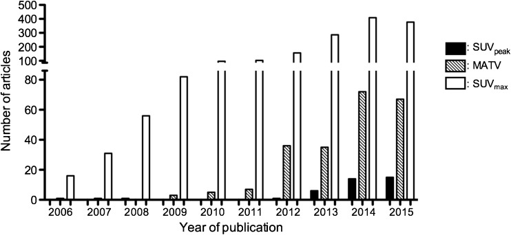
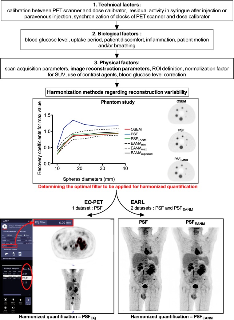
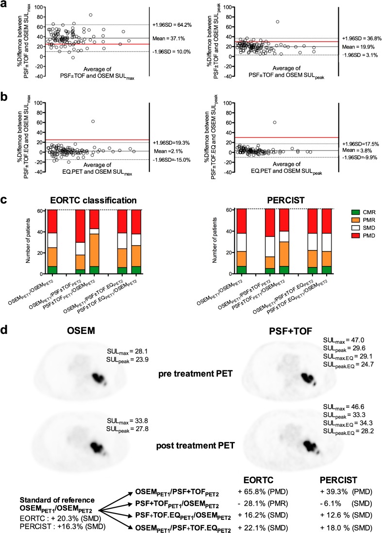
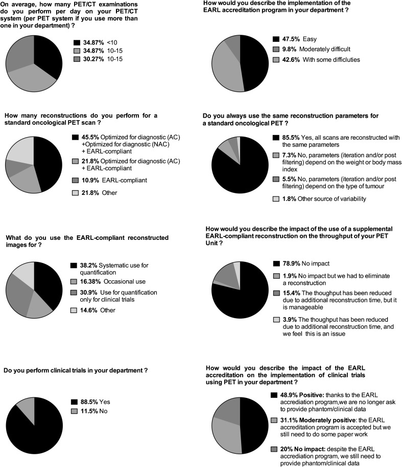
Quantitative positron emission tomography/computed tomography (PET/CT) can be used as diagnostic or prognostic tools (i.e. single measurement) or for therapy monitoring (i.e. longitudinal studies) in multicentre studies. Use of quantitative parameters, such as standardized uptake values (SUVs), metabolic active tumor volumes (MATVs) or total lesion glycolysis (TLG), in a multicenter setting requires that these parameters be comparable among patients and sites, regardless of the PET/CT system used. This review describes the motivations and the methodologies for quantitative PET/CT performance harmonization with emphasis on the EANM Research Ltd. (EARL) Fluorodeoxyglucose (FDG) PET/CT accreditation program, one of the international harmonization programs aiming at using FDG PET as a quantitative imaging biomarker. In addition, future accreditation initiatives will be discussed. The validation of the EARL accreditation program to harmonize SUVs and MATVs is described in a wide range of tumor types, with focus on therapy assessment using either the European Organization for Research and Treatment of Cancer (EORTC) criteria or PET Evaluation Response Criteria in Solid Tumors (PERCIST), as well as liver-based scales such as the Deauville score. Finally, also presented in this paper are the results from a survey across 51 EARL-accredited centers reporting how the program was implemented and its impact on daily routine and in clinical trials, harmonization of new metrics such as MATV and heterogeneity features.
Overview
The claims and statistics of this paper haven’t been checked yet.
Run the check — free, no account needed. It reads the open-access text and checks the sources it cites.
Integrity signals
Registry
Cites
Statistics
Figures
Verification
Claims the paper makes
The claims and statistics of this paper haven’t been checked yet.
Run the check — free, no account needed. It reads the open-access text and checks the sources it cites.
Claims other papers make about this one
Each is checked against this paper’s own text: does it say what the citing paper says it does?
The source backs part of the claim, not all of it — or backs it for a different population, condition or comparison.
In the citing paper
“This has undermined any attempts to establish widely adoptable SUV thresholds unless rigorous harmonization measures are followed [101,103,181].”
From Influences on PET Quantification and Interpretation (2022) · cites this paper as [181]
Experimental — accuracy not yet measured
Self-check
Percentages, sums and reported differences re-computed from the paper’s own numbers.
The self-check needs the paper’s full text.
Citations
Cites
From RECIST to PERCIST: Evolving Considerations for PET Response Criteria in Solid Tumors
Wahl, R. L., Jacene, H., Kasamon, Y., & Lodge, M. A. (2009) · Journal of Nuclear Medicine · doi:10.2967/jnumed.108.057307 · Cited by 3,777
FDG PET/CT: EANM procedure guidelines for tumour imaging: version 2.0
Boellaard, R., Delgado-Bolton, R., Oyen, W. J. G., Giammarile, F., Tatsch, K., Eschner, W., Verzijlbergen, F. J., Barrington, S. F., Pike, L. C., Weber, W. A., et al. (2015) · European Journal of Nuclear Medicine and Molecular Imaging · doi:10.1007/s00259-014-2961-x · Cited by 3,303
Self-citationFDG PET and PET/CT: EANM procedure guidelines for tumour PET imaging: version 1.0
Boellaard, R., O’Doherty, M. J., Weber, W., Mottaghy, F. M., Lonsdale, M. N., Stroobants, S. G., Oyen, W. J. G., Kotzerke, J., Hoekstra, O. S., Pruim, J., et al. (2009) · European Journal of Nuclear Medicine and Molecular Imaging · doi:10.1007/s00259-009-1297-4 · Cited by 1,329
Self-citationStandards for PET Image Acquisition and Quantitative Data Analysis
Boellaard, R. (2009) · Journal of Nuclear Medicine · doi:10.2967/jnumed.108.057182 · Cited by 906
Self-citationPerformance Measurements of the Siemens mMR Integrated Whole-Body PET/MR Scanner
Delso, G., Fürst, S., Jakoby, B., Ladebeck, R., Ganter, C., Nekolla, S. G., Schwaiger, M., & Ziegler, S. I. (2011) · Journal of Nuclear Medicine · doi:10.2967/jnumed.111.092726 · Cited by 870
Fully 3-D PET reconstruction with system matrix derived from point source measurements
Panin, V. Y., Kehren, F., Michel, C., & Casey, M. (2006) · IEEE Transactions on Medical Imaging · doi:10.1109/tmi.2006.876171 · Cited by 536
Characterization of PET/CT images using texture analysis: the past, the present… any future?
Hatt, M., Tixier, F., Pierce, L. A., Kinahan, P. E., Rest, C. C. L., & Visvikis, D. (2016) · European Journal of Nuclear Medicine and Molecular Imaging · doi:10.1007/s00259-016-3427-0 · Cited by 494
Nuyts, J. L., Dupont, P. J., Stroobants, S. G., Benninck, R., Mortelmans, L., & Suetens, P. (1999) · IEEE Transactions on Medical Imaging · doi:10.1109/42.774167 · Cited by 361
van Baardwijk, A. A. W., Bosmans, G., Boersma, L. J., Buijsen, J., Wanders, S. L., Hochstenbag, M. M. H., van Suylen, R. J., Dekker, A. L. A. J., Oberije, C., Houben, R. M. A., et al. (2007) · International Journal of Radiation Oncology*Biology*Physics · doi:10.1016/j.ijrobp.2006.12.067 · Cited by 295
Mikhaeel, N. G., Smith, D., Dunn, J., Phillips, M., Møller, H., Fields, P. A., Wrench, D., & Barrington, S. F. (2016) · European Journal of Nuclear Medicine and Molecular Imaging · doi:10.1007/s00259-016-3315-7 · Cited by 295
Research works printed but not linked: 2
- Quak E, Le Roux PY, Hofman MS, Robin P, Bourhis D, Callahan J, et al. Harmonizing FDG PET quantification while maintaining optimal lesion detection: prospective multicentre validation in 517 oncology patients. Eur J Nucl med Mol Imaging. 2015;42:2072–82. doi: 10.1007/s00259-015-3128-0 . · DOI · Paper page
- Barrington SF, Kluge R. FDG-PET for therapy monitoring in Hodgkin and non-Hodgkin lymphoma. Eur J Nucl Med Mol Imaging. 2017. doi: 10.1007/s00259-017-3690-8 . · DOI
Cited by
Sorted by citations
Items 301–344 of 344
Multimodality Medical Image Analysis in the Era of Precision Medicine
Zaidi, H. (2025) · Lecture notes in networks and systems · doi:10.1007/978-3-031-97985-9_6
Wang, J., Zhong, A., Xu, Q., Huang, H., Zhu, Y., Lu, J., Wang, M., Jiang, J., Li, C., Ni, M. A., et al. (2025) · medRxiv · doi:10.1101/2025.10.20.25338339
Li, S., Fan, X. M., & He, J. (2025) · World Journal of Clinical Oncology · doi:10.5306/wjco.v16.i11.110462
Shang, J., Duan, X., He, Y., Wu, B., Cai, Q., Cao, X., Tang, Y., Mai, J., Tan, S., Li, H., et al. (2025) · Cancer Imaging · doi:10.1186/s40644-025-00954-1
van Elferen, I., Vaz, S. C., Nelemans, P. J., Wierts, R., van de Weijer, T., Smidt, M. L., Mottaghy, F. M., & van Nijnatten, T. J. A. (2025) · Nuclear Medicine Communications · doi:10.1097/mnm.0000000000002091
Leube, J., Claeys, W., Gustafsson, J., Salas-Ramirez, M., Lassmann, M., Koole, M., & Tran‐Gia, J. (2024) · Figshare · doi:10.6084/m9.figshare.26122596
Siekkinen, R., Partanen, H., Kukola, L., Tolvanen, T., Fenwick, A., Smith, N. A. S., Teräs, M., Saraste, A., & Teuho, J. (2024) · Figshare · doi:10.6084/m9.figshare.26741114
Shekari, M., Verwer, E. E., Yaqub, M. M., Daamen, M., Buckley, C. J., Frisoni, G. B., Visser, P. J., Farrar, G., Barkhof, F., Gispert, J. D., et al. (2023) · Figshare · doi:10.6084/m9.figshare.24472521
Self-citationSiekkinen, R., Partanen, H., Kukola, L., Tolvanen, T., Fenwick, A., Smith, N. A. S., Teräs, M., Saraste, A., & Teuho, J. (2024) · Figshare · doi:10.6084/m9.figshare.26741111
Shekari, M., Verwer, E. E., Yaqub, M. M., Daamen, M., Buckley, C. J., Frisoni, G. B., Visser, P. J., Farrar, G., Barkhof, F., Gispert, J. D., et al. (2024) · Figshare · doi:10.6084/m9.figshare.24472578
Self-citationPfaehler, E. A. G., Mesotten, L., Kramer, G. M., Thomeer, M., Vanhove, K., de Jong, J., Adriaensens, P., Hoekstra, O. S., & Boellaard, R. (2021) · Figshare · doi:10.6084/m9.figshare.13532428
Self-citationAyati, N., McIntosh, L., Buteau, J. P., Alipour, R., Pudís, M., Daw, N., Jackson, P. A., & Hofman, M. S. (2024) · Figshare · doi:10.6084/m9.figshare.25762911
Pfaehler, E. A. G., Euba, D., Rinscheid, A., Hoekstra, O. S., Zijlstra, J. M., van Sluis, J., Brouwers, A. H., Lapa, C., & Boellaard, R. (2022) · Figshare · doi:10.6084/m9.figshare.20462214
Self-citationDesmonts, C., Lasnon, C., Jaudet, C., & Aide, N. (2024) · Figshare · doi:10.6084/m9.figshare.26624416
Self-citationDesmonts, C., Lasnon, C., Jaudet, C., & Aide, N. (2024) · Figshare · doi:10.6084/m9.figshare.26624413
Self-citationEnhancing PET/CT target assessment with porous 3D printed grids: a pilot study
Nalla, S. K. K., Maronnier, Q., Hazan, T. P., Kennedy, J. A., & Caselles, O. (2025) · Physical and Engineering Sciences in Medicine · doi:10.1007/s13246-025-01687-y
Nuclear Medicine in the Era of Precision Medicine
Kuber, R., Malik, M. M. U. D., & Alqahtani, M. M. (2025) · BENTHAM SCIENCE PUBLISHERS eBooks · doi:10.2174/9798898813543125010017
Liu, F., Yang, H., Zhang, J., Li, J., Qiao, Y., Chen, J., & Song, S. (2026) · EJNMMI Research · doi:10.1186/s13550-026-01402-2
Obolenski, O., Hristova, I., Munk, O. L., Shekari, M., Holstensson, M., Higuchi, T., Stokke, C., Goh, V., Kreppel, B., Gaertner, F. C., et al. (2026) · EANM Innovation · doi:10.1016/j.eanmi.2026.100164
Self-citationFumimoto, S., Sato, K., Katsumata, T., & Hanaoka, N. (2026) · Cancer Treatment and Research Communications · doi:10.1016/j.ctarc.2026.101406
[18F] Fludarabine pour l'imagerie TEP des lymphomes
Hovhannisyan, N. (2018) · doi:10.70675/d77503f2zecc0z4be9za91bz5de99d70dd63
A study of machine learning and deep learning methods and their application to medical imaging
Wallis, D. I. (2021) · theses.fr (ABES) · doi:10.70675/136c540fz5d49z41f4zb634zbd9663332582
Tatoğlu, M. T., Ökten, I. N., İbişoğlu, E., Söylemez, T., Tatoğlu, F. Y., Uslu, H., & Özülker, F. (2026) · Clinical and Translational Imaging · doi:10.1007/s40336-026-00767-7
van Geel, J., Eisses, B., Elias, S. G., Brouwers, A. H., Bensch, F., Gerritse, S. L., Angus, L., Glaudemans, A. W. J. M., Kuip, E. J. M., Jager, A., et al. (2026) · JAMA Oncology · doi:10.1001/jamaoncol.2026.0767
Self-citationEisses, B., Angus, L., Kuip, E. J. M., van Oordt, C. W. M. D. H., van der Vegt, B., Glaudemans, A. W. J. M., Brouwers, A. H., Oprea‐Lager, D. E., Oyen, W. J. G., Emmering, J., et al. (2026) · Diagnostics · doi:10.3390/diagnostics16081197
Claeys, W., Baete, K., Beels, L., Bernard, C., Danieli, R., D’Asseler, Y., Crop, A. D., Hesse, M., Nuttens, V., Vanderlinden, B., et al. (2026) · EJNMMI Physics · doi:10.1186/s40658-026-00877-1
Straightforward PET phantom preparation using 68Gallium diluted with acid
Yamao, T., Miwa, K., Hasegawa, K., Miyaji, N., Akiya, N., Wachi, K., Ukon, N., Joho, T., Shiga, T., & Ito, H. (2026) · Annals of Nuclear Medicine · doi:10.1007/s12149-026-02245-6
Arita, Y., Zheng, X., Wen, F., Fujiwara, M., Bijari, M., Glaudemans, A. W. J. M., Ruiter, S. J. S., Lütje, S., & Noordzij, W. (2026) · Abdominal Radiology · doi:10.1007/s00261-026-05593-z
Long vs. short axial field-of-view PET scanners for brain imaging: a phantom study
Providência, L., Turmacu, V., Mohr, P., van der Weijden, C. W. J., Dierckx, R., Lammertsma, A. A., & Tsoumpas, C. (2026) · Frontiers in Nuclear Medicine · doi:10.3389/fnume.2026.1856651
Splenic FDG PET uptake and CT volume as prognostic biomarkers in diffuse large B cell lymphoma
D’Argenzio, J., Rampi, N., Pozzi, J., Vespro, V., Pavan, M., Casiraghi, E., Rossi, F. G., Castello, A., Forzenigo, L. V., Castellani, M., et al. (2026) · La radiologia medica · doi:10.1007/s11547-026-02233-w
Kheruka, S. C., Al-Rashdi, S., Al-Maymani, N., Al-Balushi, A., Raii, T. A. A., Al-Makhmari, N., Jayakrishnan, V. V., Jain, A., Riyami, K. A., & Al-Sukaiti, R. (2026) · Journal of Nuclear Medicine Technology · doi:10.2967/jnmt.125.271634
Liu, H., & Liu, X. (2026) · Korean Journal of Radiology · doi:10.3348/kjr.2026.0821
Atieh, M., Pushparajah, K., Ghidaglia, J., Castilla-Lièvre, M., Deliu, D., Massaria, V., Harnais, K., Buffon, A., Beau, M., Jentreau, Y., et al. (2026) · EANM Innovation · doi:10.1016/j.eanmi.2026.100280
Hino, A., Ishiwata, Y., Kakiuchi, A., Numata, T., Kamide, H., Kurihara, H., Sekikawa, Z., & Utsunomiya, D. (2026) · Tomography · doi:10.3390/tomography12070104
Jougla, A., Nunes, P., Giraudet, A., Olivier, A., Blanc, J., Cochet, A., Ladoire, S., Vrigneaud, J., Vija-Racaru, L., & Drouet, C. (2026) · Journal of Nuclear Medicine · doi:10.2967/jnumed.126.272368
Feng, S., Xie, Y., Liu, C., Li, Y., Luo, J., Hu, X., Song, S., Wang, B., & Yang, Z. (2026) · European Journal of Nuclear Medicine and Molecular Imaging · doi:10.1007/s00259-026-08101-z
Ikari, Y., Tsuda, K., Maeda, Y., Akamatsu, G., Matsumoto, K., Daisaki, H., Hirata, K., Tateishi, U., Mori, K., Fujii, H., et al. (2026) · The EANM Journal · doi:10.1016/j.eanmj.2026.100239
Recent advances in nuclear medicine for cancer diagnosis and targeted therapy
Oisakede, E. O., Atitebi, O. F., Bamidele, D. B., Matope, E. M., Alabi, J. O., & Olawade, D. B. (2026) · Cancer Bioscience · doi:10.1016/j.cbs.2026.100006
Standardization and quality control in PET imaging: Advances, challenges, and clinical implications
Chalewska, W., Cegła, P., Sackiewicz, A., & Dedecjus, M. (2026) · Radiography · doi:10.1016/j.radi.2026.103542
18F-FDG PET/CT in Oncology Biologic, Technical, and Quantitative Foundations for Interpretation.
Hasani, N., Mohseny, A., Patel, D., Sitek, A., Ghesani, M., & Farhadi, F. (2026) · The Radiologic clinics of North America · doi:10.1016/j.rcl.2026.07.006
Chang, R., Yang, L., Gao, J., Liu, X., Wang, B., Li, Y., & Duan, X. (2026) · Frontiers in Nuclear Medicine · doi:10.3389/fnume.2026.1920721
Azimi, M. S., Cheraghi, M., & Zaidi, H. (2026) · Medical Physics · doi:10.1002/mp.70731
Moustaquim, J., van Geel, J., van Kruchten, M., de Vries, E. F. J., Stokkel, M. P. M., Glaudemans, A. W. J. M., Hospers, G. A. P., van der Vegt, B., & Schroder, C. P. (2026) · ESMO rare cancers. · doi:10.1016/j.esmorc.2026.100560
Metadata
- Authors
- Aide, N., Lasnon, C., Veit-Haibach, P., Sera, T., Sattler, B., & Boellaard, R.
- Affiliations
- Inserm (FR)
- University Hospital Leipzig (DE)
- University Medical Center Groningen (NL)
- Corresponding author
- Aide, Nicolas
- Venue
- European Journal of Nuclear Medicine and Molecular Imaging
- Published
- 2017-06-16
- Volume
- 44
- Issue
- S1
- Pages
- 17-31
- ISSN
- 1619-7070 (ISSN-L)
- 1619-7089
- PMC
- PMC5541084
- OpenAlex
- W2625583840
- Cited by
- 344
- References
- 64
- Citations by year
- 2026: 37 · 2025: 49 · 2024: 37 · 2023: 33 · 2022: 30 · 2021: 54 · 2020: 32 · 2019: 26 · 2018: 33 · 2017: 12
- FWCI
- 4.26
- Type
- journal article
- Language
- en
- Open access
- hybrid
- doi.org
- Locations
- European Journal of Nuclear Medicine and Molecular Imaging (publishedVersion, cc-by)
- Europe PMC (PubMed Central) (publishedVersion, cc-by)
- PubMed Central (publishedVersion, cc-by)
- PubMed (publishedVersion)
- Pure Amsterdam UMC (publishedVersion, other-oa)
- Amsterdam UMC (VU Amsterdam) - Institutional Repository (publishedVersion)
- University of Groningen research database (University of Groningen / Centre for Information Technology) (publishedVersion, cc-by)
- SZTE Publicatio Repozitórium (University of Szeged) (acceptedVersion, other-oa)
- Zurich Open Repository and Archive (University of Zurich) (acceptedVersion, cc-by)
- HAL (submittedVersion, other-oa)
- Zurich Open Repository and Archive (University of Zurich) (submittedVersion, cc-by)
- University of Groningen research database (University of Groningen / Centre for Information Technology) (submittedVersion, cc-by)
- Data Archiving and Networked Services (DANS) (submittedVersion)
- Zurich Open Repository and Archive (University of Zurich)
- Topics
- Health Sciences › Medicine › Radiology, Nuclear Medicine and Imaging › Medical Imaging Techniques and Applications
- Health Sciences › Medicine › Pulmonary and Respiratory Medicine › Lung Cancer Diagnosis and Treatment
- Health Sciences › Medicine › Radiology, Nuclear Medicine and Imaging › Radiomics and Machine Learning in Medical Imaging
- Keywords
- PET quantification, method harmonization, FDG PET/CT, standardized uptake value, total lesion glycolysis, therapy response assessment, EORTC criteria, PERCIST, Deauville score, imaging biomarkers
- MeSH terms
- Humans, Magnetic Resonance Imaging, Neoplasms, Reference Standards, Societies, Medical, Multicenter Studies as Topic, Positron-Emission Tomography
About these checks
Registry, retraction, Cites and statistics results are computed, not judged, with one exception: a printed reference that no index lists is read once by a model before it counts as a citation error. Claims are extracted by a model and kept only when their quote appears verbatim in the text; each cited source is then checked, with a model’s help, for whether it says what the claim relies on. Those verdicts can be wrong — read the claim’s quote and the cited source before relying on one.
Statistics and arithmetic are recomputed, not judged.
Confidence bands are shown only for fields calibrated on our annotated test set. None is calibrated yet, so no band is shown.
What counts as a claim ›How we measure confidence ›How we check this ›
Are you an author, or do you see a problem with this page? Request a review
