

The DOI matches its Crossref record; 64 of the 64 references it cites were found in a registry or an index.
EANM/EARL harmonization strategies in PET quantification: from daily practice to multicentre oncological studies
Open accessAbstract
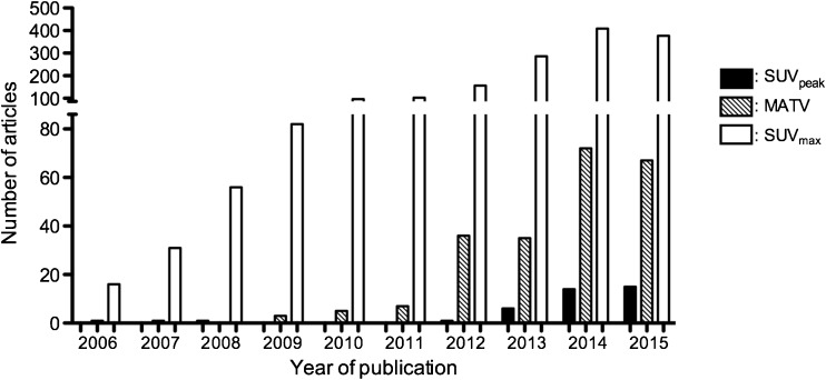
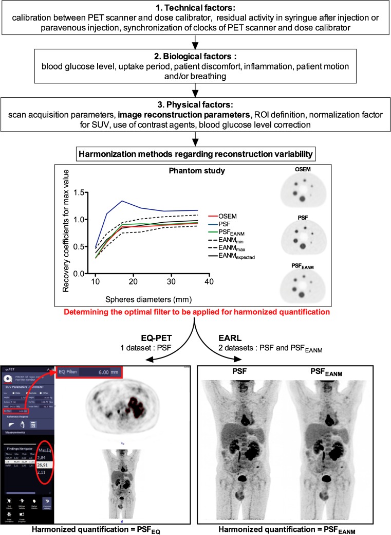
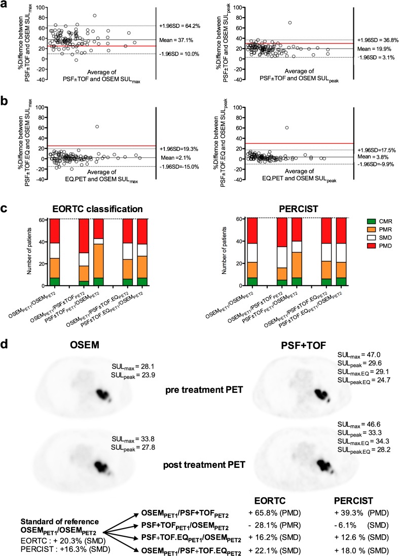
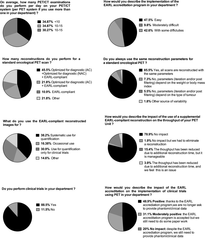
Quantitative positron emission tomography/computed tomography (PET/CT) can be used as diagnostic or prognostic tools (i.e. single measurement) or for therapy monitoring (i.e. longitudinal studies) in multicentre studies. Use of quantitative parameters, such as standardized uptake values (SUVs), metabolic active tumor volumes (MATVs) or total lesion glycolysis (TLG), in a multicenter setting requires that these parameters be comparable among patients and sites, regardless of the PET/CT system used. This review describes the motivations and the methodologies for quantitative PET/CT performance harmonization with emphasis on the EANM Research Ltd. (EARL) Fluorodeoxyglucose (FDG) PET/CT accreditation program, one of the international harmonization programs aiming at using FDG PET as a quantitative imaging biomarker. In addition, future accreditation initiatives will be discussed. The validation of the EARL accreditation program to harmonize SUVs and MATVs is described in a wide range of tumor types, with focus on therapy assessment using either the European Organization for Research and Treatment of Cancer (EORTC) criteria or PET Evaluation Response Criteria in Solid Tumors (PERCIST), as well as liver-based scales such as the Deauville score. Finally, also presented in this paper are the results from a survey across 51 EARL-accredited centers reporting how the program was implemented and its impact on daily routine and in clinical trials, harmonization of new metrics such as MATV and heterogeneity features.
Overview
The claims and statistics of this paper haven’t been checked yet.
Run the check — free, no account needed. It reads the open-access text and checks the sources it cites.
Integrity signals
Registry
Cites
Statistics
Figures
Verification
Claims the paper makes
The claims and statistics of this paper haven’t been checked yet.
Run the check — free, no account needed. It reads the open-access text and checks the sources it cites.
Claims other papers make about this one
Each is checked against this paper’s own text: does it say what the citing paper says it does?
The source backs part of the claim, not all of it — or backs it for a different population, condition or comparison.
In the citing paper
“This has undermined any attempts to establish widely adoptable SUV thresholds unless rigorous harmonization measures are followed [101,103,181].”
From Influences on PET Quantification and Interpretation (2022) · cites this paper as [181]
Experimental — accuracy not yet measured
Self-check
Percentages, sums and reported differences re-computed from the paper’s own numbers.
The self-check needs the paper’s full text.
Citations
Cites
From RECIST to PERCIST: Evolving Considerations for PET Response Criteria in Solid Tumors
Wahl, R. L., Jacene, H., Kasamon, Y., & Lodge, M. A. (2009) · Journal of Nuclear Medicine · doi:10.2967/jnumed.108.057307 · Cited by 3,777
FDG PET/CT: EANM procedure guidelines for tumour imaging: version 2.0
Boellaard, R., Delgado-Bolton, R., Oyen, W. J. G., Giammarile, F., Tatsch, K., Eschner, W., Verzijlbergen, F. J., Barrington, S. F., Pike, L. C., Weber, W. A., et al. (2015) · European Journal of Nuclear Medicine and Molecular Imaging · doi:10.1007/s00259-014-2961-x · Cited by 3,303
Self-citationFDG PET and PET/CT: EANM procedure guidelines for tumour PET imaging: version 1.0
Boellaard, R., O’Doherty, M. J., Weber, W., Mottaghy, F. M., Lonsdale, M. N., Stroobants, S. G., Oyen, W. J. G., Kotzerke, J., Hoekstra, O. S., Pruim, J., et al. (2009) · European Journal of Nuclear Medicine and Molecular Imaging · doi:10.1007/s00259-009-1297-4 · Cited by 1,329
Self-citationStandards for PET Image Acquisition and Quantitative Data Analysis
Boellaard, R. (2009) · Journal of Nuclear Medicine · doi:10.2967/jnumed.108.057182 · Cited by 906
Self-citationPerformance Measurements of the Siemens mMR Integrated Whole-Body PET/MR Scanner
Delso, G., Fürst, S., Jakoby, B., Ladebeck, R., Ganter, C., Nekolla, S. G., Schwaiger, M., & Ziegler, S. I. (2011) · Journal of Nuclear Medicine · doi:10.2967/jnumed.111.092726 · Cited by 870
Fully 3-D PET reconstruction with system matrix derived from point source measurements
Panin, V. Y., Kehren, F., Michel, C., & Casey, M. (2006) · IEEE Transactions on Medical Imaging · doi:10.1109/tmi.2006.876171 · Cited by 536
Characterization of PET/CT images using texture analysis: the past, the present… any future?
Hatt, M., Tixier, F., Pierce, L. A., Kinahan, P. E., Rest, C. C. L., & Visvikis, D. (2016) · European Journal of Nuclear Medicine and Molecular Imaging · doi:10.1007/s00259-016-3427-0 · Cited by 494
Nuyts, J. L., Dupont, P. J., Stroobants, S. G., Benninck, R., Mortelmans, L., & Suetens, P. (1999) · IEEE Transactions on Medical Imaging · doi:10.1109/42.774167 · Cited by 361
van Baardwijk, A. A. W., Bosmans, G., Boersma, L. J., Buijsen, J., Wanders, S. L., Hochstenbag, M. M. H., van Suylen, R. J., Dekker, A. L. A. J., Oberije, C., Houben, R. M. A., et al. (2007) · International Journal of Radiation Oncology*Biology*Physics · doi:10.1016/j.ijrobp.2006.12.067 · Cited by 295
Mikhaeel, N. G., Smith, D., Dunn, J., Phillips, M., Møller, H., Fields, P. A., Wrench, D., & Barrington, S. F. (2016) · European Journal of Nuclear Medicine and Molecular Imaging · doi:10.1007/s00259-016-3315-7 · Cited by 295
Research works printed but not linked: 2
- Quak E, Le Roux PY, Hofman MS, Robin P, Bourhis D, Callahan J, et al. Harmonizing FDG PET quantification while maintaining optimal lesion detection: prospective multicentre validation in 517 oncology patients. Eur J Nucl med Mol Imaging. 2015;42:2072–82. doi: 10.1007/s00259-015-3128-0 . · DOI · Paper page
- Barrington SF, Kluge R. FDG-PET for therapy monitoring in Hodgkin and non-Hodgkin lymphoma. Eur J Nucl Med Mol Imaging. 2017. doi: 10.1007/s00259-017-3690-8 . · DOI
Cited by
Sorted by citations
FDG PET/CT for assessing tumour response to immunotherapy
Aide, N., Hicks, R. J., Tourneau, C. L., Lheureux, S., Fanti, S., & Lopci, E. (2018) · European Journal of Nuclear Medicine and Molecular Imaging · doi:10.1007/s00259-018-4171-4 · Cited by 266
Self-citationPapadimitroulas, P., Brocki, L., Christopher Chung, N., Marchadour, W., Vermet, F., Gaubert, L., Eleftheriadis, V., Plachouris, D., Visvikis, D., Kagadis, G. C., et al. (2021) · Physica Medica · doi:10.1016/j.ejmp.2021.03.009 · Cited by 226
Quantification, improvement, and harmonization of small lesion detection with state-of-the-art PET
van der Vos, C. S., Koopman, D., Rijnsdorp, S., Arends, A. J., Boellaard, R., van Dalen, J. A., Lubberink, M., Willemsen, A. T. M., & Visser, E. P. (2017) · European Journal of Nuclear Medicine and Molecular Imaging · doi:10.1007/s00259-017-3727-z · Cited by 206
Self-citationHarmonization strategies for multicenter radiomics investigations
Da‐ano, R., Visvikis, D., & Hatt, M. (2020) · Physics in Medicine and Biology · doi:10.1088/1361-6560/aba798 · Cited by 167
Albert, N. L., Galldiks, N., Ellingson, B. M., van den Bent, M. J., Chang, S. M., Cicone, F., de Groot, J., Koh, E., Law, I. G., Rhun, É. L., et al. (2024) · The Lancet Oncology · doi:10.1016/s1470-2045(23)00525-9 · Cited by 198
Grubmüller, B., Senn, D., Kramer, G., Baltzer, P., D’Andrea, D., Grubmüller, K. H., Mitterhauser, M., Eidherr, H., Haug, A. R., Wadsak, W., et al. (2018) · European Journal of Nuclear Medicine and Molecular Imaging · doi:10.1007/s00259-018-4236-4 · Cited by 153
deSouza, N. M., Liu, Y., Chiti, A., Oprea‐Lager, D. E., Gebhart, G., BEERS, B. E. V., Herrmann, K., & Lecouvet, F. E. (2018) · European Journal of Cancer · doi:10.1016/j.ejca.2017.12.012 · Cited by 138
Eertink, J. J., van de Brug, T., Wiegers, S. E., Zwezerijnen, G. J. C., Pfaehler, E. A. G., Lugtenburg, P. J., van der Holt, B., de Vet, H. C. W., Hoekstra, O. S., Boellaard, R., et al. (2021) · European Journal of Nuclear Medicine and Molecular Imaging · doi:10.1007/s00259-021-05480-3 · Cited by 137
Self-citationReview of Gallium-68 PSMA PET/CT Imaging in the Management of Prostate Cancer
Lenzo, N. P., Meyrick, D. P., & Turner, J. H. (2018) · Diagnostics · doi:10.3390/diagnostics8010016 · Cited by 136
Seban, R., Mezquita, L., Berenbaum, A., Dercle, L., Botticella, A., Pechoux, C. L., Caramella, C., Deutsch, É., Grimaldi, S., Adam, J., et al. (2019) · European Journal of Nuclear Medicine and Molecular Imaging · doi:10.1007/s00259-019-04615-x · Cited by 139
Baum, R. P., Zhang, J., Schuchardt, C., Müller, D., & Mäcke, H. (2021) · Journal of Nuclear Medicine · doi:10.2967/jnumed.120.258889 · Cited by 127
SNMMI Procedure Standard/EANM Practice Guideline on Pediatric18F-FDG PET/CT for Oncology 1.0
Vali, R., Alessio, A. M., Balza, R., Borgwardt, L., Bar‐Sever, Z., Czachowski, M. R., Jehanno, N., Kurch, L., Pandit-Taskar, N. D., Parisi, M. T., et al. (2020) · Journal of Nuclear Medicine · doi:10.2967/jnumed.120.254110 · Cited by 125
Diagnostic Imaging in Vascular Graft Infection: A Systematic Review and Meta-Analysis
Folmer, E. I. R., von Meijenfeldt, G. C. I., van der Laan, M. J., Glaudemans, A. W. J. M., Slart, R. H. J. A., Saleem, B. R., & Zeebregts, C. J. (2018) · European Journal of Vascular and Endovascular Surgery · doi:10.1016/j.ejvs.2018.07.010 · Cited by 128
Correlated Multimodal Imaging in Life Sciences: Expanding the Biomedical Horizon
Walter, A., Paul‐Gilloteaux, P., Plochberger, B., Šefc, L., Verkade, P., Mannheim, J. G., Slezak, P., Unterhuber, A., Marchetti‐Deschmann, M., Ogris, M., et al. (2020) · Frontiers in Physics · doi:10.3389/fphy.2020.00047 · Cited by 130
Slart, R. H. J. A., Glaudemans, A. W. J. M., Gheysens, O. S., Lubberink, M., Kero, T., Dweck, M. R., Habib, G., Gaemperli, O., Saraste, A., Gimelli, A., et al. (2020) · European Journal of Nuclear Medicine and Molecular Imaging · doi:10.1007/s00259-020-05066-5 · Cited by 118
Radiomics: Data Are Also Images
Hatt, M., Rest, C. C. L., Tixier, F., Badic, B., Schick, U., & Visvikis, D. (2019) · Journal of Nuclear Medicine · doi:10.2967/jnumed.118.220582 · Cited by 125
Lopci, E., Hicks, R. J., Dimitrakopoulou‐Strauss, A., Dercle, L., Iravani, A., Seban, R., Sachpekidis, C., Humbert, O., Gheysens, O. S., Glaudemans, A. W. J. M., et al. (2022) · European Journal of Nuclear Medicine and Molecular Imaging · doi:10.1007/s00259-022-05780-2 · Cited by 119
Self-citationBarrington, S. F., Zwezerijnen, G. J. C., de Vet, H. C. W., Heymans, M. W., Mikhaeel, N. G., Burggraaff, C. N., Eertink, J. J., Pike, L. C., Hoekstra, O. S., Zijlstra, J. M., et al. (2020) · Journal of Nuclear Medicine · doi:10.2967/jnumed.119.238923 · Cited by 112
Self-citationJoint EANM-SNMMI guideline on the role of 2-[18F]FDG PET/CT in no special type breast cancer
Vaz, S. C., Woll, J. P. P., Cardoso, F., Groheux, D., Cook, G. J. R., Ulaner, G., Jacene, H., Rubio, I. T., Schoones, J., Peeters, M.-J. V., et al. (2024) · European Journal of Nuclear Medicine and Molecular Imaging · doi:10.1007/s00259-024-06696-9 · Cited by 102
European Society of Radiology, deSouza, N. M., Achten, E., Alberich‐Bayarri, Á., Bamberg, F., Boellaard, R., Clémеnt, O., Fournier, L. S., Gallagher, F. A., Golay, X., et al. (2019) · Insights into Imaging · doi:10.1186/s13244-019-0764-0 · Cited by 108
Self-citationKurland, B. F., Wiggins, J. R., Coche, A., Fontan, C., Bouvet, Y., Webner, P., Divgi, C. R., & Linden, H. M. (2020) · The Oncologist · doi:10.1634/theoncologist.2019-0967 · Cited by 102
Duclos, V., Iep, A., Gomez, L., Goldfarb, L., & Besson, F. L. (2021) · International Journal of Molecular Sciences · doi:10.3390/ijms22084159 · Cited by 95
Aide, N., Fruchart, C., Nganoa, C., Gac, A., & Lasnon, C. (2020) · European Radiology · doi:10.1007/s00330-020-06815-8 · Cited by 92
Self-citationHelsen, N., WYNGAERT, T. V. D., Carp, L., & Stroobants, S. G. (2018) · European Journal of Nuclear Medicine and Molecular Imaging · doi:10.1007/s00259-018-3978-3 · Cited by 96
Clinical insignificance of [18F]PSMA-1007 avid non-specific bone lesions: a retrospective evaluation
Arnfield, E. G., Thomas, P. A., Roberts, M. J., Pelecanos, A. M., Ramsay, S. C., Lin, C. Y., Latter, M. J., Garcia, P. L., & Pattison, D. A. (2021) · European Journal of Nuclear Medicine and Molecular Imaging · doi:10.1007/s00259-021-05456-3 · Cited by 87
Nuclear medicine and molecular imaging advances in the 21st century
Vaz, S. C., Oliveira, F., Herrmann, K., & Veit-Haibach, P. (2020) · The British Journal of Radiology · doi:10.1259/bjr.20200095 · Cited by 86
Self-citationIntegrating molecular nuclear imaging in clinical research to improve anticancer therapy
de Vries, E. G. E., de Ruijter, L. K., Hooge, M. N. L., Dierckx, R. A. J. O., Elias, S. G., & Oosting, S. F. (2018) · Nature Reviews Clinical Oncology · doi:10.1038/s41571-018-0123-y · Cited by 82
Radiomics in PET/CT: Current Status and Future AI-Based Evolutions
Hatt, M., Rest, C. C. L., Antonorsi, N., Tixier, F., Tankyevych, O., Jaouen, V., Lucia, F., Bourbonne, V., Schick, U., Badic, B., et al. (2020) · Seminars in Nuclear Medicine · doi:10.1053/j.semnuclmed.2020.09.002 · Cited by 73
New PET technologies – embracing progress and pushing the limits
Aide, N., Lasnon, C., Kesner, A., Levin, C. S., Buvat, I., Iagaru, A., Hermann, K., Badawi, R. D., Cherry, S. R., Bradley, K. M., et al. (2021) · European Journal of Nuclear Medicine and Molecular Imaging · doi:10.1007/s00259-021-05390-4 · Cited by 73
Self-citationKircher, M., Tran‐Gia, J., Kemmer, L., Zhang, X., Schirbel, A., Werner, R. A., Buck, A. K., Wester, H., Hacker, M., Lapa, C., et al. (2019) · Journal of Nuclear Medicine · doi:10.2967/jnumed.119.234484 · Cited by 72
Albert, N. L., Preusser, M., Traub‐Weidinger, T., Tolboom, N., Law, I. G., Palmer, J. D., Guedj, É., Furtner, J., Fraioli, F., Huang, R. Y., et al. (2024) · European Journal of Nuclear Medicine and Molecular Imaging · doi:10.1007/s00259-024-06783-x · Cited by 70
Grubmüller, B., Rasul, S., Baltzer, P., Fajković, H., D’Andrea, D., Berndl, F., Maj‐Hes, A., Grubmüller, K. H., Mitterhauser, M., Wadsak, W., et al. (2019) · The Prostate · doi:10.1002/pros.23919 · Cited by 69
Bolton, R. C. D., Aide, N., Colletti, P. M., Ferrero, A., Páez, D., Skanjeti, A., & Giammarile, F. (2021) · European Journal of Nuclear Medicine and Molecular Imaging · doi:10.1007/s00259-021-05450-9 · Cited by 69
Self-citationvan Geel, J., Boers, J., Elias, S. G., Glaudemans, A. W. J. M., de Vries, E. F. J., Hospers, G. A. P., van Kruchten, M., Kuip, E. J. M., Jager, A., van Oordt, C. W. M. D. H., et al. (2022) · Journal of Clinical Oncology · doi:10.1200/jco.22.00400 · Cited by 71
Weyts, K., Lasnon, C., Ciappuccini, R., Lequesne, J., Corroyer-Dulmont, A., Quak, E., Clarisse, B., Roussel, L., Bardet, S., & Jaudet, C. (2022) · European Journal of Nuclear Medicine and Molecular Imaging · doi:10.1007/s00259-022-05800-1 · Cited by 59
Self-citationRadiomics Analysis for Clinical Decision Support in Nuclear Medicine
Ibrahim, A., Vallières, M., Woodruff, H. C., Primakov, S. P., Beheshti, M., Keek, S. A., Refaee, T., Sanduleanu, S., Walsh, S., Morin, O., et al. (2019) · Seminars in Nuclear Medicine · doi:10.1053/j.semnuclmed.2019.06.005 · Cited by 61
Lactate and Lactate Transporters as Key Players in the Maintenance of the Warburg Effect
Pereira‐Nunes, A., Afonso, J., Granja, S., & Baltazar, F. (2020) · Advances in experimental medicine and biology · doi:10.1007/978-3-030-34025-4_3 · Cited by 63
Wang, Q., Li, Y., Li, Y., Hua, F., Wang, Q.-S., Zhang, X.-L., Cheng, C., Wu, H., Yao, Z., Zhang, W., et al. (2018) · European Journal of Nuclear Medicine and Molecular Imaging · doi:10.1007/s00259-018-4121-1 · Cited by 59
Fournier, L., de Geus-Oei, L. D., Regge, D., Oprea-Lager, D., D'Anastasi, M., Bidaut, L., Bäuerle, T., Lopci, E., Cappello, G., Lecouvet, F., et al. (2022) · Frontiers in Oncology · doi:10.3389/fonc.2021.800547 · Cited by 49
Vaz, S. C., Adam, J. A., Bolton, R. C. D., Véra, P., van Elmpt, W., Herrmann, K., Hicks, R. J., Lievens, Y. N., Santos, A., Schöder, H. M., et al. (2022) · European Journal of Nuclear Medicine and Molecular Imaging · doi:10.1007/s00259-021-05624-5 · Cited by 61
Applications of Artificial Intelligence in PSMA PET/CT for Prostate Cancer Imaging
Belal, S. L., Frantz, S., Minarik, D., Enqvist, O., Wikström, E., Edenbrandt, L., & Trägårdh, E. (2023) · Seminars in Nuclear Medicine · doi:10.1053/j.semnuclmed.2023.06.001 · Cited by 63
Advances in PET/CT Technology: An Update
Aide, N., Lasnon, C., Desmonts, C., Armstrong, I. S., Walker, M. D., & McGowan, D. R. (2021) · Seminars in Nuclear Medicine · doi:10.1053/j.semnuclmed.2021.10.005 · Cited by 55
Self-citationLanguage Analysis of Speakers with Dementia of the Alzheimer’s Type
Guinn, C. I., & Habash, A. (2012) · National Conference on Artificial Intelligence · Cited by 53
Influences on PET Quantification and Interpretation
Rogasch, J. M. M., Hofheinz, F., van Heek, L., Voltin, C.-A., Boellaard, R., & Kobe, C. (2022) · Diagnostics · doi:10.3390/diagnostics12020451 · Cited by 57
Self-citationAbbreviated scan protocols to capture 18F-FDG kinetics for long axial FOV PET scanners
Viswanath, V., Sari, H., Pantel, A., Conti, M., Daube-Witherspoon, M., Mingels, C., Alberts, I., Eriksson, L., Shi, K., Rominger, A., et al. (2022) · European Journal of Nuclear Medicine and Molecular Imaging · doi:10.1007/s00259-022-05747-3 · Cited by 43
[18F]FDG PET/CT: EANM procedure guidelines for tumour imaging: version 3.0
Boellaard, R., Herrmann, K., Barrington, S. F., Cranston, I. C., Mottaghy, F. M., Hoekstra, C., Beyer, T., Pike, L., Willemsen, A., Graham, M. M., et al. (2025) · The EANM Journal · doi:10.1016/j.eanmj.2025.100006 · Cited by 43
Self-citationAide, N., Talbot, M., Fruchart, C., Damaj, G. L., & Lasnon, C. (2017) · European Journal of Nuclear Medicine and Molecular Imaging · doi:10.1007/s00259-017-3899-6 · Cited by 48
Self-citationte Riet, J., Rijnsdorp, S., Roef, M. J., & Arends, A. J. (2019) · EJNMMI Physics · doi:10.1186/s40658-019-0262-y · Cited by 46
Dercle, L., Henry, T., Carré, A., Nikos Paragios, A., Deutsch, É., & Robert, C. (2020) · Methods · doi:10.1016/j.ymeth.2020.07.003 · Cited by 47
Prigent, K., Lasnon, C., Ezine, E., Janson, M., Coudrais, N., Joly, E., Césaire, L., Stefan, A., Depontville, M., & Aide, N. (2021) · European Journal of Nuclear Medicine and Molecular Imaging · doi:10.1007/s00259-020-05103-3 · Cited by 46
Self-citationGuo, R., Hu, X., Song, H., Xu, P., Xu, H., Rominger, A., Lin, X., Menze, B. H., Li, B., & Shi, K. (2021) · European Journal of Nuclear Medicine and Molecular Imaging · doi:10.1007/s00259-021-05232-3 · Cited by 46
Takahashi, M. E. S., Mosci, C., de Souza, E. M., Brunetto, S. Q., Etchebehere, E., Santos, A. O., Camacho, M., MIRANDA, E. C. M., Lima, M., Amorim, B. J., et al. (2019) · Scientific Reports · doi:10.1038/s41598-019-52740-2 · Cited by 43
Valkema, M. J., Noordman, B. J., Wijnhoven, B. P. L., Spaander, M. C. W., Biermann, K., Lagarde, S. M., Bennink, R. J., Schreurs, W. M. J., Roef, M. J., Hobbelink, M. G. G., et al. (2019) · Journal of Nuclear Medicine · doi:10.2967/jnumed.118.224196 · Cited by 42
Production of Ga-68 with a General Electric PETtrace cyclotron by liquid target
Riga, S., Cicoria, G., Pancaldi, D., Zagni, F., Vichi, S., Dassenno, M., Mora, L., Lodi, F., Morigi, M. P., & Marengo, M. (2018) · Physica Medica · doi:10.1016/j.ejmp.2018.10.018 · Cited by 40
Eertink, J. J., Zwezerijnen, G. J. C., Heymans, M. W., Pieplenbosch, S., Wiegers, S. E., Dührsen, U., Hüttmann, A., Kurch, L., Hanoun, C., Lugtenburg, P. J., et al. (2023) · Blood · doi:10.1182/blood.2022018558 · Cited by 39
Self-citationHarmonization of brain PET images in multi-center PET studies using Hoffman phantom scan
Shekari, M., Verwer, E. E., Yaqub, M. M., Daamen, M., Buckley, C. J., Frisoni, G. B., Visser, P. J., Farrar, G., Barkhof, F., Gispert, J. D., et al. (2023) · EJNMMI Physics · doi:10.1186/s40658-023-00588-x · Cited by 39
Self-citationNon-negative matrix factorisation improves Centiloid robustness in longitudinal studies
Bourgeat, P., Dóre, V., Doecke, J., Ames, D., Masters, C., Rowe, C., Fripp, J., & Villemagne, V. (2020) · NeuroImage · doi:10.1016/j.neuroimage.2020.117593 · Cited by 39
Reproducibility of [18F]FDG PET/CT liver SUV as reference or normalisation factor
Zwezerijnen, G., Eertink, J. J., Ferrández, M. C., Wiegers, S., Burggraaff, C., Lugtenburg, P., Heymans, M., de Vet, H. D., Zijlstra, J., & Boellaard, R. (2022) · European Journal of Nuclear Medicine and Molecular Imaging · doi:10.1007/s00259-022-05977-5 · Cited by 35
Self-citationSafarian, A., Mirshahvalad, S. A., Nasrollahi, H., Jung, T., Pirich, C., Arabi, H., & Beheshti, M. (2025) · Seminars in Nuclear Medicine · doi:10.1053/j.semnuclmed.2025.02.006 · Cited by 40
Standardization of Small Animal Imaging—Current Status and Future Prospects
Mannheim, J. G., Kara, F., Doorduin, J., Fuchs, K., Reischl, G., Liang, S., Verhoye, M., Gremse, F., Mezzanotte, L., & Huisman, M. C. (2017) · Molecular Imaging and Biology · doi:10.1007/s11307-017-1126-2 · Cited by 45
Barrington, S. F., Sulkin, T., Forbes, A., & Johnson, P. (2017) · European Journal of Nuclear Medicine and Molecular Imaging · doi:10.1007/s00259-017-3893-z · Cited by 38
PET/CT variants and pitfalls in malignant melanoma
Aide, N., Iravani, A., Prigent, K., Kottler, D., Alipour, R., & Hicks, R. J. (2022) · Cancer Imaging · doi:10.1186/s40644-021-00440-4 · Cited by 38
Self-citationβ-amyloid PET harmonisation across longitudinal studies: Application to AIBL, ADNI and OASIS3
Bourgeat, P., Doré, V., Burnham, S. C., Benzinger, T. L. S., Tosun, D., Li, S., Goyal, M. S., LaMontagne, P., Jin, L., Rowe, C. C., et al. (2022) · NeuroImage · doi:10.1016/j.neuroimage.2022.119527 · Cited by 37
Devriese, J., Beels, L., Maes, A., de Wiele, C. V., & Pottel, H. (2018) · EJNMMI Physics · doi:10.1186/s40658-018-0235-6 · Cited by 37
Kurland, B. F., Peterson, L. M., Shields, A. T., Lee, J. H., Byrd, D. W., Novakova-Jiresova, A., Muzi, M., Specht, J. M., Mankoff, D. A., Linden, H. M., et al. (2018) · Journal of Nuclear Medicine · doi:10.2967/jnumed.118.209544 · Cited by 36
van den Bosch, S., Doornaert, P., Dijkema, T., Zwijnenburg, E. M., Verhoef, L. C. G., Hoeben, B. A. W., Kasperts, N., Smid, E. J., Terhaard, C. H. J., & Kaanders, J. H. A. M. (2019) · Radiotherapy and Oncology · doi:10.1016/j.radonc.2019.07.025 · Cited by 36
Iqbal, R., Yaqub, M. M., Bektas, H. O., Oprea‐Lager, D. E., de Vries, E. G. E., Glaudemans, A. W. J. M., Aftimos, P. G., Gebhart, G., Beelen, A. P., Schuit, R. C., et al. (2023) · Clinical Cancer Research · doi:10.1158/1078-0432.ccr-22-2720 · Cited by 36
Self-citationEmerging role of 18F-FDG PET/CT in Castleman disease: a review
Koa, B., Borja, A. J., Aly, M., Padmanabhan, S., Tran, J., Zhang, V., Rojulpote, C., Pierson, S. K., Tamakloe, M.-A., Khor, J. S., et al. (2021) · Insights into Imaging · doi:10.1186/s13244-021-00963-1 · Cited by 35
Monaco, L., Gemelli, M., Gotuzzo, I., Bauckneht, M., Crivellaro, C., Genova, C., Cortinovis, D. L., Zullo, L., Ammoni, L. C., Bernasconi, D. P., et al. (2021) · Cancers · doi:10.3390/cancers13071634 · Cited by 35
The value of the Standardized Uptake Value (SUV) and Metabolic Tumor Volume (MTV) in lung cancer.
Hicks, R. (2022) · Seminars in nuclear medicine · doi:10.1053/j.semnuclmed.2022.04.007 · Cited by 33
EANM guidelines for PET-CT and PET-MR routine quality control
Koole, M. J. B., Armstrong, I. S., Krizsan, A. K., Stromvall, A., Visvikis, D., Sattler, B., Nekolla, S. G., & Dickson, J. C. (2022) · Zeitschrift für Medizinische Physik · doi:10.1016/j.zemedi.2022.08.003 · Cited by 34
Self-citationLee, K., Le, T. K., Hau, E. K. C., Hanna, G. G., Gee, H. E., Vinod, S. K., Dammak, S., Palma, D. A., Ong, A., Yeghiaian‐Alvandi, R., et al. (2021) · International Journal of Radiation Oncology*Biology*Physics · doi:10.1016/j.ijrobp.2021.11.027 · Cited by 34
Fonti, R., Pellegrino, S., Catalano, L., Pane, F., Vecchio, S. D., & Pace, L. (2019) · Annals of Hematology · doi:10.1007/s00277-019-03852-2 · Cited by 33
He, M., Zha, W., Tan, F. Z., Rankine, L. J., Fain, S. B., & Driehuys, B. (2018) · Academic Radiology · doi:10.1016/j.acra.2018.08.015 · Cited by 32
Ly, J., Minarik, D., Edenbrandt, L., Wollmer, P., & Trägårdh, E. (2019) · EJNMMI Research · doi:10.1186/s13550-019-0536-3 · Cited by 31
Bellèvre, D., Bailliez, A., Delelis, F., Blaire, T., Agostini, D., Mouquet, F., Maréchaux, S., & Manrique, A. (2020) · Journal of Nuclear Cardiology · doi:10.1007/s12350-020-02274-2 · Cited by 31
Repeatability of two semi-automatic artificial intelligence approaches for tumor segmentation in PET
Pfaehler, E. A. G., Mesotten, L., Kramer, G. M., Thomeer, M., Vanhove, K., de Jong, J., Adriaensens, P., Hoekstra, O. S., & Boellaard, R. (2021) · EJNMMI Research · doi:10.1186/s13550-020-00744-9 · Cited by 30
Self-citationChauvie, S., Mazzoni, L., & O'Doherty, J. (2023) · Tomography · doi:10.3390/tomography9050149 · Cited by 25
Feasibility of PET/CT system performance harmonisation for quantitative multicentre 89Zr studies
on behalf of, E. A. R. L., Kaalep, A., EATRIS, Huisman, M. C., Séra, T., Vugts, D. J., & Boellaard, R. (2018) · EJNMMI Physics · doi:10.1186/s40658-018-0226-7 · Cited by 29
Self-citationHoudu, B., Lasnon, C., Licaj, I., Thomas, G., Do, P., Guizard, A.-V., Desmonts, C., & Aide, N. (2019) · European Journal of Nuclear Medicine and Molecular Imaging · doi:10.1007/s00259-018-4151-8 · Cited by 37
Self-citationRole of Fludeoxyglucose in Breast Cancer
Groheux, D. (2018) · PET Clinics · doi:10.1016/j.cpet.2018.02.003 · Cited by 27
Measuring PET Spatial Resolution Using a Cylinder Phantom Positioned at an Oblique Angle
Lodge, M. A., Leal, J. P., Rahmim, A., Sunderland, J. J., & Frey, E. C. (2018) · Journal of Nuclear Medicine · doi:10.2967/jnumed.118.209593 · Cited by 28
Lasnon, C., Quak, E., Roux, P. L., Robin, P., Hofman, M. S., Bourhis, D., Callahan, J., Binns, D., Desmonts, C., Salaün, P., et al. (2017) · EJNMMI Physics · doi:10.1186/s40658-017-0185-4 · Cited by 27
Self-citationDoes PET Reconstruction Method Affect Deauville Score in Lymphoma Patients?
Enilorac, B., Lasnon, C., Nganoa, C., Fruchart, C., Gac, A., Damaj, G. L., & Aide, N. (2017) · Journal of Nuclear Medicine · doi:10.2967/jnumed.117.202721 · Cited by 27
Self-citationPET radiomics in lung cancer: advances and translational challenges
Zhang, Y., Huang, W., Jiao, H., & Kang, L. (2024) · EJNMMI Physics · doi:10.1186/s40658-024-00685-5 · Cited by 27
Prenosil, G. A., Weitzel, T., Fürstner, M., Hentschel, M., Krause, T., Cumming, P. D., Rominger, A., & Klaeser, B. (2020) · PLoS ONE · doi:10.1371/journal.pone.0229560 · Cited by 25
Imaging of Invasive Fungal Infections- The Role of PET/CT
Ankrah, A. O., Lawal, I. O., Dierckx, R., Sathekge, M. M., & Glaudemans, A. W. J. M. (2022) · Seminars in Nuclear Medicine · doi:10.1053/j.semnuclmed.2022.07.003 · Cited by 29
Exterkate, L., Hermsen, R., Küsters‐Vandevelde, H. V. N., Prette, J. F., Baas, D. J. H., Somford, D. M., & van Basten, J. A. (2023) · European Urology Oncology · doi:10.1016/j.euo.2023.04.006 · Cited by 25
ESMO/ASCO Recommendations for a Global Curriculum in Medical Oncology Edition 2023
Čufer, T., Kosty, M. P., & on behalf of the Curriculum Development Subgroup—ESMO/ASCO Global Curriculum Working Group (2023) · JCO Global Oncology · doi:10.1200/go.23.00277 · Cited by 26
Azimi, M.-S., Rahmim, A., Arabi, H., Sanaat, A., Zeraatkar, N., Bouchareb, Y., Liu, C., Alavi, A. A., King, M. A., Boellaard, R., et al. (2025) · European Journal of Nuclear Medicine and Molecular Imaging · doi:10.1007/s00259-025-07612-5 · Cited by 25
Self-citationENETS consensus guidelines for synoptic reporting of molecular imaging studies
Hicks, R., Dromain, C., de Herder, W. D., Costa, F., Deroose, C., Frilling, A., Koumarianou, A., Krenning, E., Raymond, E., Bodei, L., et al. (2021) · Journal of neuroendocrinology · doi:10.1111/jne.13040 · Cited by 20
Meyer, M., Allenbach, G., Lalonde, M. N., Schaefer, N. G., Prior, J. O., & Gnesin, S. (2020) · Scientific Reports · doi:10.1038/s41598-019-57313-x · Cited by 26
Prenosil, G. A., Hentschel, M., Weitzel, T., Sari, H., Shi, K., Afshar‐Oromieh, A., & Rominger, A. (2022) · EJNMMI Physics · doi:10.1186/s40658-022-00455-1 · Cited by 23
Prigent, K., & Aide, N. (2019) · PET Clinics · doi:10.1016/j.cpet.2019.08.006 · Cited by 24
Self-citationZimmermann, P.-A., Houdu, B., Césaire, L., Nakouri, I., Pontville, M. D., Lasnon, C., & Aide, N. (2021) · Annals of Nuclear Medicine · doi:10.1007/s12149-021-01608-5 · Cited by 22
Self-citationSilvoniemi, A., Laine, J., Aro, K. L. S., Nissi, L., Bäck, L. J. J., Schildt, J., Hirvonen, J., Hagström, J., Irjala, H., Aaltonen, L., et al. (2023) · Cancers · doi:10.3390/cancers15153970 · Cited by 22
on behalf of the SPUtNIk investigators, Weir-McCall, J. R., Harris, S., Miles, K. A., Qureshi, N. R., Rintoul, R. C., Dizdarevic, S., Pike, L. C., Cheow, H., & Gilbert, F. J. (2020) · European Journal of Nuclear Medicine and Molecular Imaging · doi:10.1007/s00259-020-05089-y · Cited by 22
Daisaki, H., Kitajima, K., Nakajo, M., Watabe, T., Ito, K., Sakamoto, F., Nakahara, T., Ishibashi, M., & Toriihara, A. (2021) · Scientific Reports · doi:10.1038/s41598-021-87942-0 · Cited by 21
Miwa, K., Wagatsuma, K., Iimori, T., Sawada, K., Kamiya, T., Sakurai, M., Miyaji, N., Murata, T., & Sato, E. (2018) · Physica Medica · doi:10.1016/j.ejmp.2018.07.001 · Cited by 20
Kitajima, K., Watabe, T., Nakajo, M., Ishibashi, M., Daisaki, H., Soeda, F., Tanemura, A., Kanekura, T., Yamazaki, N., & Ito, K. (2021) · Japanese Journal of Radiology · doi:10.1007/s11604-021-01174-w · Cited by 17
Metadata
- Authors
- Aide, N., Lasnon, C., Veit-Haibach, P., Sera, T., Sattler, B., & Boellaard, R.
- Affiliations
- Inserm (FR)
- University Hospital Leipzig (DE)
- University Medical Center Groningen (NL)
- Corresponding author
- Aide, Nicolas
- Venue
- European Journal of Nuclear Medicine and Molecular Imaging
- Published
- 2017-06-16
- Volume
- 44
- Issue
- S1
- Pages
- 17-31
- ISSN
- 1619-7070 (ISSN-L)
- 1619-7089
- PMC
- PMC5541084
- OpenAlex
- W2625583840
- Cited by
- 344
- References
- 64
- Citations by year
- 2026: 37 · 2025: 49 · 2024: 37 · 2023: 33 · 2022: 30 · 2021: 54 · 2020: 32 · 2019: 26 · 2018: 33 · 2017: 12
- FWCI
- 4.26
- Type
- journal article
- Language
- en
- Open access
- hybrid
- doi.org
- Locations
- European Journal of Nuclear Medicine and Molecular Imaging (publishedVersion, cc-by)
- Europe PMC (PubMed Central) (publishedVersion, cc-by)
- PubMed Central (publishedVersion, cc-by)
- PubMed (publishedVersion)
- Pure Amsterdam UMC (publishedVersion, other-oa)
- Amsterdam UMC (VU Amsterdam) - Institutional Repository (publishedVersion)
- University of Groningen research database (University of Groningen / Centre for Information Technology) (publishedVersion, cc-by)
- SZTE Publicatio Repozitórium (University of Szeged) (acceptedVersion, other-oa)
- Zurich Open Repository and Archive (University of Zurich) (acceptedVersion, cc-by)
- HAL (submittedVersion, other-oa)
- Zurich Open Repository and Archive (University of Zurich) (submittedVersion, cc-by)
- University of Groningen research database (University of Groningen / Centre for Information Technology) (submittedVersion, cc-by)
- Data Archiving and Networked Services (DANS) (submittedVersion)
- Zurich Open Repository and Archive (University of Zurich)
- Topics
- Health Sciences › Medicine › Radiology, Nuclear Medicine and Imaging › Medical Imaging Techniques and Applications
- Health Sciences › Medicine › Pulmonary and Respiratory Medicine › Lung Cancer Diagnosis and Treatment
- Health Sciences › Medicine › Radiology, Nuclear Medicine and Imaging › Radiomics and Machine Learning in Medical Imaging
- Keywords
- PET quantification, method harmonization, FDG PET/CT, standardized uptake value, total lesion glycolysis, therapy response assessment, EORTC criteria, PERCIST, Deauville score, imaging biomarkers
- MeSH terms
- Humans, Magnetic Resonance Imaging, Neoplasms, Reference Standards, Societies, Medical, Multicenter Studies as Topic, Positron-Emission Tomography
About these checks
Registry, retraction, Cites and statistics results are computed, not judged, with one exception: a printed reference that no index lists is read once by a model before it counts as a citation error. Claims are extracted by a model and kept only when their quote appears verbatim in the text; each cited source is then checked, with a model’s help, for whether it says what the claim relies on. Those verdicts can be wrong — read the claim’s quote and the cited source before relying on one.
Statistics and arithmetic are recomputed, not judged.
Confidence bands are shown only for fields calibrated on our annotated test set. None is calibrated yet, so no band is shown.
What counts as a claim ›How we measure confidence ›How we check this ›
Are you an author, or do you see a problem with this page? Request a review
