

The DOI matches its Crossref record; 201 of the 203 references it cites were found in a registry or an index.
Propagation of alpha-synuclein pathology: hypotheses, discoveries, and yet unresolved questions from experimental and human brain studies
Open accessAbstract
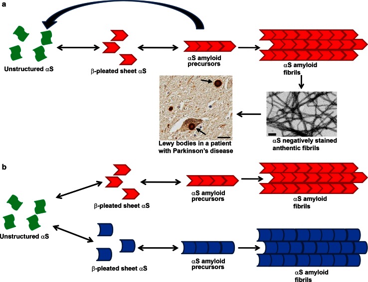
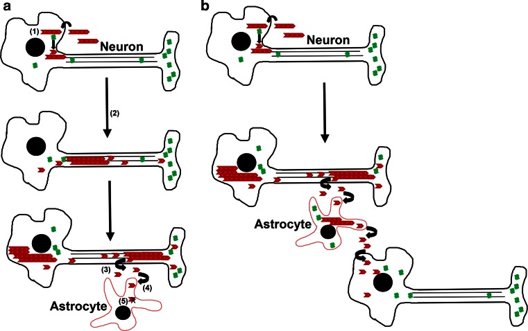
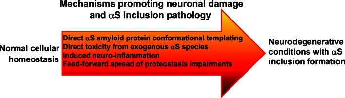
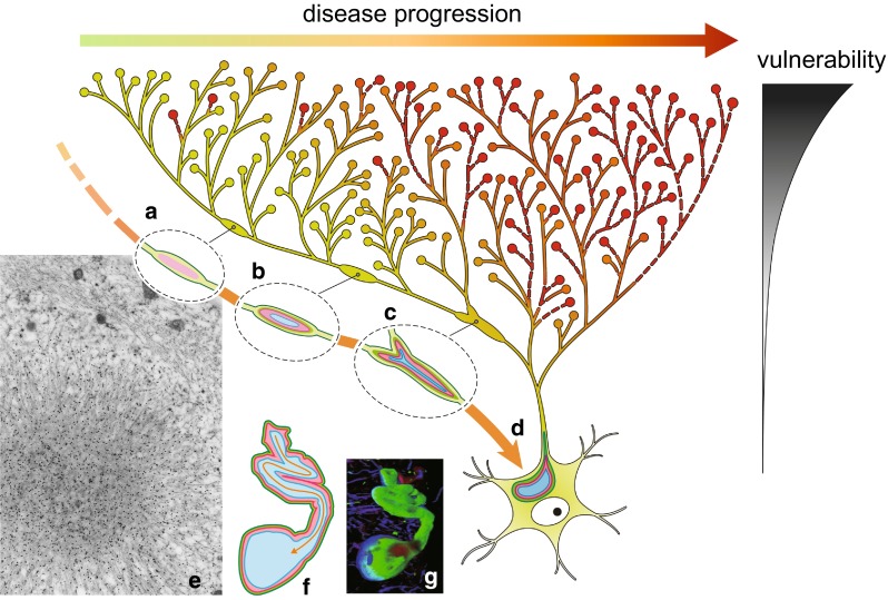
Progressive aggregation of alpha-synuclein (αS) through formation of amorphous pale bodies to mature Lewy bodies or in neuronal processes as Lewy neurites may be the consequence of conformational protein changes and accumulations, which structurally represents "molecular template". Focal initiation and subsequent spread along anatomically connected structures embody "structural template". To investigate the hypothesis that both processes might be closely associated and involved in the progression of αS pathology, which can be observed in human brains, αS amyloidogenic precursors termed "seeds" were experimentally injected into the brain or peripheral nervous system of animals. Although these studies showed that αS amyloidogenic seeds can induce αS pathology, which can spread in the nervous system, the findings are still not unequivocal in demonstrating predominant transsynaptic or intraneuronal spreads either in anterograde or retrograde directions. Interpretation of some of these studies is further complicated by other concurrent aberrant processes including neuroimmune activation, injury responses and/or general perturbation of proteostasis. In human brain, αS deposition and neuronal degeneration are accentuated in distal axon/synapse. Hyperbranching of axons is an anatomical commonality of Lewy-prone systems, providing a structural basis for abundance in distal axons and synaptic terminals. This neuroanatomical feature also can contribute to such distal accentuation of vulnerability in neuronal demise and the formation of αS inclusion pathology. Although retrograde progression of αS aggregation in hyperbranching axons may be a consistent feature of Lewy pathology, the regional distribution and gradient of Lewy pathology are not necessarily compatible with a predictable pattern such as upward progression from lower brainstem to cerebral cortex. Furthermore, "focal Lewy body disease" with the specific isolated involvement of autonomic, olfactory or cardiac systems suggests that spread of αS pathology is not always consistent. In many instances, the regional variability of Lewy pathology in human brain cannot be explained by a unified hypothesis such as transsynaptic spread. Thus, the distribution of Lewy pathology in human brain may be better explained by variable combinations of independent focal Lewy pathology to generate "multifocal Lewy body disease" that could be coupled with selective but variable neuroanatomical spread of αS pathology. More flexible models are warranted to take into account the relative propensity to develop Lewy pathology in different Lewy-prone systems, even without interconnections, compatible with the expanding clinicopathological spectra of Lewy-related disorders. These revised models are useful to better understand the mechanisms underlying the variable progression of Lewy body diseases so that diagnostic and therapeutic strategies are improved.
Overview
The claims and statistics of this paper haven’t been checked yet.
Run the check — free, no account needed. It reads the open-access text and checks the sources it cites.
Integrity signals
Registry
Cites
Statistics
Figures
Verification
Claims the paper makes
The claims and statistics of this paper haven’t been checked yet.
Run the check — free, no account needed. It reads the open-access text and checks the sources it cites.
Claims other papers make about this one
Each is checked against this paper’s own text: does it say what the citing paper says it does?
In the citing paper
“In the human brain, synuclein deposition and neuronal degeneration are accentuated in distal regions23.”
From Loss of autophagy in dopaminergic neurons causes Lewy pathology and motor dysfunction in aged mice (2018) · cites this paper as [23]
Self-check
Percentages, sums and reported differences re-computed from the paper’s own numbers.
The self-check needs the paper’s full text.
Citations
Cites
Staging of brain pathology related to sporadic Parkinson’s disease
Braak, H., Tredici, K. D., Rüb, U., de Vos, R. A. I., Jansen Steur, E. N. H., & Braak, E. (2003) · Neurobiology of Aging · doi:10.1016/s0197-4580(02)00065-9 · Cited by 10,777
Spillantini, M. G., Schmidt, M. L., Lee, V. M.-Y., Trojanowski, J. Q., Jakes, R., & Goedert, M. (1997) · Nature · doi:10.1038/42166 · Cited by 8,382
Mutation in the α-Synuclein Gene Identified in Families with Parkinson's Disease
Polymeropoulos, M. H., Lavedan, C., Leroy, E., Ide, S. E., Dehejia, A., Dutra, A., Pike, B., Root, H., Rubenstein, J., Boyer, R., et al. (1997) · Science · doi:10.1126/science.276.5321.2045 · Cited by 8,240
α-Synuclein Locus Triplication Causes Parkinson's Disease
Singleton, A. B., Farrer, M., Johnson, J., Singleton, A., Hague, S., Kachergus, J., Hulihan, M., Peuralinna, T., Dutra, A., Nussbaum, R., et al. (2003) · Science · doi:10.1126/science.1090278 · Cited by 4,383
Common structure of soluble amyloid oligomers implies common mechanism of pathogenesis
Kayed, R., Head, E., Thompson, J. L., McIntire, T. M., Milton, S. C., Cotman, C. W., & Glabe, C. G. (2003) · Science · doi:10.1126/science.1079469 · Cited by 4,064
AlaSOPro mutation in the gene encoding α-synuclein in Parkinson's disease
Krüger, R., Kuhn, W., Müller, T., Woitalla, D., Graeber, M., Kösel, S., Przuntek, H., Epplen, J. T., Schols, L., & Riess, O. (1998) · Nature Genetics · doi:10.1038/ng0298-106 · Cited by 3,836
The new mutation, E46K, of α‐synuclein causes parkinson and Lewy body dementia
Zarranz, J. J., Alegre, J., Gómez‐Esteban, J. C., Lezcano, E., Ros, R., Ampuero, I., Vidal, L., Hoenicka, J., Rodriguez, O., Atarés, B., et al. (2004) · Annals of Neurology · doi:10.1002/ana.10795 · Cited by 2,737
Luk, K. C., Kehm, V., Carroll, J., Zhang, B., O’Brien, P., Trojanowski, J. Q., & Lee, V. M.-Y. (2012) · Science · doi:10.1126/science.1227157 · Cited by 2,563
α-Synuclein is phosphorylated in synucleinopathy lesions
Fujiwara, H., Hasegawa, M., Dohmae, N., Kawashima, A., Masliah, E., Goldberg, M. S., Shen, J., Takio, K., & Iwatsubo, T. (2002) · Nature Cell Biology · doi:10.1038/ncb748 · Cited by 2,174
α-synuclein locus duplication as a cause of familial Parkinson's disease
Chartier-Harlin, M.-C., Kachergus, J., Roumier, C., Mouroux, V., Douay, X., Lincoln, S., Levecque, C., Larvor, L., Andrieux, J., Hulihan, M., et al. (2004) · The Lancet · doi:10.1016/s0140-6736(04)17103-1 · Cited by 2,029
Cited by
Sorted by citations
Selective neuronal vulnerability in Parkinson disease
Surmeier, D. J., Obeso, J. A., & Halliday, G. M. (2017) · Nature Reviews Neuroscience · doi:10.1038/nrn.2016.178 · Cited by 1,116
Brain-first versus body-first Parkinson’s disease: a multimodal imaging case-control study
Horsager, J., Andersen, K. B., Knudsen, K., Skjærbæk, C., Fedorova, T. D., Okkels, N., Schaeffer, E., Bonkat, S. K., Geday, J., Otto, M., et al. (2020) · Brain · doi:10.1093/brain/awaa238 · Cited by 782
Bedarf, J. R., Hildebrand, F., Coelho, L. P., Sunagawa, S., Bahram, M., Goeser, F., Bork, P., & Wüllner, U. (2017) · Genome Medicine · doi:10.1186/s13073-017-0428-y · Cited by 642
Determinants of dopaminergic neuron loss in Parkinson's disease
Surmeier, D. J. (2018) · FEBS Journal · doi:10.1111/febs.14607 · Cited by 483
Brain-First versus Gut-First Parkinson’s Disease: A Hypothesis
Borghammer, P., & Van Den Berge, N. (2019) · Journal of Parkinson’s Disease · doi:10.3233/jpd-191721 · Cited by 419
Review: Sporadic Parkinson's disease: development and distribution ofα‐synuclein pathology
Tredici, K. D., & Braak, H. (2015) · Neuropathology and Applied Neurobiology · doi:10.1111/nan.12298 · Cited by 416
Uemura, N., Yagi, H., Uemura, M. T., Hatanaka, Y., Yamakado, H., & Takahashi, R. (2018) · Molecular Neurodegeneration · doi:10.1186/s13024-018-0257-5 · Cited by 328
Kovács, G. G. (2016) · International Journal of Molecular Sciences · doi:10.3390/ijms17020189 · Cited by 346
Knudsen, K., Fedorova, T. D., Hansen, A. K., Sommerauer, M., Otto, M., Svendsen, K. B., Nahimi, A., Stokholm, M. G., Pavese, N., Beier, C. P., et al. (2018) · The Lancet Neurology · doi:10.1016/s1474-4422(18)30162-5 · Cited by 309
Kaufman, S. K., Del Tredici, K., Thomas, T. L., Braak, H., & Diamond, M. I. (2018) · Acta Neuropathologica · doi:10.1007/s00401-018-1855-6 · Cited by 259
Borghammer, P. (2021) · Journal of Parkinson's Disease · doi:10.3233/jpd-202481 · Cited by 259
Molecular biomarkers of Alzheimer's disease: progress and prospects
Lashley, T., Schott, J. M., Weston, P. S. J., Murray, C. E., Wellington, H., Keshavan, A., Foti, S. C., Foiani, M. S., Toombs, J., Rohrer, J. D., et al. (2018) · Disease Models & Mechanisms · doi:10.1242/dmm.031781 · Cited by 251
Neuropathological evidence of body-first vs. brain-first Lewy body disease
Borghammer, P., Horsager, J., Andersen, K. B., Berge, N. V. D., Raunio, A., Murayama, S., Parkkinen, L., & Myllykangas, L. (2021) · Neurobiology of Disease · doi:10.1016/j.nbd.2021.105557 · Cited by 237
Parkinson's Disease Is Not Simply a Prion Disorder
Surmeier, D. J., Obeso, J. A., & Halliday, G. M. (2017) · Journal of Neuroscience · doi:10.1523/jneurosci.1787-16.2017 · Cited by 197
From the prion-like propagation hypothesis to therapeutic strategies of anti-tau immunotherapy
Colin, M., Dujardin, S., Schraen-Maschke, S., Meno-Tetang, G., Duyckaerts, C., Courade, J.-P., & Buée, L. (2020) · Acta Neuropathologica · doi:10.1007/s00401-019-02087-9 · Cited by 187
Sorrentino, Z. A., Giasson, B. I., & Chakrabarty, P. (2019) · Acta Neuropathologica · doi:10.1007/s00401-019-01977-2 · Cited by 179
Self-citationBrain-to-stomach transfer of α-synuclein via vagal preganglionic projections
Ulusoy, A., Phillips, R. J., Helwig, M., Klinkenberg, M., Powley, T. L., & Di Monte, D. A. (2017) · Acta Neuropathologica · doi:10.1007/s00401-016-1661-y · Cited by 179
The emerging role of α-synuclein truncation in aggregation and disease
Sorrentino, Z. A., & Giasson, B. I. (2020) · Journal of Biological Chemistry · doi:10.1074/jbc.rev120.011743 · Cited by 184
Self-citationA Cortical Pathogenic Theory of Parkinson’s Disease
Foffani, G., & Obeso, J. A. (2018) · Neuron · doi:10.1016/j.neuron.2018.07.028 · Cited by 167
Magalhães, P., & Lashuel, H. A. (2022) · npj Parkinson s Disease · doi:10.1038/s41531-022-00357-0 · Cited by 167
Propagation of α-Synuclein Strains within Human Reconstructed Neuronal Network
Gribaudo, S., Tixador, P., Bousset, L., Fenyi, A., Lino, P., Melki, R., Peyrin, J., & Perrier, A. L. (2019) · Stem Cell Reports · doi:10.1016/j.stemcr.2018.12.007 · Cited by 161
Blood–brain barrier alterations and their impact on Parkinson’s disease pathogenesis and therapy
Lau, K., Kotzur, R., & Richter, F. (2024) · Translational Neurodegeneration · doi:10.1186/s40035-024-00430-z · Cited by 174
How does parkinson's disease begin? Perspectives on neuroanatomical pathways, prions, and histology
Borghammer, P. (2017) · Movement Disorders · doi:10.1002/mds.27138 · Cited by 143
Adamowicz, D. H., Roy, S., Salmon, D. P., Galasko, D. R., Hansen, L. A., Masliah, E., & Gage, F. H. (2016) · Journal of Neuroscience · doi:10.1523/jneurosci.3047-16.2016 · Cited by 133
Loss of autophagy in dopaminergic neurons causes Lewy pathology and motor dysfunction in aged mice
Sato, S., Uchihara, T., Fukuda, T., Noda, S., Kondo, H., Saiki, S., Komatsu, M., Uchiyama, Y., Tanaka, K., & Hattori, N. (2018) · Scientific Reports · doi:10.1038/s41598-018-21325-w · Cited by 125
Self-citationThe Prion-Like Spreading of Alpha-Synuclein in Parkinson’s Disease: Update on Models and Hypotheses
Jan, A., Gonçalves, N. P., Vægter, C. B., Jensen, P. H., & Ferreira, N. (2021) · International Journal of Molecular Sciences · doi:10.3390/ijms22158338 · Cited by 133
α-Synuclein in Parkinson’s disease: causal or bystander?
Riederer, P. F., Berg, D., Casadei, N., Cheng, F., Claßen, J., Dresel, C., Jost, W. H., Krüger, R., Müller, T. H., Reichmann, H., et al. (2019) · Journal of Neural Transmission · doi:10.1007/s00702-019-02025-9 · Cited by 122
Ageing promotes pathological alpha-synuclein propagation and autonomic dysfunction in wild-type rats
Berge, N. V. D., Ferreira, N., Mikkelsen, T. W., Alstrup, A. K. O., Tamgüney, G., Karlsson, P., Terkelsen, A. J., Nyengaard, J. R., Jensen, P. H., & Borghammer, P. (2021) · Brain · doi:10.1093/brain/awab061 · Cited by 126
Dura mater is a potential source of Aβ seeds
Kovács, G. G., Lutz, M. I., Ricken, G., Ströbel, T., Höftberger, R., Preusser, M., Regelsberger, G., Hönigschnabl, S., Reiner, A., Fischer, P. A., et al. (2016) · Acta Neuropathologica · doi:10.1007/s00401-016-1565-x · Cited by 110
Current Treatments and New, Tentative Therapies for Parkinson’s Disease
Pardo-Moreno, T., García‐Morales, V., Suleiman‐Martos, N., Rivas-Domínguez, A., Mohamed-Mohamed, H., Ramos‐Rodríguez, J. J., Melguizo‐Rodríguez, L., & González‐Acedo, A. (2023) · Pharmaceutics · doi:10.3390/pharmaceutics15030770 · Cited by 116
Diagnostic Criteria for Dementia with Lewy Bodies: Updates and Future Directions
Yamada, M., Komatsu, J., Nakamura, K., Sakai, K., Samuraki-Yokohama, M., Nakajima, K., & Yoshita, M. (2019) · Journal of Movement Disorders · doi:10.14802/jmd.19052 · Cited by 110
Mezias, C., Rey, N. L., Brundin, P., & Raj, A. (2019) · Neurobiology of Disease · doi:10.1016/j.nbd.2019.104623 · Cited by 105
The brain-first vs. body-first model of Parkinson’s disease with comparison to alternative models
Borghammer, P. (2023) · Journal of Neural Transmission · doi:10.1007/s00702-023-02633-6 · Cited by 115
TIA1 regulates the generation and response to toxic tau oligomers
Jiang, L., Ash, P. E. A., Maziuk, B. F., Ballance, H. I., Boudeau, S., Abdullatif, A. A., Orlando, M., Petrucelli, L., Ikezu, T., & Wolozin, B. L. (2018) · Acta Neuropathologica · doi:10.1007/s00401-018-1937-5 · Cited by 100
The Contribution of Iron to Protein Aggregation Disorders in the Central Nervous System
Joppe, K., Roser, A., Maass, F., & Lingor, P. (2019) · Frontiers in Neuroscience · doi:10.3389/fnins.2019.00015 · Cited by 103
The 3D structure of lipidic fibrils of α-synuclein
Frieg, B., Antonschmidt, L., Dienemann, C., Geraets, J. A., Najbauer, E. E., Matthes, D., de Groot, B. L., Andreas, L. B., Becker, S., Griesinger, C., et al. (2022) · Nature Communications · doi:10.1038/s41467-022-34552-7 · Cited by 100
Ayers, J. I., Brooks, M., Rutherford, N. J., Howard, J. K., Sorrentino, Z. A., Riffe, C. J., & Giasson, B. I. (2016) · Journal of Virology · doi:10.1128/jvi.02095-16 · Cited by 97
Self-citationCalcium, mitochondrial dysfunction and slowing the progression of Parkinson's disease
Surmeier, D. J., Halliday, G. M., & Simuni, T. (2017) · Experimental Neurology · doi:10.1016/j.expneurol.2017.08.001 · Cited by 103
Sorrentino, Z. A., Vijayaraghavan, N., Gorion, K.-M. M., Riffe, C. J., Strang, K. H., Caldwell, J. D., & Giasson, B. I. (2018) · Journal of Biological Chemistry · doi:10.1074/jbc.ra118.005603 · Cited by 97
Self-citationFrom Cerebrospinal Fluid to Blood: The Third Wave of Fluid Biomarkers for Alzheimer’s Disease
Zetterberg, H., & Blennow, K. (2018) · Journal of Alzheimer s Disease · doi:10.3233/jad-179926 · Cited by 98
Sorrentino, Z. A., Goodwin, M. S., Riffe, C. J., Dhillon, J.-K. S., Xia, Y., Gorion, K.-M. M., Vijayaraghavan, N., McFarland, K. N., Golbe, L. I., Yachnis, A. T., et al. (2019) · Acta Neuropathologica Communications · doi:10.1186/s40478-019-0787-2 · Cited by 86
Self-citationThe Potential Role of Gut-Derived Inflammation in Multiple System Atrophy
Engen, P. A., Dodiya, H. B., Naqib, A. D., Forsyth, C. B., Green, S. J., Voigt, R. M., Kordower, J. H., Mutlu, E., Shannon, K. M., & Keshavarzian, A. (2017) · Journal of Parkinson s Disease · doi:10.3233/jpd-160991 · Cited by 84
Sorrentino, Z. A., Brooks, M. M. T., Hudson, V., Rutherford, N. J., Golde, T. E., Giasson, B. I., & Chakrabarty, P. (2017) · Molecular Neurodegeneration · doi:10.1186/s13024-017-0182-z · Cited by 79
Self-citationDesigner protein disaggregases to counter neurodegenerative disease
Shorter, J. (2017) · Current Opinion in Genetics & Development · doi:10.1016/j.gde.2017.01.008 · Cited by 75
Proteolysis of α‐synuclein fibrils in the lysosomal pathway limits induction of inclusion pathology
Sacino, A. N., Brooks, M., Chakrabarty, P., Saha, K., Khoshbouei, H., Golde, T. E., & Giasson, B. I. (2016) · Journal of Neurochemistry · doi:10.1111/jnc.13743 · Cited by 71
Self-citationSchaser, A. J., Stackhouse, T. L., Weston, L. J., Kerstein, P. C., Osterberg, V. R., López, C. S., Dickson, D. W., Luk, K. C., Meshul, C. K., Woltjer, R. L., et al. (2020) · Acta Neuropathologica Communications · doi:10.1186/s40478-020-01026-0 · Cited by 63
Henrich, M. T., Geibl, F. F., Lee, B., Chiu, W., Koprich, J. B., Brotchie, J. M., Timmermann, L., Decher, N., Matschke, L. A., & Oertel, W. H. (2018) · Acta Neuropathologica Communications · doi:10.1186/s40478-018-0541-1 · Cited by 70
Jan, A., Jansonius, B., Delaidelli, A., Bhanshali, F., An, Y. A., Ferreira, N., Smits, L. M., Negri, G. L., Schwamborn, J. C., Jensen, P. H., et al. (2018) · Acta Neuropathologica Communications · doi:10.1186/s40478-018-0554-9 · Cited by 66
Carbonell, F., Iturria‐Medina, Y., & Evans, A. C. (2018) · Frontiers in Neurology · doi:10.3389/fneur.2018.00037 · Cited by 65
Chartier, S., & Duyckaerts, C. J. (2018) · Cell and Tissue Research · doi:10.1007/s00441-018-2854-6 · Cited by 71
Lashuel, H. A., Mahul‐Mellier, A., Novello, S., Hegde, R. N., Jasiqi, Y., Altay, M. F., Donzelli, S., DeGuire, S. M., Burai, R., Magalhães, P., et al. (2022) · npj Parkinson s Disease · doi:10.1038/s41531-022-00388-7 · Cited by 60
Applying fluid biomarkers to Alzheimer's disease
Zetterberg, H. (2017) · American Journal of Physiology-Cell Physiology · doi:10.1152/ajpcell.00007.2017 · Cited by 63
Cervical skin denervation associates with alpha‐synuclein aggregates in Parkinson disease
Melli, G., Vacchi, E., Biemmi, V., Galati, S., Staedler, C., Ambrosini, R., & Kaelin‐Lang, A. (2018) · Annals of Clinical and Translational Neurology · doi:10.1002/acn3.669 · Cited by 63
Metagenomics of the Gut Microbiome in Parkinson's Disease: Prodromal Changes
Palacios, N., Wilkinson, J., Bjornevik, K., Schwarzschild, M. A., McIver, L., Ascherio, A., & Huttenhower, C. (2023) · Annals of Neurology · doi:10.1002/ana.26719 · Cited by 59
Rutherford, N. J., Brooks, M., & Giasson, B. I. (2016) · Acta Neuropathologica Communications · doi:10.1186/s40478-016-0357-9 · Cited by 55
Self-citationA novel panel of α-synuclein antibodies reveal distinctive staining profiles in synucleinopathies
Dhillon, J.-K. S., Riffe, C. J., Moore, B. D., Ran, Y., Chakrabarty, P., Golde, T. E., & Giasson, B. I. (2017) · PLoS ONE · doi:10.1371/journal.pone.0184731 · Cited by 57
Self-citationPathogenic Mutations Differentially Regulate Cell-to-Cell Transmission of α-Synuclein
Guan, Y., Zhao, X., Liu, F., Yan, S., Wang, Y., Du, C., Cui, X., Li, R., & Zhang, C. X. (2020) · Frontiers in Cellular Neuroscience · doi:10.3389/fncel.2020.00159 · Cited by 54
Uchihara, T. (2016) · Neuropathology · doi:10.1111/neup.12348 · Cited by 55
Self-citationUchihara, T., Giasson, B. I., & Paulus, W. (2015) · Acta Neuropathologica · doi:10.1007/s00401-015-1517-x · Cited by 55
Self-citationKoller, E. J., Brooks, M., Golde, T. E., Giasson, B. I., & Chakrabarty, P. (2017) · Molecular Neurodegeneration · doi:10.1186/s13024-016-0142-z · Cited by 55
Self-citationMotor neuron loss and neuroinflammation in a model of α-synuclein-induced neurodegeneration
Sorrentino, Z. A., Xia, Y., Funk, C., Riffe, C. J., Rutherford, N. J., Ceballos Diaz, C., Sacino, A. N., Price, N. D., Golde, T. E., Giasson, B. I., et al. (2018) · Neurobiology of Disease · doi:10.1016/j.nbd.2018.09.005 · Cited by 54
Self-citationMicrobes Tickling Your Tummy: the Importance of the Gut-Brain Axis in Parkinson’s Disease
Perez‐Pardo, P., Hartog, M., Garssen, J., & Kraneveld, A. D. (2017) · Current Behavioral Neuroscience Reports · doi:10.1007/s40473-017-0129-2 · Cited by 53
Vacchi, E., Kaelin‐Lang, A., & Melli, G. (2020) · International Journal of Molecular Sciences · doi:10.3390/ijms21145030 · Cited by 51
Gastrointestinal disorders in Parkinson’s disease and other Lewy body diseases
Hirayama, M., Nishiwaki, H., Hamaguchi, T., & Ohno, K. (2023) · npj Parkinson s Disease · doi:10.1038/s41531-023-00511-2 · Cited by 51
Cholinergic changes in Lewy body disease: implications for presentation, progression and subtypes
Okkels, N., Grothe, M. J., Taylor, J., Hasselbalch, S. G., Fedorova, T. D., Knudsen, K., van der Zee, S., van Laar, T., Bohnen, N. I., Borghammer, P., et al. (2024) · Brain · doi:10.1093/brain/awae069 · Cited by 53
Parkinson's disease: Is it a consequence of human brain evolution?
Diederich, N. J., Surmeier, D. J., Uchihara, T., Grillner, S. E., & Goetz, C. G. (2019) · Movement Disorders · doi:10.1002/mds.27628 · Cited by 48
Self-citationNeuropathology of Nonmotor Symptoms of Parkinson's Disease
Jellinger, K. A. (2017) · International review of neurobiology · doi:10.1016/bs.irn.2017.05.005 · Cited by 50
Vascellari, S., Melis, M., Palmas, V., Pisanu, S., Serra, A., Perra, D., Santoru, M. L., Oppo, V., Cusano, R., Uva, P., et al. (2021) · Biomolecules · doi:10.3390/biom11020144 · Cited by 52
Extracellular Alpha-Synuclein: Mechanisms for Glial Cell Internalization and Activation
Chavarría, C., Ivagnes, R., & Souza, J. M. (2022) · Biomolecules · doi:10.3390/biom12050655 · Cited by 47
Chib, S., & Singh, S. (2022) · Neurotoxicology and Teratology · doi:10.1016/j.ntt.2022.107124 · Cited by 47
Dhillon, J.-K. S., Trejo‐Lopez, J. A., Riffe, C. J., Levites, Y., Sacino, A. N., Borchelt, D., Yachnis, A. T., & Giasson, B. I. (2019) · Acta Neuropathologica Communications · doi:10.1186/s40478-019-0733-3 · Cited by 47
Self-citationDagra, A., Miller, D. R., Lin, M., Gopinath, A., Shaerzadeh, F., Harris, S., Sorrentino, Z. A., Støier, J. F., Velasco, S., Azar, J., et al. (2021) · npj Parkinson s Disease · doi:10.1038/s41531-021-00210-w · Cited by 51
Self-citationAyers, J. I., Riffe, C. J., Sorrentino, Z. A., Diamond, J., Fagerli, E., Brooks, M., Galaleldeen, A., Hart, P. J., & Giasson, B. I. (2018) · Journal of Virology · doi:10.1128/jvi.00586-18 · Cited by 40
Self-citationA new alpha-synuclein missense variant (Thr72Met) in two Turkish families with Parkinson's disease
Fevga, C., Park, Y., Lohmann, E., Kievit, A. J. A., Breedveld, G. J., Ferraro, F., de Boer, L., van Minkelen, R., Hanagasi, H. A., Boon, A. J. W., et al. (2021) · Parkinsonism & Related Disorders · doi:10.1016/j.parkreldis.2021.06.023 · Cited by 39
Weil, R. S., Lashley, T., Brás, J., Schrag, A. E., & Schott, J. M. (2017) · F1000Research · doi:10.12688/f1000research.11725.1 · Cited by 41
Focused ultrasound in Parkinson's disease: A twofold path toward disease modification
Foffani, G., Trigo‐Damas, I., Pineda‐Pardo, J. A., Blesa, J., Rodríguez‐Rojas, R., Martínez‐Fernández, R., & Obeso, J. A. (2019) · Movement Disorders · doi:10.1002/mds.27805 · Cited by 45
Motor Progression and Nigrostriatal Neurodegeneration in Parkinson Disease
Furukawa, K., Shima, A., Kambe, D., Nishida, A., Wada, I., Sakamaki‐Tsukita, H., Yoshimura, K., Terada, Y., Sakato, Y., Mitsuhashi, M., et al. (2022) · Annals of Neurology · doi:10.1002/ana.26373 · Cited by 41
Impact of aging on animal models of Parkinson's disease
Klæstrup, I. H., Just, M. K., Holm, K. L., Alstrup, A. K. O., Romero‐Ramos, M., Borghammer, P., & Berge, N. V. D. (2022) · Frontiers in Aging Neuroscience · doi:10.3389/fnagi.2022.909273 · Cited by 44
The Evolution-Driven Signature of Parkinson’s Disease
Diederich, N. J., Uchihara, T., Grillner, S. E., & Goetz, C. G. (2020) · Trends in Neurosciences · doi:10.1016/j.tins.2020.05.001 · Cited by 39
Self-citationModeling Parkinson's disease‐related symptoms in alpha‐synuclein overexpressing mice
Aniszewska, A., Bergström, J. J. E., Ingelsson, M., & Ekmark‐Lewén, S. (2022) · Brain and Behavior · doi:10.1002/brb3.2628 · Cited by 39
Glucocerebrosidase haploinsufficiency in A53T α-synuclein mice impacts disease onset and course
Tayebi, N., Parisiadou, L., Berhe, B. A., Gonzalez, A. N., Serra‐Vinardell, J., Tamargo, R., Maniwang, E., Sorrentino, Z. A., Fujiwara, H., Grey, R. J., et al. (2017) · Molecular Genetics and Metabolism · doi:10.1016/j.ymgme.2017.11.001 · Cited by 39
Self-citationα-Synuclein pathology in Parkinson disease activates homeostatic NRF2 anti-oxidant response
Delaidelli, A., Richner, M., Jiang, L., van der Laan, A., Christiansen, I. B. J., Ferreira, N., Nyengaard, J. R., Vægter, C. B., Jensen, P. H., Mackenzie, I. R., et al. (2021) · Acta Neuropathologica Communications · doi:10.1186/s40478-021-01209-3 · Cited by 39
Microbiome–gut–brain dysfunction in prodromal and symptomatic Lewy body diseases
Ryman, S. G., Vakhtin, A. A., Richardson, S. P., & Lin, H. C. (2022) · Journal of Neurology · doi:10.1007/s00415-022-11461-9 · Cited by 39
Differentiating Dementia with Lewy Bodies and Alzheimer's Disease by Deep Learning to Structural MRI
Nemoto, K., Sakaguchi, H., Kasai, W., Hotta, M., Kamei, R., Noguchi, T., Minamimoto, R., Arai, T., & Asada, T. (2021) · Journal of Neuroimaging · doi:10.1111/jon.12835 · Cited by 37
Reconstruction of directed neuronal networks in a microfluidic device with asymmetric microchannels
Courte, J., Renault, R., Jan, A., Viovy, J., Peyrin, J., & Villard, C. (2018) · Methods in cell biology · doi:10.1016/bs.mcb.2018.07.002 · Cited by 37
Bidirectional Relationship Between Sleep Disturbances and Parkinson's Disease
Minakawa, E. N. (2022) · Frontiers in Neurology · doi:10.3389/fneur.2022.927994 · Cited by 38
Rutherford, N. J., Dhillon, J.-K. S., Riffe, C. J., Howard, J. K., Brooks, M., & Giasson, B. I. (2017) · Human Molecular Genetics · doi:10.1093/hmg/ddx371 · Cited by 33
Self-citationHass, E. W., Sorrentino, Z. A., Xia, Y., Lloyd, G. M., Trojanowski, J. Q., Prokop, S., & Giasson, B. I. (2021) · Acta Neuropathologica Communications · doi:10.1186/s40478-021-01242-2 · Cited by 34
Self-citationMild cognitive impairment in Parkinson's disease: current view
Jellinger, K. A. (2024) · Frontiers in Cognition · doi:10.3389/fcogn.2024.1369538 · Cited by 40
Λεάνδρου, Ε., Chalatsa, I., Anagnostou, D., Machalia, C., Semitekolou, M., Filippa, V., Makridakis, M., Vlahou, A., Anastasiadou, E., Vekrellis, K., et al. (2024) · Translational Neurodegeneration · doi:10.1186/s40035-024-00401-4 · Cited by 33
Familial Mutations May Switch Conformational Preferences in α-Synuclein Fibrils
Xu, L., Ma, B., Nussinov, R., & Thompson, D. (2017) · ACS Chemical Neuroscience · doi:10.1021/acschemneuro.6b00406 · Cited by 31
Current Management and Emerging Therapies in Multiple System Atrophy
Burns, M. R., & McFarland, N. R. (2020) · Neurotherapeutics · doi:10.1007/s13311-020-00890-x · Cited by 34
Hass, E. W., Sorrentino, Z. A., Lloyd, G. M., McFarland, N. R., Prokop, S., & Giasson, B. I. (2021) · Acta Neuropathologica Communications · doi:10.1186/s40478-021-01173-y · Cited by 31
Self-citationConte, C., Ingrassia, A. M. T., Brevé, J. J. P., Bol, J. G. J. M., Huisman, E., van Dam, A., Beccari, T., & van de Berg, W. D. J. (2023) · Cells · doi:10.3390/cells12101368 · Cited by 33
Effect of Levodopa Initiation on the Gut Microbiota in Parkinson's Disease
Palacios, N., Hannoun, A., Flahive, J., Ward, D. V., Goostrey, K. J., Deb, A., & Smith, K. M. (2021) · Frontiers in Neurology · doi:10.3389/fneur.2021.574529 · Cited by 33
Dysfunction of motor cortices in Parkinson’s disease
Chu, H., Smith, Y., Lytton, W. W., Grafton, S. T., Villalba, R. M., Masilamoni, G. J., & Wichmann, T. (2024) · Cerebral Cortex · doi:10.1093/cercor/bhae294 · Cited by 32
Sekiya, H., Matsubara, T., Deture, M. A., & Dickson, D. W. (2025) · Molecular Neurodegeneration · doi:10.1186/s13024-025-00900-6 · Cited by 35
Histone Deacetylase 6 and the Disease Mechanisms of α-Synucleinopathies
Lemos, M., & Stefanova, N. (2020) · Frontiers in Synaptic Neuroscience · doi:10.3389/fnsyn.2020.586453 · Cited by 29
Lloyd, G. M., Sorrentino, Z. A., Quintin, S. S., Gorion, K.-M. M., Bell, B. M., Paterno, G., Long, B., Prokop, S., & Giasson, B. I. (2022) · Acta Neuropathologica · doi:10.1007/s00401-022-02425-4 · Cited by 28
Self-citationThe Irony of Iron: The Element with Diverse Influence on Neurodegenerative Diseases
Lee, S., & Kovács, G. G. (2024) · International Journal of Molecular Sciences · doi:10.3390/ijms25084269 · Cited by 30
Metadata
- Authors
- Uchihara, T., & Giasson, B. I.
- Affiliations
- Tokyo Metropolitan Institute of Medical Science (JP)
- University of Florida (US)
- Corresponding author
- Uchihara, Toshiki
- Giasson, Benoit I.
- Venue
- Acta Neuropathologica
- Published
- 2015-10-07
- Volume
- 131
- Issue
- 1
- Pages
- 49-73
- ISSN
- 0001-6322 (ISSN-L)
- 1432-0533
- PMC
- PMC4698305
- OpenAlex
- W1854508876
- Cited by
- 249
- References
- 203
- Citations by year
- 2026: 8 · 2025: 21 · 2024: 37 · 2023: 19 · 2022: 24 · 2021: 28 · 2020: 28 · 2019: 22 · 2018: 27 · 2017: 23 · 2016: 10 · 2015: 2
- FWCI
- 2.72
- Type
- journal article
- Language
- en
- Open access
- hybrid
- doi.org
- Locations
- Acta Neuropathologica (publishedVersion, cc-by)
- Europe PMC (PubMed Central) (publishedVersion, cc-by)
- PubMed Central (publishedVersion, cc-by)
- PubMed (publishedVersion)
- Funders
- Japan Society for the Promotion of Science (25430057)
- Japan foundation for Neuroscience and Mental Health
- Mitsui Life Social Welfare Foundation
- Tokyo Metropolitan Institute of Medical Science
- National Institute of Neurological Disorders and Stroke (NS089622)
- National Parkinson Foundation (NPF-UN203)
- Topics
- Health Sciences › Medicine › Clinical Neurology › Parkinson's Disease Mechanisms and Treatments
- Life Sciences › Neuroscience › Cellular and Molecular Neuroscience › Alzheimer's disease research and treatments
- Life Sciences › Neuroscience › Neurology › Neuroinflammation and Neurodegeneration Mechanisms
- Keywords
- alpha-synuclein, Lewy bodies, Lewy neurites, neurodegeneration, neuroimmune activation, proteostasis, Parkinson's disease, dementia with Lewy bodies, multiple system atrophy, synucleinopathies, neuroinflammation
- MeSH terms
- Animals, Axons, Brain, Humans, Parkinson Disease, Lewy Bodies, Lewy Body Disease, alpha-Synuclein
About these checks
Registry, retraction, Cites and statistics results are computed, not judged, with one exception: a printed reference that no index lists is read once by a model before it counts as a citation error. Claims are extracted by a model and kept only when their quote appears verbatim in the text; each cited source is then checked, with a model’s help, for whether it says what the claim relies on. Those verdicts can be wrong — read the claim’s quote and the cited source before relying on one.
Statistics and arithmetic are recomputed, not judged.
Confidence bands are shown only for fields calibrated on our annotated test set. None is calibrated yet, so no band is shown.
What counts as a claim ›How we measure confidence ›How we check this ›
Are you an author, or do you see a problem with this page? Request a review
