

The DOI matches its Crossref record; 44 of the 44 references it cites were found in a registry or an index.
Distinct amyloid-β and tau-associated microglia profiles in Alzheimer’s disease
Open accessAbstract
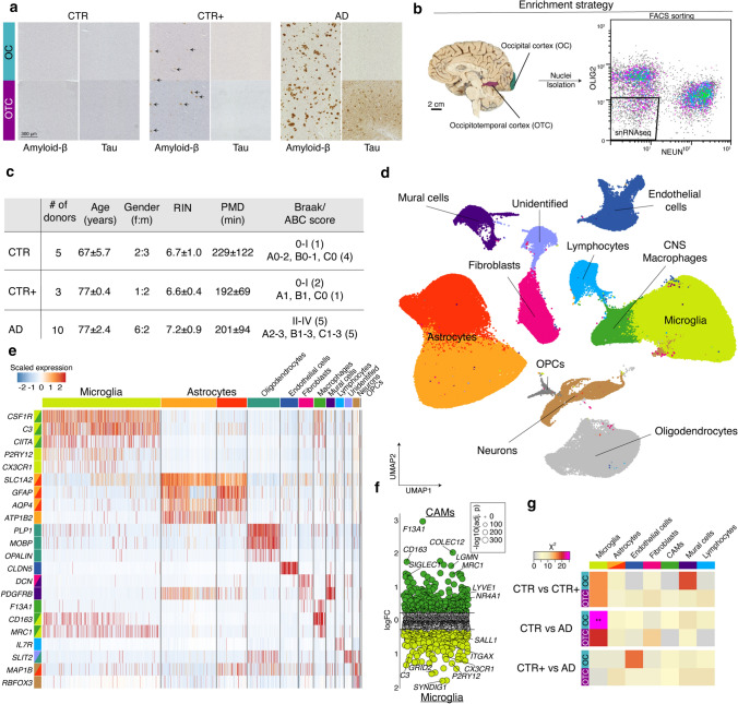
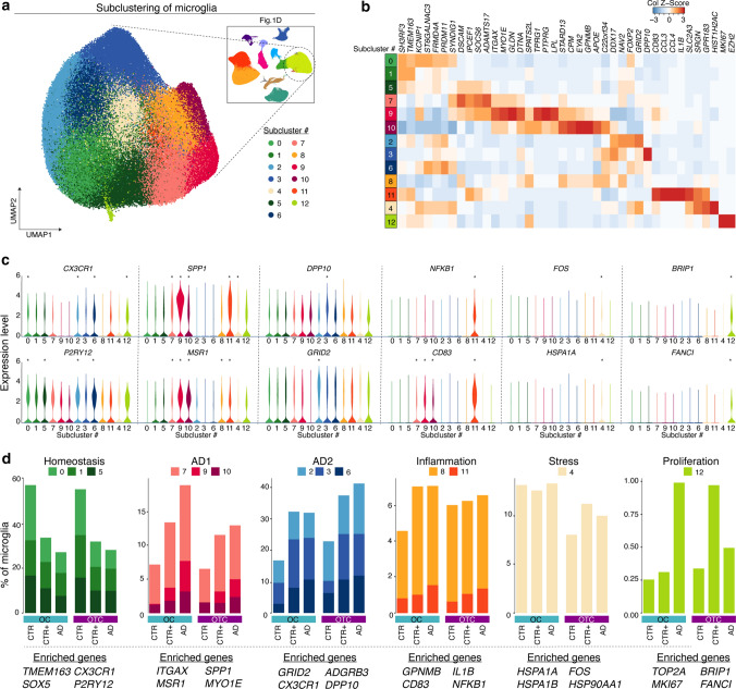
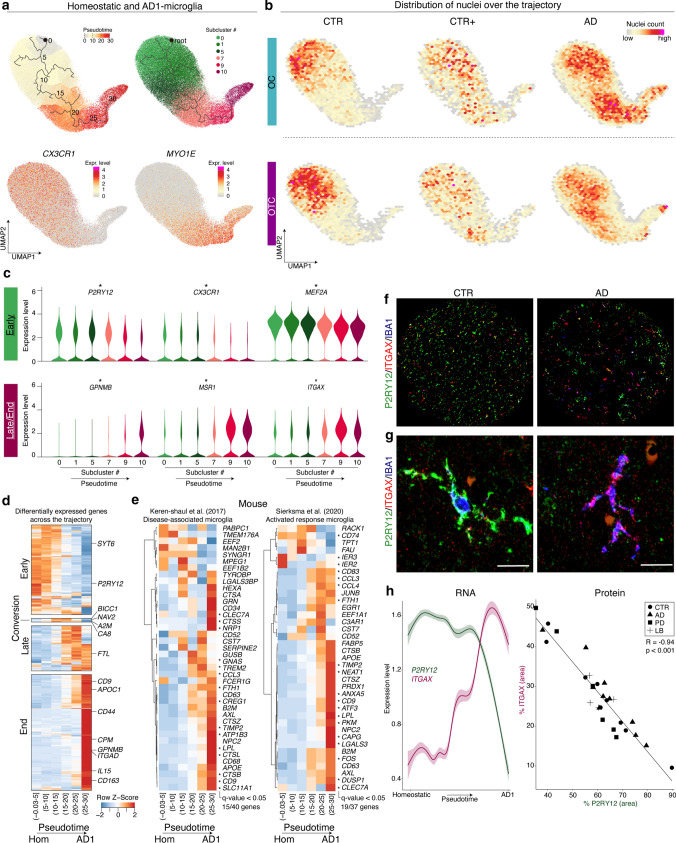
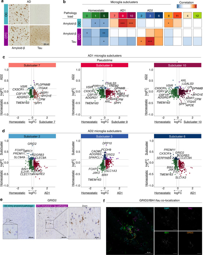
Alzheimer’s disease (AD) is the most prevalent form of dementia and is characterized by abnormal extracellular aggregates of amyloid-β and intraneuronal hyperphosphorylated tau tangles and neuropil threads. Microglia, the tissue-resident macrophages of the central nervous system (CNS), are important for CNS homeostasis and implicated in AD pathology. In amyloid mouse models, a phagocytic/activated microglia phenotype has been identified. How increasing levels of amyloid-β and tau pathology affect human microglia transcriptional profiles is unknown. Here, we performed snRNAseq on 482,472 nuclei from non-demented control brains and AD brains containing only amyloid-β plaques or both amyloid-β plaques and tau pathology. Within the microglia population, distinct expression profiles were identified of which two were AD pathology-associated. The phagocytic/activated AD1-microglia population abundance strongly correlated with tissue amyloid-β load and localized to amyloid-β plaques. The AD2-microglia abundance strongly correlated with tissue phospho-tau load and these microglia were more abundant in samples with overt tau pathology. This full characterization of human disease-associated microglia phenotypes provides new insights in the pathophysiological role of microglia in AD and offers new targets for microglia-state-specific therapeutic strategies.
Overview
The claims and statistics of this paper haven’t been checked yet.
Run the check — free, no account needed. It reads the open-access text and checks the sources it cites.
Integrity signals
Registry
Cites
Statistics
Figures
Verification
Claims the paper makes
The claims and statistics of this paper haven’t been checked yet.
Run the check — free, no account needed. It reads the open-access text and checks the sources it cites.
Claims other papers make about this one
Each is checked against this paper’s own text: does it say what the citing paper says it does?
The source backs part of the claim, not all of it — or backs it for a different population, condition or comparison.
In the citing paper
“a population of clusters, namely AD1, closely reassembled the mouse DAM/MGnD phenotype, it was only abundant in human samples containing Aβ and not tau”
From Microglia and monocytes in inflammatory CNS disease: integrating phenotype and function (2022) · cites this paper as [119]
Experimental — accuracy not yet measuredThe source backs part of the claim, not all of it — or backs it for a different population, condition or comparison.
In the citing paper
“Together this work demonstrates, firstly, that there exist other microglial subsets in AD besides the DAM/MGnD phenotype, and secondly, that human microglia are heterogenous, thus limiting the translation of the DAM signature from animal models.”
From Microglia and monocytes in inflammatory CNS disease: integrating phenotype and function (2022) · cites this paper as [119]
Experimental — accuracy not yet measuredThe source backs part of the claim, not all of it — or backs it for a different population, condition or comparison.
In the citing paper
“These findings were not formally published, however two recent reports suggest the presence of B cells in the brains of patients with AD [81,82].”
From Is there a role for B cells in Alzheimer's disease? (2026) · cites this paper as [82]
Experimental — accuracy not yet measuredThe source backs part of the claim, not all of it — or backs it for a different population, condition or comparison.
In the citing paper
“A study using singlenucleus RNA sequencing identified a cluster of lymphocytes in AD brains enriched for the expression of IL-7R and MS4A1 genes, which encode the interleukin-7 receptor and CD20 respectively, both estab lished markers of B lymphocytes [82].”
From Is there a role for B cells in Alzheimer's disease? (2026) · cites this paper as [82]
Experimental — accuracy not yet measuredThe cited source says what the claim attributes to it.
In the citing paper
“There was considerable overlap between the Micro_D profile and previously reported microglial states, such as the MIMS-foamy state reported in ref. 11, amyloid-associated microglia in Alzheimer’s disease43,46,47 and lipid-associated macrophages (LAMs) in obesity48 (Extended Data Fig. 8).”
From Foamy microglia link oxylipins to disease progression in multiple sclerosis (2026) · cites this paper as [46]
The cited source says what the claim attributes to it.
In the citing paper
“The foamy, GPNMB+ microglial phenotype in MS lesions bears striking transcriptional similarity to LAMs in obesity, amyloid-responsive microglia in Alzheimer’s disease and foam cells in atherosclerosis18,46,48,50,51.”
From Foamy microglia link oxylipins to disease progression in multiple sclerosis (2026) · cites this paper as [46]
The cited source says what the claim attributes to it.
In the citing paper
“single nuclei RNA sequencing on AD brains showed that out of seven cell types, gene expression changes were shown only in microglia [119]”
From Microglia and monocytes in inflammatory CNS disease: integrating phenotype and function (2022) · cites this paper as [119]
The cited source says what the claim attributes to it.
In the citing paper
“AD2 did not overlap with AD1 and was more frequently observed in human brain samples showing both tau and amyloid pathology [119]”
From Microglia and monocytes in inflammatory CNS disease: integrating phenotype and function (2022) · cites this paper as [119]
The cited source says what the claim attributes to it.
In the citing paper
“single cell nuclei extraction from frozen human brain tissue revealed 13 distinct microglial clusters [119]”
From Microglia and monocytes in inflammatory CNS disease: integrating phenotype and function (2022) · cites this paper as [119]
In the citing paper
“Moreover, distinct Aβ and tau-associated microglia signatures have been discovered in AD patients.”
From Microglia in neurodegenerative diseases: mechanism and potential therapeutic targets (2023) · cites this paper as [15]
Self-check
Percentages, sums and reported differences re-computed from the paper’s own numbers.
The self-check needs the paper’s full text.
Citations
Cites
Moderated estimation of fold change and dispersion for RNA-seq data with DESeq2
Love, M. I., Huber, W., & Anders, S. (2014) · Genome Biology · doi:10.1186/s13059-014-0550-8 · Cited by 95,106
Robinson, M. D., McCarthy, D. J., & Smyth, G. K. (2010) · Bioinformatics · doi:10.1093/bioinformatics/btp616 · Cited by 41,364
HTSeq—a Python framework to work with high-throughput sequencing data
Anders, S., Pyl, P. T., & Huber, W. (2015) · Bioinformatics · doi:10.1093/bioinformatics/btu638 · Cited by 21,464
Neuropathological stageing of Alzheimer-related changes.
Braak, H., & Braak, E. (1991) · Acta Neuropathologica · doi:10.1007/bf00308809 · Cited by 16,123
Comprehensive Integration of Single-Cell Data
Stuart, T., Butler, A., Hoffman, P., Hafemeister, C., Papalexi, E., Mauck, W. M., Hao, Y., Stoeckius, M., Smibert, P., & Satija, R. (2019) · Cell · doi:10.1016/j.cell.2019.05.031 · Cited by 15,453
A Unique Microglia Type Associated with Restricting Development of Alzheimer’s Disease
Keren-Shaul, H., Spinrad, A., Weiner, A., Matcovitch-Natan, O., Dvir-Szternfeld, R., Ulland, T. K., David, E., Baruch, K., Lara-Astaiso, D., Toth, B., et al. (2017) · Cell · doi:10.1016/j.cell.2017.05.018 · Cited by 5,493
Reversed graph embedding resolves complex single-cell trajectories
Qiu, X., Mao, Q., Tang, Y., Wang, L., Chawla, R., Pliner, H. A., & Trapnell, C. (2017) · Nature Methods · doi:10.1038/nmeth.4402 · Cited by 5,070
Buniello, A., MacArthur, J. A. L., Cerezo, M., Harris, L. W., Hayhurst, J., Malangone, C., McMahon, A., Morales, J., Mountjoy, E., Sollis, E., et al. (2019) · Nucleic Acids Research · doi:10.1093/nar/gky1120 · Cited by 4,000
Complement and microglia mediate early synapse loss in Alzheimer mouse models
Hong, S., Beja-Glasser, V. F., Nfonoyim, B. M., Frouin, A., Li, S., Ramakrishnan, S., Merry, K. M., Shi, Q., Rosenthal, A., Barres, B. A., et al. (2016) · Science · doi:10.1126/science.aad8373 · Cited by 3,343
Braak, H., Alafuzoff, I., Arzberger, T., Kretzschmar, H., & Del Tredici, K. (2006) · Acta Neuropathologica · doi:10.1007/s00401-006-0127-z · Cited by 3,172
Cited by
Sorted by citations
Microglia states and nomenclature: A field at its crossroads
Paolicelli, R. C., Sierra, A., Stevens, B., Tremblay, M.-E., Aguzzi, A., Ajami, B., Amit, I., Audinat, E., Bechmann, I., Bennett, M., et al. (2022) · Neuron · doi:10.1016/j.neuron.2022.10.020 · Cited by 1,973
Self-citationMicroglia in neurodegenerative diseases: mechanism and potential therapeutic targets
Gao, C., Jiang, J., Tan, Y., & Chen, S. (2023) · Signal Transduction and Targeted Therapy · doi:10.1038/s41392-023-01588-0 · Cited by 1,616
Microbiota–gut–brain axis and its therapeutic applications in neurodegenerative diseases
Loh, J. S., Mak, W. Q., Tan, L. K. S., Ng, C. X., Chan, H. H., Yeow, S. H., Foo, J. B., Ong, Y. S., How, C. W., & Khaw, K. Y. (2024) · Signal Transduction and Targeted Therapy · doi:10.1038/s41392-024-01743-1 · Cited by 1,193
Microglia: Immune and non-immune functions
Borst, K., Dumas, A. A., & Prinz, M. (2021) · Immunity · doi:10.1016/j.immuni.2021.09.014 · Cited by 804
hdWGCNA identifies co-expression networks in high-dimensional transcriptomics data
Morabito, S., Reese, F., Rahimzadeh, N., Miyoshi, E., & Swarup, V. (2023) · Cell Reports Methods · doi:10.1016/j.crmeth.2023.100498 · Cited by 773
Metadata
- Authors
- Gerrits, E., Brouwer, N., Kooistra, S. M., Woodbury, M. E., Vermeiren, Y., Lambourne, M., Mulder, J., Kummer, M., Möller, T., Biber, K., Dunnen, W. F. A. D., De Deyn, P. P., Eggen, B. J. L., & Boddeke, E. W. G. M.
- Affiliations
- University Medical Center Groningen (NL)
- AbbVie (United States) (US)
- Karolinska Institutet (SE)
- AbbVie (Germany) (DE)
- University of Copenhagen (DK)
- Corresponding author
- Boddeke, Erik W. G. M.
- ORCID
- Venue
- Acta Neuropathologica
- Published
- 2021-02-20
- Volume
- 141
- Issue
- 5
- Pages
- 681-696
- ISSN
- 0001-6322 (ISSN-L)
- 1432-0533
- PMC
- PMC8043951
- OpenAlex
- W3129453843
- Cited by
- 441
- References
- 42
- Citations by year
- 2026: 69 · 2025: 80 · 2024: 124 · 2023: 53 · 2022: 87 · 2021: 27
- FWCI
- 30.05
- Type
- journal article
- Language
- en
- Open access
- hybrid
- doi.org
- Locations
- Acta Neuropathologica (publishedVersion, cc-by)
- PubMed Central (publishedVersion, cc-by)
- PubMed (publishedVersion)
- Research at the University of Copenhagen (University of Copenhagen) (publishedVersion, cc-by)
- University of Groningen research database (University of Groningen / Centre for Information Technology) (publishedVersion, cc-by)
- Socio-Environmental Systems Modeling (acceptedVersion, cc-by)
- University of Groningen research database (University of Groningen / Centre for Information Technology) (submittedVersion, cc-by)
- Data Archiving and Networked Services (DANS) (submittedVersion, cc-by)
- Funders
- Rijksuniversiteit Groningen
- Stichting MS Research
- Fonds Wetenschappelijk Onderzoek (12Z1620N)
- Universitair Medisch Centrum Groningen
- Vlaamse Instelling voor Technologisch Onderzoek
- Alzheimer Nederland
- Vlaamse regering
- Topics
- Life Sciences › Neuroscience › Neurology › Neuroinflammation and Neurodegeneration Mechanisms
- Life Sciences › Neuroscience › Cellular and Molecular Neuroscience › Alzheimer's disease research and treatments
- Health Sciences › Medicine › Psychiatry and Mental health › Dementia and Cognitive Impairment Research
- Keywords
- Alzheimer's disease, microglia, amyloid plaques, tau pathology, microglial activation, microglial phenotype, single-nucleus RNA sequencing, phosphorylated tau, disease-associated microglia, microglia-targeted therapy
- MeSH terms
- Aged, Aged, 80 and over, Alzheimer Disease, Brain, Female, Humans, Male, Amyloid beta-Peptides, tau Proteins, Microglia
About these checks
Registry, retraction, Cites and statistics results are computed, not judged, with one exception: a printed reference that no index lists is read once by a model before it counts as a citation error. Claims are extracted by a model and kept only when their quote appears verbatim in the text; each cited source is then checked, with a model’s help, for whether it says what the claim relies on. Those verdicts can be wrong — read the claim’s quote and the cited source before relying on one.
Statistics and arithmetic are recomputed, not judged.
Confidence bands are shown only for fields calibrated on our annotated test set. None is calibrated yet, so no band is shown.
What counts as a claim ›How we measure confidence ›How we check this ›
Are you an author, or do you see a problem with this page? Request a review
