

The DOI matches its Crossref record; 44 of the 44 references it cites were found in a registry or an index.
Distinct amyloid-β and tau-associated microglia profiles in Alzheimer’s disease
Open accessAbstract
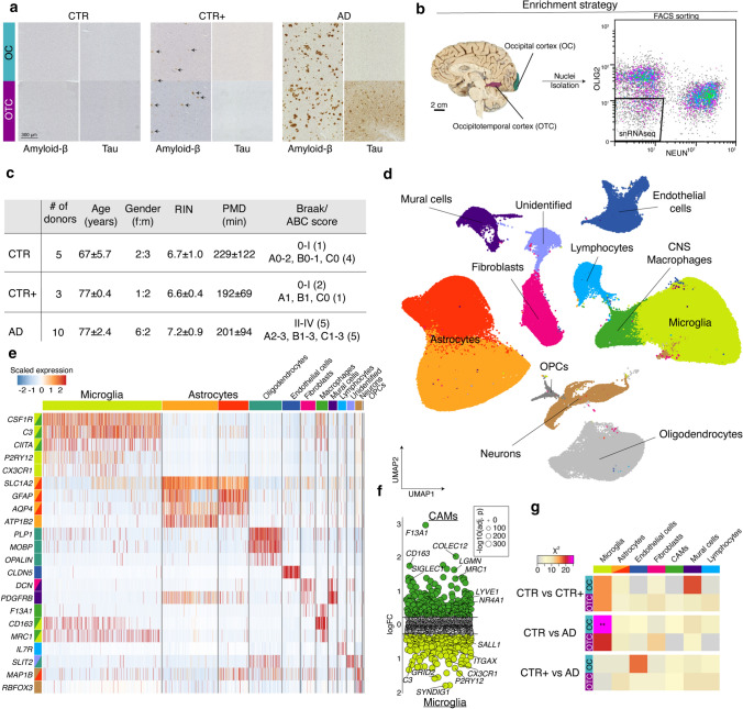
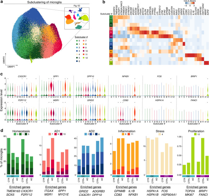
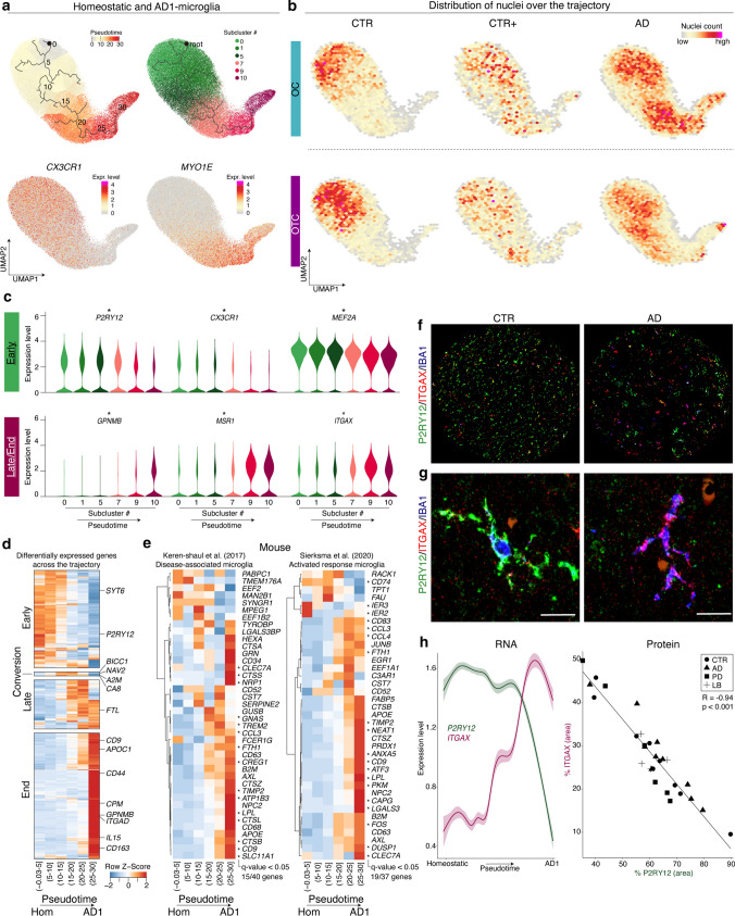
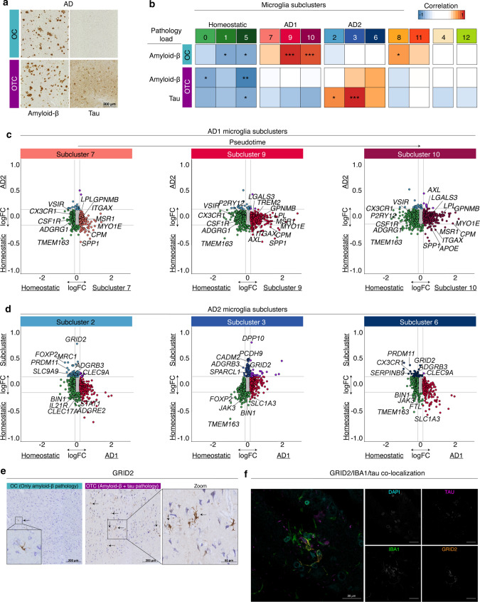
Alzheimer’s disease (AD) is the most prevalent form of dementia and is characterized by abnormal extracellular aggregates of amyloid-β and intraneuronal hyperphosphorylated tau tangles and neuropil threads. Microglia, the tissue-resident macrophages of the central nervous system (CNS), are important for CNS homeostasis and implicated in AD pathology. In amyloid mouse models, a phagocytic/activated microglia phenotype has been identified. How increasing levels of amyloid-β and tau pathology affect human microglia transcriptional profiles is unknown. Here, we performed snRNAseq on 482,472 nuclei from non-demented control brains and AD brains containing only amyloid-β plaques or both amyloid-β plaques and tau pathology. Within the microglia population, distinct expression profiles were identified of which two were AD pathology-associated. The phagocytic/activated AD1-microglia population abundance strongly correlated with tissue amyloid-β load and localized to amyloid-β plaques. The AD2-microglia abundance strongly correlated with tissue phospho-tau load and these microglia were more abundant in samples with overt tau pathology. This full characterization of human disease-associated microglia phenotypes provides new insights in the pathophysiological role of microglia in AD and offers new targets for microglia-state-specific therapeutic strategies.
Overview
The claims and statistics of this paper haven’t been checked yet.
Run the check — free, no account needed. It reads the open-access text and checks the sources it cites.
Integrity signals
Registry
Cites
Statistics
Figures
Verification
Claims the paper makes
The claims and statistics of this paper haven’t been checked yet.
Run the check — free, no account needed. It reads the open-access text and checks the sources it cites.
Claims other papers make about this one
Each is checked against this paper’s own text: does it say what the citing paper says it does?
The source backs part of the claim, not all of it — or backs it for a different population, condition or comparison.
In the citing paper
“a population of clusters, namely AD1, closely reassembled the mouse DAM/MGnD phenotype, it was only abundant in human samples containing Aβ and not tau”
From Microglia and monocytes in inflammatory CNS disease: integrating phenotype and function (2022) · cites this paper as [119]
Experimental — accuracy not yet measuredThe source backs part of the claim, not all of it — or backs it for a different population, condition or comparison.
In the citing paper
“Together this work demonstrates, firstly, that there exist other microglial subsets in AD besides the DAM/MGnD phenotype, and secondly, that human microglia are heterogenous, thus limiting the translation of the DAM signature from animal models.”
From Microglia and monocytes in inflammatory CNS disease: integrating phenotype and function (2022) · cites this paper as [119]
Experimental — accuracy not yet measuredThe source backs part of the claim, not all of it — or backs it for a different population, condition or comparison.
In the citing paper
“These findings were not formally published, however two recent reports suggest the presence of B cells in the brains of patients with AD [81,82].”
From Is there a role for B cells in Alzheimer's disease? (2026) · cites this paper as [82]
Experimental — accuracy not yet measuredThe source backs part of the claim, not all of it — or backs it for a different population, condition or comparison.
In the citing paper
“A study using singlenucleus RNA sequencing identified a cluster of lymphocytes in AD brains enriched for the expression of IL-7R and MS4A1 genes, which encode the interleukin-7 receptor and CD20 respectively, both estab lished markers of B lymphocytes [82].”
From Is there a role for B cells in Alzheimer's disease? (2026) · cites this paper as [82]
Experimental — accuracy not yet measuredThe cited source says what the claim attributes to it.
In the citing paper
“There was considerable overlap between the Micro_D profile and previously reported microglial states, such as the MIMS-foamy state reported in ref. 11, amyloid-associated microglia in Alzheimer’s disease43,46,47 and lipid-associated macrophages (LAMs) in obesity48 (Extended Data Fig. 8).”
From Foamy microglia link oxylipins to disease progression in multiple sclerosis (2026) · cites this paper as [46]
The cited source says what the claim attributes to it.
In the citing paper
“The foamy, GPNMB+ microglial phenotype in MS lesions bears striking transcriptional similarity to LAMs in obesity, amyloid-responsive microglia in Alzheimer’s disease and foam cells in atherosclerosis18,46,48,50,51.”
From Foamy microglia link oxylipins to disease progression in multiple sclerosis (2026) · cites this paper as [46]
The cited source says what the claim attributes to it.
In the citing paper
“single nuclei RNA sequencing on AD brains showed that out of seven cell types, gene expression changes were shown only in microglia [119]”
From Microglia and monocytes in inflammatory CNS disease: integrating phenotype and function (2022) · cites this paper as [119]
The cited source says what the claim attributes to it.
In the citing paper
“AD2 did not overlap with AD1 and was more frequently observed in human brain samples showing both tau and amyloid pathology [119]”
From Microglia and monocytes in inflammatory CNS disease: integrating phenotype and function (2022) · cites this paper as [119]
The cited source says what the claim attributes to it.
In the citing paper
“single cell nuclei extraction from frozen human brain tissue revealed 13 distinct microglial clusters [119]”
From Microglia and monocytes in inflammatory CNS disease: integrating phenotype and function (2022) · cites this paper as [119]
In the citing paper
“Moreover, distinct Aβ and tau-associated microglia signatures have been discovered in AD patients.”
From Microglia in neurodegenerative diseases: mechanism and potential therapeutic targets (2023) · cites this paper as [15]
Self-check
Percentages, sums and reported differences re-computed from the paper’s own numbers.
The self-check needs the paper’s full text.
Citations
Cites
Moderated estimation of fold change and dispersion for RNA-seq data with DESeq2
Love, M. I., Huber, W., & Anders, S. (2014) · Genome Biology · doi:10.1186/s13059-014-0550-8 · Cited by 95,106
Robinson, M. D., McCarthy, D. J., & Smyth, G. K. (2010) · Bioinformatics · doi:10.1093/bioinformatics/btp616 · Cited by 41,364
HTSeq—a Python framework to work with high-throughput sequencing data
Anders, S., Pyl, P. T., & Huber, W. (2015) · Bioinformatics · doi:10.1093/bioinformatics/btu638 · Cited by 21,464
Neuropathological stageing of Alzheimer-related changes.
Braak, H., & Braak, E. (1991) · Acta Neuropathologica · doi:10.1007/bf00308809 · Cited by 16,123
Comprehensive Integration of Single-Cell Data
Stuart, T., Butler, A., Hoffman, P., Hafemeister, C., Papalexi, E., Mauck, W. M., Hao, Y., Stoeckius, M., Smibert, P., & Satija, R. (2019) · Cell · doi:10.1016/j.cell.2019.05.031 · Cited by 15,453
A Unique Microglia Type Associated with Restricting Development of Alzheimer’s Disease
Keren-Shaul, H., Spinrad, A., Weiner, A., Matcovitch-Natan, O., Dvir-Szternfeld, R., Ulland, T. K., David, E., Baruch, K., Lara-Astaiso, D., Toth, B., et al. (2017) · Cell · doi:10.1016/j.cell.2017.05.018 · Cited by 5,493
Reversed graph embedding resolves complex single-cell trajectories
Qiu, X., Mao, Q., Tang, Y., Wang, L., Chawla, R., Pliner, H. A., & Trapnell, C. (2017) · Nature Methods · doi:10.1038/nmeth.4402 · Cited by 5,070
Buniello, A., MacArthur, J. A. L., Cerezo, M., Harris, L. W., Hayhurst, J., Malangone, C., McMahon, A., Morales, J., Mountjoy, E., Sollis, E., et al. (2019) · Nucleic Acids Research · doi:10.1093/nar/gky1120 · Cited by 4,000
Complement and microglia mediate early synapse loss in Alzheimer mouse models
Hong, S., Beja-Glasser, V. F., Nfonoyim, B. M., Frouin, A., Li, S., Ramakrishnan, S., Merry, K. M., Shi, Q., Rosenthal, A., Barres, B. A., et al. (2016) · Science · doi:10.1126/science.aad8373 · Cited by 3,343
Braak, H., Alafuzoff, I., Arzberger, T., Kretzschmar, H., & Del Tredici, K. (2006) · Acta Neuropathologica · doi:10.1007/s00401-006-0127-z · Cited by 3,172
Cited by
Sorted by citations
Microglia states and nomenclature: A field at its crossroads
Paolicelli, R. C., Sierra, A., Stevens, B., Tremblay, M.-E., Aguzzi, A., Ajami, B., Amit, I., Audinat, E., Bechmann, I., Bennett, M., et al. (2022) · Neuron · doi:10.1016/j.neuron.2022.10.020 · Cited by 1,973
Self-citationMicroglia in neurodegenerative diseases: mechanism and potential therapeutic targets
Gao, C., Jiang, J., Tan, Y., & Chen, S. (2023) · Signal Transduction and Targeted Therapy · doi:10.1038/s41392-023-01588-0 · Cited by 1,616
Microbiota–gut–brain axis and its therapeutic applications in neurodegenerative diseases
Loh, J. S., Mak, W. Q., Tan, L. K. S., Ng, C. X., Chan, H. H., Yeow, S. H., Foo, J. B., Ong, Y. S., How, C. W., & Khaw, K. Y. (2024) · Signal Transduction and Targeted Therapy · doi:10.1038/s41392-024-01743-1 · Cited by 1,193
Microglia: Immune and non-immune functions
Borst, K., Dumas, A. A., & Prinz, M. (2021) · Immunity · doi:10.1016/j.immuni.2021.09.014 · Cited by 804
hdWGCNA identifies co-expression networks in high-dimensional transcriptomics data
Morabito, S., Reese, F., Rahimzadeh, N., Miyoshi, E., & Swarup, V. (2023) · Cell Reports Methods · doi:10.1016/j.crmeth.2023.100498 · Cited by 773
ApoE in Alzheimer’s disease: pathophysiology and therapeutic strategies
Raulin, A.-C., Doss, S. V., Trottier, Z. A., Ikezu, T. C., Bu, G., & Liu, C.-C. (2022) · Molecular Neurodegeneration · doi:10.1186/s13024-022-00574-4 · Cited by 756
Microglia in Neuroinflammation and Neurodegeneration: From Understanding to Therapy
Muzio, L., Viotti, A., & Martino, G. (2021) · Frontiers in Neuroscience · doi:10.3389/fnins.2021.742065 · Cited by 581
Kamath, T., Abdulraouf, A., Burris, S. J., Langlieb, J., Gazestani, V., Nadaf, N. M., Balderrama, K., Vanderburg, C., & Macosko, E. Z. (2022) · Nature Neuroscience · doi:10.1038/s41593-022-01061-1 · Cited by 552
Human microglial state dynamics in Alzheimer’s disease progression
Sun, N., Victor, M. B., Park, Y. P., Xiong, X., Scannail, A. N., Leary, N., Prosper, S., Viswanathan, S., Luna, X., Boix, C. A., et al. (2023) · Cell · doi:10.1016/j.cell.2023.08.037 · Cited by 462
Sadick, J. S., O’Dea, M. R., Hasel, P., Dykstra, T., Faustin, A., & Liddelow, S. A. (2022) · Neuron · doi:10.1016/j.neuron.2022.03.008 · Cited by 415
Integrated multimodal cell atlas of Alzheimer’s disease
Gabitto, M. I., Travaglini, K. J., Rachleff, V. M., Kaplan, E. S., Long, B., Ariza, J., Ding, Y., Mahoney, J. T., Dee, N., Goldy, J., et al. (2024) · Nature Neuroscience · doi:10.1038/s41593-024-01774-5 · Cited by 397
Microglia in Alzheimer's disease at single-cell level. Are there common patterns in humans and mice?
Chen, Y., & Colonna, M. (2021) · Journal of Experimental Medicine · doi:10.1084/jem.20202717 · Cited by 339
The effects of microglia-associated neuroinflammation on Alzheimer’s disease
Wang, C., Zong, S., Cui, X., Wang, X., Wu, S., Wang, L., Liu, Y., & Lu, Z. (2023) · Frontiers in Immunology · doi:10.3389/fimmu.2023.1117172 · Cited by 331
Three-Dimensional Cell Cultures: The Bridge between In Vitro and In Vivo Models
Urzì, O., Gasparro, R., Costanzo, E., Luca, A. D., Giavaresi, G., Fontana, S., & Alessandro, R. (2023) · International Journal of Molecular Sciences · doi:10.3390/ijms241512046 · Cited by 263
Mitochondrial dysfunction in microglia: a novel perspective for pathogenesis of Alzheimer’s disease
Li, Y., Xia, X., Wang, Y., & Zheng, J. (2022) · Journal of Neuroinflammation · doi:10.1186/s12974-022-02613-9 · Cited by 231
Microglia and monocytes in inflammatory CNS disease: integrating phenotype and function
Spiteri, A. G., Wishart, C. L., Pamphlett, R., Locatelli, G., & King, N. J. C. (2022) · Acta Neuropathologica · doi:10.1007/s00401-021-02384-2 · Cited by 216
Cell type‐specific roles of APOE4 in Alzheimer disease
Blumenfeld, J., Yip, O., Kim, M. J., & Huang, Y. (2024) · Nature Reviews Neuroscience · doi:10.1038/s41583-023-00776-9 · Cited by 202
Early Alzheimer’s disease pathology in human cortex involves transient cell states
Gazestani, V., Kamath, T., Nadaf, N. M., Dougalis, A., Burris, S. J., Rooney, B., Junkkari, A., Vanderburg, C., Pelkonen, A., Gomez-Budia, M., et al. (2023) · Cell · doi:10.1016/j.cell.2023.08.005 · Cited by 209
Diverse human astrocyte and microglial transcriptional responses to Alzheimer’s pathology
Smith, A. M., Davey, K., Tsartsalis, S., Khozoie, C., Fancy, N., Tang, S. S., Liaptsi, E., Weinert, M., McGarry, A., Muirhead, R. C. J., et al. (2022) · Acta Neuropathologica · doi:10.1007/s00401-021-02372-6 · Cited by 209
“We confirmed that transcripts positively differentially expressed in microglia were significantly enriched in nuclei with human AD pathology in previously published snRNASeq studies [16, 18, 36, 76] (Supplementary Table 3, online resource).”
BackgroundMethodResultInfluentialCellular communities reveal trajectories of brain ageing and Alzheimer’s disease
Green, G. S., Fujita, M., Yang, H.-S., Taga, M., Cain, A., McCabe, C., Comandante-Lou, N., White, C. C., Schmidtner, A. K., Zeng, L., et al. (2024) · Nature · doi:10.1038/s41586-024-07871-6 · Cited by 204
Autophagy enables microglia to engage amyloid plaques and prevents microglial senescence
Choi, I., Wang, M., Yoo, S., Xu, P., Seegobin, S. P., Li, X., Han, X., Wang, Q., Peng, J., Zhang, B., et al. (2023) · Nature Cell Biology · doi:10.1038/s41556-023-01158-0 · Cited by 202
Microglia regulation of central nervous system myelin health and regeneration
Kent, S. A., & Miron, V. E. (2024) · Nature Reviews Immunology · doi:10.1038/s41577-023-00907-4 · Cited by 183
Dolan, M.-J., Therrien, M., Jereb, S., Kamath, T., Gazestani, V., Atkeson, T., Marsh, S. E., Goeva, A., Lojek, N. M., Murphy, S., et al. (2023) · Nature Immunology · doi:10.1038/s41590-023-01558-2 · Cited by 185
Sabogal-Guáqueta, A. M., Marmolejo-Garza, A., Trombetta-Lima, M., Oun, A., Hunneman, J., Chen, T., Koıstınaho, J., Lehtonen, Š., Kortholt, A., Wolters, J. C., et al. (2023) · Nature Communications · doi:10.1038/s41467-023-42096-7 · Cited by 180
Self-citationA multi-pronged human microglia response to Alzheimer’s disease Aβ pathology
Mancuso, R., Fattorelli, N., Martinez-Muriana, A., Davis, E., Wolfs, L., Van Den Daele, J., Geric, I., Preman, P., Serneels, L., Poovathingal, S., et al. (2022) · doi:10.1101/2022.07.07.499139 · Cited by 169
Deregulated mitochondrial microRNAs in Alzheimer's disease: Focus on synapse and mitochondria
Gowda, P., Reddy, P. H., & Kumar, S. (2021) · Ageing Research Reviews · doi:10.1016/j.arr.2021.101529 · Cited by 170
Human Microglia Show Unique Transcriptional Changes in Alzheimer's Disease
Prater, K. E., Green, K. J., Mamde, S., Sun, W., Cochoit, A., Smith, C. L., Chiou, K. L., Heath, L., Rose, S. E., Wiley, J., et al. (2023) · Nature Aging · doi:10.1038/s43587-023-00424-y · Cited by 164
Microglial activation states and their implications for Alzheimer's Disease
Valiukas, Z., Tangalakis, K., Apostolopoulos, V., & Feehan, J. (2025) · The Journal of Prevention of Alzheimer s Disease · doi:10.1016/j.tjpad.2024.100013 · Cited by 167
Insights into Alzheimer’s disease from single-cell genomic approaches
Murdock, M. H., & Tsai, L.-H. (2023) · Nature Neuroscience · doi:10.1038/s41593-022-01222-2 · Cited by 148
Microglial mechanisms drive amyloid-β clearance in immunized patients with Alzheimer’s disease
van Olst, L., Simonton, B., Edwards, A. J., Forsyth, A. V., Boles, J. S., Jamshidi, P., Watson, T. H., Shepard, N., Krainc, T., Argue, B. M., et al. (2025) · Nature Medicine · doi:10.1038/s41591-025-03574-1 · Cited by 164
Astrocyte transcriptomic changes along the spatiotemporal progression of Alzheimer’s disease
Serrano-Pozo, A., Li, H., Li, Z., Muñoz-Castro, C., Jaisa-aad, M., Healey, M. A., Welikovitch, L. A., Jayakumar, R., Bryant, A. G., Noori, A., et al. (2024) · Nature Neuroscience · doi:10.1038/s41593-024-01791-4 · Cited by 140
Self-citationMartins-Ferreira, R., Calafell-Segura, J., Leal, B., Rodríguez-Ubreva, J., Martínez-Saez, E., Mereu, E., Pinho E Costa, P., Laguna, A., & Ballestar, E. (2025) · Nature Communications · doi:10.1038/s41467-025-56124-1 · Cited by 123
MicroRNAs: pioneering regulators in Alzheimer’s disease pathogenesis, diagnosis, and therapy
Li, Y.-B., Fu, Q., Guo, M., Du, Y., Chen, Y., & Cheng, Y. (2024) · Translational Psychiatry · doi:10.1038/s41398-024-03075-8 · Cited by 134
Yin, Z., Herron, S., Silveira, S., Kleemann, K., Gauthier, C., Mallah, D., Cheng, Y., Margeta, M. A., Pitts, K. M., Barry, J.-L., et al. (2023) · Nature Neuroscience · doi:10.1038/s41593-023-01355-y · Cited by 120
Puntambekar, S. S., Moutinho, M., Lin, P. B.-C., Jadhav, V., Tumbleson-Brink, D., Balaji, A., Benito, M. A., Xu, G., Oblak, A., Lasagna-Reeves, C. A., et al. (2022) · Molecular Neurodegeneration · doi:10.1186/s13024-022-00545-9 · Cited by 113
Kapoor, M., & Chinnathambi, S. (2023) · Journal of Neuroinflammation · doi:10.1186/s12974-023-02751-8 · Cited by 114
Wang, M., Song, W., Chen, M., Wang, Q., Zhou, X., Xu, P., Krek, A., Yoon, Y., Ho, L., Orr, M. E., et al. (2022) · Molecular Neurodegeneration · doi:10.1186/s13024-022-00517-z · Cited by 104
A neurodegenerative cellular stress response linked to dark microglia and toxic lipid secretion
Flury, A. G., Aljayousi, L., Park, H.-J., Khakpour, M., Mechler, J., Aziz, S., McGrath, J. D., Deme, P., Sandberg, C., Ibáñez, F. G., et al. (2024) · Neuron · doi:10.1016/j.neuron.2024.11.018 · Cited by 102
Tsartsalis, S., Sleven, H., Fancy, N., Wessely, F., Smith, A. M., Willumsen, N., Cheung, T. K. D., Rokicki, M. J., Chau, V., Ifie, E., et al. (2024) · Nature Communications · doi:10.1038/s41467-024-46630-z · Cited by 95
Apolipoprotein E in Alzheimer’s disease: molecular insights and therapeutic opportunities
Belaidi, A. A., Bush, A. I., & Ayton, S. (2025) · Molecular Neurodegeneration · doi:10.1186/s13024-025-00843-y · Cited by 91
Ostkamp, P., Deffner, M., Schulte‐Mecklenbeck, A., Wünsch, C., Lu, I., Wu, G. F., Goelz, S. E., Jager, P. L. D., Kuhlmann, T., Groß, C. C., et al. (2022) · Science Translational Medicine · doi:10.1126/scitranslmed.adc9778 · Cited by 93
The emerging tale of microglia in psychiatric disorders
Rahimian, R., Wakid, M., O’Leary, L. A., & Mechawar, N. (2021) · Neuroscience & Biobehavioral Reviews · doi:10.1016/j.neubiorev.2021.09.023 · Cited by 96
White matter microglia heterogeneity in the CNS
Amor, S., McNamara, N. B., Gerrits, E., Marzin, M. C., Kooistra, S. M., Miron, V. E., & Nutma, E. (2022) · Acta Neuropathologica · doi:10.1007/s00401-021-02389-x · Cited by 89
Self-citationMicroglia heterogeneity, modeling and cell-state annotation in development and neurodegeneration
Fumagalli, L., Nazlie Mohebiany, A., Premereur, J., Polanco Miquel, P., Bijnens, B., Van de Walle, P., Fattorelli, N., & Mancuso, R. (2025) · Nature Neuroscience · doi:10.1038/s41593-025-01931-4 · Cited by 90
Noteworthy perspectives on microglia in neuropsychiatric disorders
Zhu, H., Guan, A., Liu, J., Li, P., Zhang, Z., & Wang, S. (2023) · Journal of Neuroinflammation · doi:10.1186/s12974-023-02901-y · Cited by 91
Rosenzweig, N., Kleemann, K. L., Rust, T., Carpenter, M., Grucci, M., Aronchik, M., Brouwer, N., Valenbreder, I., Cooper-Hohn, J., Iyer, M., et al. (2024) · Nature Medicine · doi:10.1038/s41591-024-03122-3 · Cited by 90
Self-citationGerrits, E., Giannini, L. A. A., Brouwer, N., Melhem, S., Seilhean, D., Le Ber, I., The Brainbank Neuro-CEB Neuropathology Network, Kamermans, A., Kooij, G., de Vries, H. E., et al. (2022) · Nature Neuroscience · doi:10.1038/s41593-022-01124-3 · Cited by 88
Self-citationMicroglia transcriptional states and their functional significance: Context drives diversity
Depp, C., Doman, J. L., Hingerl, M., Xia, J., & Stevens, B. (2025) · Immunity · doi:10.1016/j.immuni.2025.04.009 · Cited by 84
Bryant, A. G., Li, Z., Jayakumar, R., Serrano‐Pozo, A., Woost, B., Hu, M., Woodbury, M. E., Wachter, A., Lin, G., Kwon, T., et al. (2023) · Journal of Neuroscience · doi:10.1523/jneurosci.0237-23.2023 · Cited by 84
Self-citationTuddenham, J. F., Taga, M. F., Haage, V. C., Marshe, V. S., Roostaei, T., White, C. C., Lee, A., Fujita, M., Khairallah, A., Zhang, Y., et al. (2024) · Nature Neuroscience · doi:10.1038/s41593-024-01764-7 · Cited by 79
Immune Activation in Alzheimer Disease
Mary, A., Mancuso, R., & Heneka, M. T. (2024) · Annual Review of Immunology · doi:10.1146/annurev-immunol-101921-035222 · Cited by 79
The link between amyloid β and ferroptosis pathway in Alzheimer’s disease progression
Majerníková, N., Marmolejo-Garza, A., Salinas, C. S., Luu, M. D. A., Zhang, Y., Trombetta-Lima, M., Tomin, T., Birner-Gruenberger, R., Lehtonen, Š., Koistinaho, J., et al. (2024) · Cell Death & Disease · doi:10.1038/s41419-024-07152-0 · Cited by 73
Self-citationLodder, C., Scheyltjens, I., Stancu, I., Lucena, P. B., de Ravé, M. G., Vanherle, S., Vanmierlo, T., Cremers, N., Vanrusselt, H., Brône, B., et al. (2021) · Acta Neuropathologica Communications · doi:10.1186/s40478-021-01204-8 · Cited by 71
Seeker, L. A., Bestard-Cuche, N., Jäkel, S., Kazakou, N.-L., Bøstrand, S. M. K., Wagstaff, L. J., Cholewa-Waclaw, J., Kilpatrick, A. M., van Bruggen, D., Kabbe, M., et al. (2023) · Acta Neuropathologica Communications · doi:10.1186/s40478-023-01568-z · Cited by 70
Muñoz-Castro, C., Mejias-Ortega, M., Sanchez-Mejias, E., Navarro, V., Trujillo-Estrada, L., Jimenez, S., Garcia-Leon, J. A., Fernandez-Valenzuela, J. J., Sanchez-Mico, M. V., Romero-Molina, C., et al. (2023) · Acta Neuropathologica Communications · doi:10.1186/s40478-023-01530-z · Cited by 66
Kim, D. W., Tu, K. J., Wei, A., Lau, A., Gonzalez-Gil, A., Cao, T., Braunstein, K. E., Ling, J. P., Troncoso, J. C., Wong, P. C., et al. (2022) · Molecular Neurodegeneration · doi:10.1186/s13024-022-00589-x · Cited by 66
Miedema, A., Gerrits, E., Brouwer, N., Jiang, Q., Kracht, L., Meijer, M., Nutma, E., Peferoen-Baert, R., Pijnacker, A. T. E., Wesseling, E. M., et al. (2022) · Acta Neuropathologica Communications · doi:10.1186/s40478-021-01306-3 · Cited by 65
Self-citationXu, Y., Au, N. P. B., & Ma, C. H. E. (2022) · Frontiers in Aging Neuroscience · doi:10.3389/fnagi.2022.896852 · Cited by 68
Microglial Activation and Connectivity in Alzheimer Disease and Aging
Rauchmann, B., Brendel, M., Franzmeier, N., Trappmann, L., Zaganjori, M., Ersoezlue, E., Morenas‐Rodriguez, E., Guersel, S., Burow, L., Kurz, C., et al. (2022) · Annals of Neurology · doi:10.1002/ana.26465 · Cited by 61
“Within the microglia population, two distinct expression profiles were recently identified, associated with either Aβ load or tissue phosphorylated tau (ptau).(11) Colocalization of tau and activated microglia was demonstrated,(12,13) and Aβ plaque-dependent microglia was identified.”
Towards PET imaging of the dynamic phenotypes of microglia
Beaino, W., Janssen, B., Vugts, D. J., de Vries, H. E., & Windhorst, A. D. (2021) · Clinical & Experimental Immunology · doi:10.1111/cei.13649 · Cited by 63
Wang, Z., Liu, J., Han, J., Zhang, T., Li, S., Hou, Y., Su, H., Han, F., & Zhang, C. (2024) · Journal of Neuroinflammation · doi:10.1186/s12974-024-03166-9 · Cited by 60
Microglia Heterogeneity in Alzheimer’s Disease: Insights From Single-Cell Technologies
Wang, H. (2021) · Frontiers in Synaptic Neuroscience · doi:10.3389/fnsyn.2021.773590 · Cited by 62
Patel, T., Carnwath, T., Wang, X., Allen, M., Lincoln, S., Lewis‐Tuffin, L. J., Quicksall, Z., Lin, S., Tutor‐New, F. Q., Ho, C. C. G., et al. (2022) · Aging Cell · doi:10.1111/acel.13606 · Cited by 61
Cell type-specific changes identified by single-cell transcriptomics in Alzheimer’s disease
Luquez, T., Gaur, P., Kosater, I. M., Lam, M., Lee, D. I., Mares, J. A., Paryani, F., Yadav, A., & Menon, V. (2022) · Genome Medicine · doi:10.1186/s13073-022-01136-5 · Cited by 64
Majerníková, N., den Dunnen, W. F. A., & Dolga, A. M. (2021) · Frontiers in Aging Neuroscience · doi:10.3389/fnagi.2021.745046 · Cited by 56
Self-citationExperimental animal models of chronic inflammation
Rafiyan, M., Sadeghmousavi, S., Akbarzadehmoallemkolaei, M., & Rezaei, N. (2023) · Current Research in Immunology · doi:10.1016/j.crimmu.2023.100063 · Cited by 54
Ferroptosis mechanism and Alzheimer's disease
Feng, L., Sun, J. J., Xia, L., Shi, Q., Hou, Y., Zhang, L., Li, M., Fan, C., & Sun, B. (2023) · Neural Regeneration Research · doi:10.4103/1673-5374.389362 · Cited by 56
Podleśny-Drabiniok, A., Novikova, G., Liu, Y., Dunst, J., Temizer, R., Giannarelli, C., Marro, S., Kreslavsky, T., Marcora, E., & Goate, A. M. (2024) · Nature Communications · doi:10.1038/s41467-024-46315-7 · Cited by 52
Harnessing human iPSC-microglia for CNS-wide delivery of disease-modifying proteins
Chadarevian, J. P., Davtyan, H., Chadarevian, A. L., Nguyen, J., Capocchi, J. K., Le, L., Escobar, A., Chadarevian, T., Mansour, K., Deynega, E., et al. (2025) · Cell Stem Cell · doi:10.1016/j.stem.2025.03.009 · Cited by 60
Green, G. S., Yang, H., Fujita, M., Taga, M., Regev, A., Menon, V., Bennett, D. A. A., Habib, N., & De Jager, P. L. (2023) · Alzheimer's & Dementia · doi:10.1002/alz.083212 · Cited by 51
Neurodegeneration cell per cell
Balusu, S., Praschberger, R., Lauwers, E., Strooper, B. D., & Verstreken, P. (2023) · Neuron · doi:10.1016/j.neuron.2023.01.016 · Cited by 53
The multiple faces of extracellular vesicles released by microglia: Where are we 10 years after?
Gabrielli, M., Raffaele, S., Fumagalli, M., & Verderio, C. (2022) · Frontiers in Cellular Neuroscience · doi:10.3389/fncel.2022.984690 · Cited by 55
Tracking cell-type-specific temporal dynamics in human and mouse brains
Lu, Z., Zhang, M., Lee, J., Sziraki, A., Anderson, S., Zhang, Z., Xu, Z., Jiang, W., Ge, S., Nelson, P. T., et al. (2023) · Cell · doi:10.1016/j.cell.2023.08.042 · Cited by 51
Kuntić, M., Hahad, O., Münzel, T. F., & Daiber, A. (2024) · Antioxidants · doi:10.3390/antiox13030266 · Cited by 53
İş, Ö., Wang, X., Reddy, J. S., Min, Y., Yılmaz, E., Bhattarai, P., Patel, T., Bergman, J., Quicksall, Z., Heckman, M. G., et al. (2024) · Nature Communications · doi:10.1038/s41467-024-48926-6 · Cited by 55
Alzheimer’s disease-related transcriptional sex differences in myeloid cells
Coales, I., Tsartsalis, S., Fancy, N., Weinert, M., Clode, D., Owen, D., & Matthews, P. M. (2022) · Journal of Neuroinflammation · doi:10.1186/s12974-022-02604-w · Cited by 49
Repeated head trauma causes neuron loss and inflammation in young athletes
Butler, M. L. M. D., Pervaiz, N., Breen, K., Calderazzo, S. M., Ypsilantis, P., Wang, Y., Breda, J. C., Mazzilli, S. A., Nicks, R., Spurlock, E., et al. (2025) · Nature · doi:10.1038/s41586-025-09534-6 · Cited by 57
Microglia in Health and Diseases: Integrative Hubs of the Central Nervous System (CNS)
Sierra, A., Miron, V. E., Paolicelli, R. C., & Ransohoff, R. M. (2024) · Cold Spring Harbor Perspectives in Biology · doi:10.1101/cshperspect.a041366 · Cited by 47
Identification of distinct and age‐dependent p16High microglia subtypes
Talma, N., Gerrits, E., Wang, B., Eggen, B. J. L., & Demaria, M. (2021) · Aging Cell · doi:10.1111/acel.13450 · Cited by 46
Self-citationOverlapping Neuroimmune Mechanisms and Therapeutic Targets in Neurodegenerative Disorders
Marchi, F. D., Munitić, I., Vidatic, L., Papić, E., Rački, V., Nimac, J., Jurak, I., Novotni, G., Rogelj, B., Vuletić, V., et al. (2023) · Biomedicines · doi:10.3390/biomedicines11102793 · Cited by 47
Rao, A. T., Chen, N., Kim, M. J., Blumenfeld, J. R., Yip, O., Liang, Z., Shostak, D. M., Hao, Y., Nelson, M. R., Koutsodendris, N., et al. (2024) · Cell stem cell · doi:10.1016/j.stem.2024.10.005 · Cited by 48
Central Nervous System Macrophages in Health and Disease
Hove, H. V., Feo, D. D., Greter, M., & Becher, B. (2025) · Annual Review of Immunology · doi:10.1146/annurev-immunol-082423-041334 · Cited by 53
Das, S., Li, Z., Wachter, A., Alla, S., Noori, A., Abdourahman, A., Tamm, J. A., Woodbury, M. E., Talanian, R. V., Biber, K., et al. (2024) · Alzheimer's & Dementia · doi:10.1002/alz.13387 · Cited by 46
Self-citationShi, H., Yin, Z., Koronyo, Y., Fuchs, D.-T. S., Sheyn, J., Davis, M. R., Wilson, J. W., Margeta, M. A., Pitts, K. M., Herron, S., et al. (2022) · Acta Neuropathologica Communications · doi:10.1186/s40478-022-01439-z · Cited by 45
Zhang, P., Gao, C., Guo, Q., Yang, D., Zhang, G., Lü, H., Zhang, L., Zhang, G., & Li, D. (2024) · Journal of Neuroinflammation · doi:10.1186/s12974-024-03113-8 · Cited by 44
Gao, C., Shen, X., Tan, Y., & Chen, S. (2022) · Journal of Neuroinflammation · doi:10.1186/s12974-022-02580-1 · Cited by 48
An emerging role for microglia in stress‐effects on memory
Sanguino‐Gómez, J., Buurstede, J. C., Abiega, O., Fitzsimons, C. P., Lucassen, P. J., Eggen, B. J. L., Lesuis, S. L., Meijer, O. C., & Krugers, H. J. (2021) · European Journal of Neuroscience · doi:10.1111/ejn.15188 · Cited by 43
Self-citationBouvier, D. S., Fixemer, S., Heurtaux, T., Jeannelle, F., Frauenknecht, K. B. M., & Mittelbronn, M. G. A. (2022) · Frontiers in Physiology · doi:10.3389/fphys.2022.814889 · Cited by 43
Muñoz-Castro, C., Noori, A., Magdamo, C. G., Li, Z., Marks, J. D., Frosch, M. P., Das, S., Hyman, B. T., & Serrano-Pozo, A. (2022) · Journal of Neuroinflammation · doi:10.1186/s12974-022-02383-4 · Cited by 41
Qiu, Y., Hou, Y., Gohel, D., Zhou, Y., Xu, J., Bykova, M., Yang, Y., Leverenz, J. B., Pieper, A. A., Nussinov, R., et al. (2024) · Cell Reports · doi:10.1016/j.celrep.2024.114128 · Cited by 40
Ardura-Fabregat, A., Bosch, L. F. P., Wogram, E., Mossad, O., Sankowski, R., Aktories, P., Kieger, L., Cook, J., Hasavci, D., Ulupinar, H., et al. (2025) · Nature Neuroscience · doi:10.1038/s41593-025-02006-0 · Cited by 42
Zhou, Y., Xu, J., Hou, Y., Bekris, L., Leverenz, J. B., Pieper, A. A., Cummings, J., & Cheng, F. (2022) · Alzheimer's & Dementia: Translational Research & Clinical Interventions · doi:10.1002/trc2.12350 · Cited by 39
Multimodal Omics Approaches to Aging and Age-Related Diseases
Ji, Q., Jiang, X., Wang, M., Xin, Z., Zhang, W., Qu, J. T., & Liu, G. (2024) · Phenomics · doi:10.1007/s43657-023-00125-x · Cited by 39
Monocyte-derived macrophages act as reinforcements when microglia fall short in Alzheimer’s disease
Abellanas, M. A., Purnapatre, M., Burgaletto, C., & Schwartz, M. (2025) · Nature Neuroscience · doi:10.1038/s41593-024-01847-5 · Cited by 40
Utilising Induced Pluripotent Stem Cells in Neurodegenerative Disease Research: Focus on Glia
Albert, K., Niskanen, J., Kälvälä, S., & Lehtonen, Š. (2021) · International Journal of Molecular Sciences · doi:10.3390/ijms22094334 · Cited by 40
Spatially resolved gene signatures of white matter lesion progression in multiple sclerosis
Alsema, A. M., Wijering, M. H. C., Miedema, A., Kotah, J. M., Koster, M., Rijnsburger, M., van Weering, H. R. J., de Vries, H. E., Baron, W., Kooistra, S. M., et al. (2024) · Nature Neuroscience · doi:10.1038/s41593-024-01765-6 · Cited by 38
Self-citationSynapses, Microglia, and Lipids in Alzheimer’s Disease
Paasila, P. J., Aramideh, J. A., Sutherland, G. T., & Graeber, M. B. (2022) · Frontiers in Neuroscience · doi:10.3389/fnins.2021.778822 · Cited by 40
The Roles of Tetramethylpyrazine During Neurodegenerative Disease
Meng, Z., Chen, H., & Meng, S. (2021) · Neurotoxicity Research · doi:10.1007/s12640-021-00398-y · Cited by 37
Decoding Aging Hallmarks at the Single-Cell Level
Ma, S., Chi, X., Cai, Y., Ji, Z., Wang, S., Ren, J., & Liu, G. (2023) · Annual Review of Biomedical Data Science · doi:10.1146/annurev-biodatasci-020722-120642 · Cited by 37
Microglial phagocytosis in Alzheimer disease
Brown, G. C., St George-Hyslop, P., Paolicelli, R. C., & Lemke, G. (2026) · Nature Reviews Neurology · doi:10.1038/s41582-025-01162-y · Cited by 36
Metadata
- Authors
- Gerrits, E., Brouwer, N., Kooistra, S. M., Woodbury, M. E., Vermeiren, Y., Lambourne, M., Mulder, J., Kummer, M., Möller, T., Biber, K., Dunnen, W. F. A. D., De Deyn, P. P., Eggen, B. J. L., & Boddeke, E. W. G. M.
- Affiliations
- University Medical Center Groningen (NL)
- AbbVie (United States) (US)
- Karolinska Institutet (SE)
- AbbVie (Germany) (DE)
- University of Copenhagen (DK)
- Corresponding author
- Boddeke, Erik W. G. M.
- ORCID
- Venue
- Acta Neuropathologica
- Published
- 2021-02-20
- Volume
- 141
- Issue
- 5
- Pages
- 681-696
- ISSN
- 0001-6322 (ISSN-L)
- 1432-0533
- PMC
- PMC8043951
- OpenAlex
- W3129453843
- Cited by
- 441
- References
- 42
- Citations by year
- 2026: 69 · 2025: 80 · 2024: 124 · 2023: 53 · 2022: 87 · 2021: 27
- FWCI
- 30.05
- Type
- journal article
- Language
- en
- Open access
- hybrid
- doi.org
- Locations
- Acta Neuropathologica (publishedVersion, cc-by)
- PubMed Central (publishedVersion, cc-by)
- PubMed (publishedVersion)
- Research at the University of Copenhagen (University of Copenhagen) (publishedVersion, cc-by)
- University of Groningen research database (University of Groningen / Centre for Information Technology) (publishedVersion, cc-by)
- Socio-Environmental Systems Modeling (acceptedVersion, cc-by)
- University of Groningen research database (University of Groningen / Centre for Information Technology) (submittedVersion, cc-by)
- Data Archiving and Networked Services (DANS) (submittedVersion, cc-by)
- Funders
- Rijksuniversiteit Groningen
- Stichting MS Research
- Fonds Wetenschappelijk Onderzoek (12Z1620N)
- Universitair Medisch Centrum Groningen
- Vlaamse Instelling voor Technologisch Onderzoek
- Alzheimer Nederland
- Vlaamse regering
- Topics
- Life Sciences › Neuroscience › Neurology › Neuroinflammation and Neurodegeneration Mechanisms
- Life Sciences › Neuroscience › Cellular and Molecular Neuroscience › Alzheimer's disease research and treatments
- Health Sciences › Medicine › Psychiatry and Mental health › Dementia and Cognitive Impairment Research
- Keywords
- Alzheimer's disease, microglia, amyloid plaques, tau pathology, microglial activation, microglial phenotype, single-nucleus RNA sequencing, phosphorylated tau, disease-associated microglia, microglia-targeted therapy
- MeSH terms
- Aged, Aged, 80 and over, Alzheimer Disease, Brain, Female, Humans, Male, Amyloid beta-Peptides, tau Proteins, Microglia
About these checks
Registry, retraction, Cites and statistics results are computed, not judged, with one exception: a printed reference that no index lists is read once by a model before it counts as a citation error. Claims are extracted by a model and kept only when their quote appears verbatim in the text; each cited source is then checked, with a model’s help, for whether it says what the claim relies on. Those verdicts can be wrong — read the claim’s quote and the cited source before relying on one.
Statistics and arithmetic are recomputed, not judged.
Confidence bands are shown only for fields calibrated on our annotated test set. None is calibrated yet, so no band is shown.
What counts as a claim ›How we measure confidence ›How we check this ›
Are you an author, or do you see a problem with this page? Request a review
