

The DOI matches its Crossref record; 97 of the 98 references it cites were found in a registry or an index.
Distribution of axon diameters in cortical white matter: an electron-microscopic study on three human brains and a macaque.
Open accessAbstract
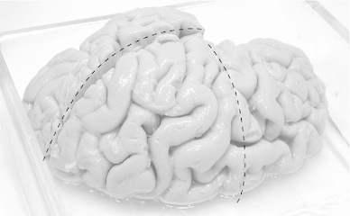
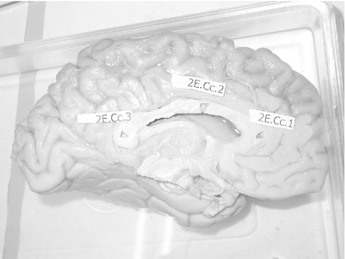
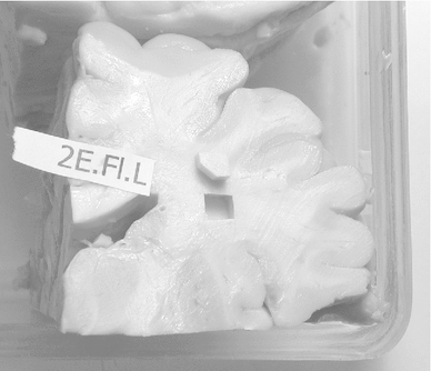
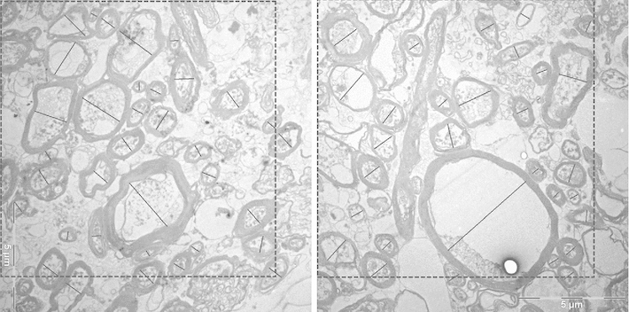
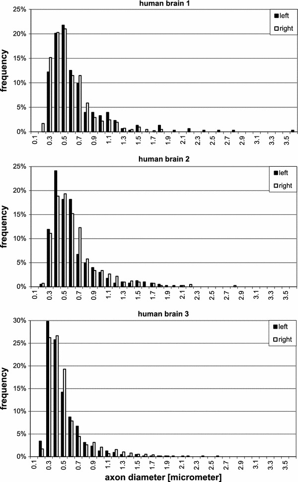
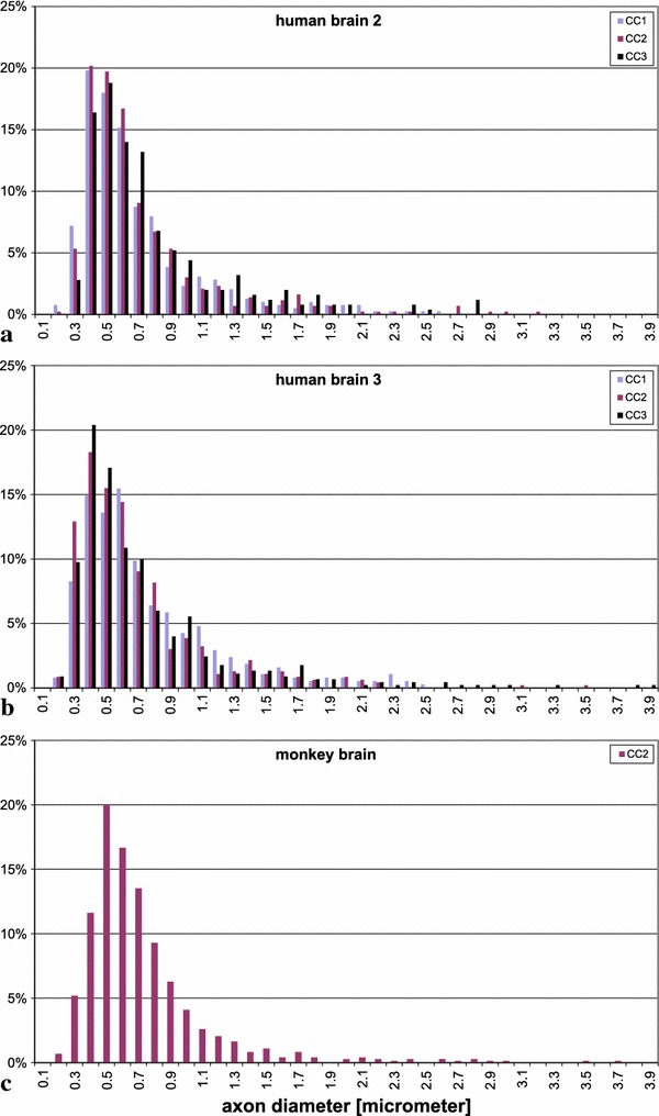
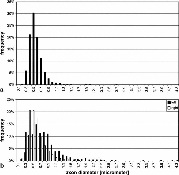
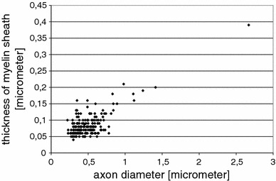
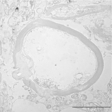
The aim of this study was to obtain information on the axonal diameters of cortico-cortical fibres in the human brain, connecting distant regions of the same hemisphere via the white matter. Samples for electron microscopy were taken from the region of the superior longitudinal fascicle and from the transitional white matter between temporal and frontal lobe where the uncinate and inferior occipitofrontal fascicle merge. We measured the inner diameter of cross sections of myelinated axons. For comparison with data from the literature on the human corpus callosum, we also took samples from that region. For comparison with well-fixed material, we also included samples from corresponding regions of a monkey brain (Macaca mulatta). Fibre diameters in human brains ranged from 0.16 to 9 μm. Distributions of diameters were similar in the three systems of cortico-cortical fibres investigated, both in humans and the monkey, with most of the average values below 1 μm diameter and a small population of much thicker fibres. Within individual human brains, the averages were larger in the superior longitudinal fascicle than in the transitional zone between temporal and frontal lobe. An asymmetry between left and right could be found in one of the human brains, as well as in the monkey brain. A correlation was also found between the thickness of the myelin sheath and the inner axon diameter for axons whose calibre was greater than about 0.6 μm. The results are compared to white matter data in other mammals and are discussed with respect to conduction velocity, brain size, cognition, as well as diffusion weighted imaging studies.
Overview
The claims and statistics of this paper haven’t been checked yet.
Run the check — free, no account needed. It reads the open-access text and checks the sources it cites.
Integrity signals
Registry
Cites
Statistics
Figures
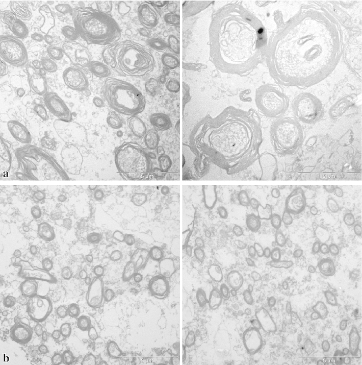
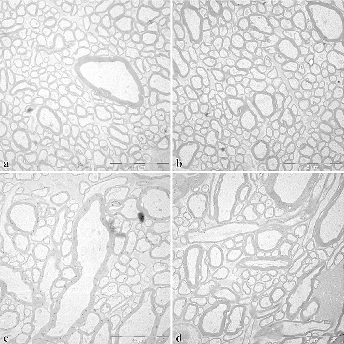
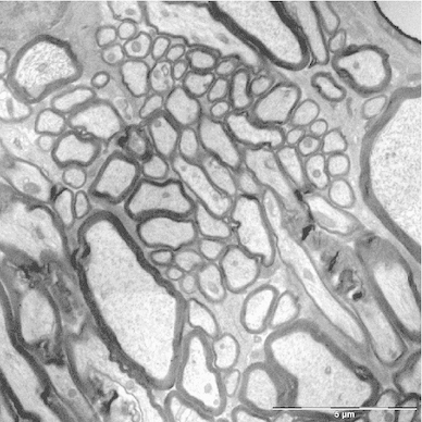
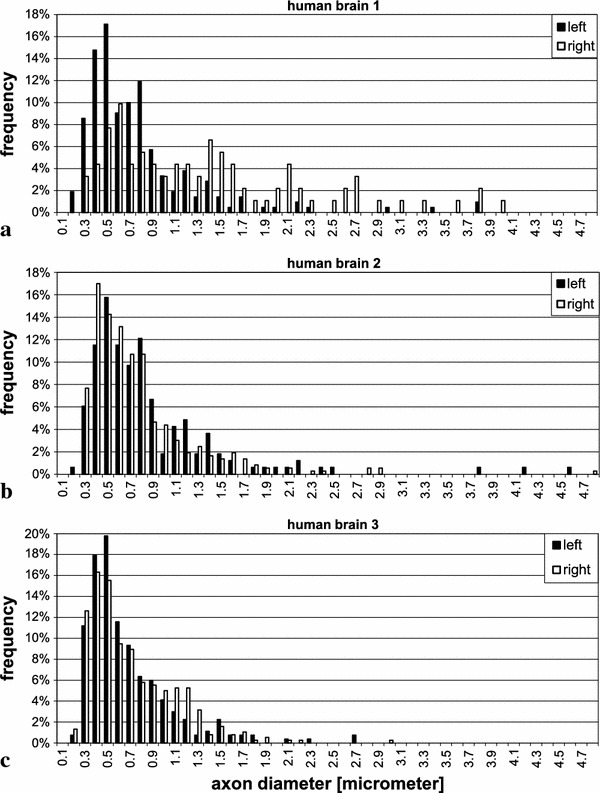
Verification
Claims the paper makes
The claims and statistics of this paper haven’t been checked yet.
Run the check — free, no account needed. It reads the open-access text and checks the sources it cites.
Claims other papers make about this one
Each is checked against this paper’s own text: does it say what the citing paper says it does?
The source backs part of the claim, not all of it — or backs it for a different population, condition or comparison.
In the citing paper
“For example, certain axonal and dendritic domains are less than 0.50 μm in diameter (Shepherd and Harris, 1998), and myelinated axons at three different sites of the corpus callosum in the human brain are estimated to have axon diameters of ≥0.50 μm, in 70–90% of cases (range 0.16–3.73 μm, mean 0.73 ± 0.55 μm) (Liewald et al., 2014).”
From Human Brain/Cloud Interface (2019) · cites this paper as Liewald et al., 2014
Experimental — accuracy not yet measured
Self-check
Percentages, sums and reported differences re-computed from the paper’s own numbers.
The self-check needs the paper’s full text.
Citations
Cites
The structure of the nervous system of the nematode Caenorhabditis elegans
White, J. G., Southgate, E., Thomson, J. N., & Brenner, S. (1986) · Philosophical Transactions of the Royal Society of London. B, Biological Sciences · doi:10.1098/rstb.1986.0056 · Cited by 5,589
Fiber Tract–based Atlas of Human White Matter Anatomy
Wakana, S., Jiang, H., Nagae‐Poetscher, L. M., van Zijl, P. C. M., & Mori, S. (2004) · Radiology · doi:10.1148/radiol.2301021640 · Cited by 2,023
The Fine Structure of the Nervous System.
Tennyson, V. M. (1970) · Archives of Neurology · doi:10.1001/archneur.1970.00480240094019 · Cited by 1,946
Lectu re Notes in Biomathematics
Levin, S. A. (1983) · Cited by 1,568
Fiber tract-based atlas of human white matter anatomy
Wakana, S. (2006) · Medical Entomology and Zoology · Cited by 1,531
Fiber composition of the human corpus callosum
Aboitiz, F., Scheibel, A. B., Fisher, R. S., & Eran Zaidel, B. (1992) · Brain Research · doi:10.1016/0006-8993(92)90178-c · Cited by 1,467
(2009) · Elsevier eBooks · doi:10.1016/c2009-1-03742-3 · Cited by 1,335
The Human Central Nervous System
Nieuwenhuys, R., Voogd, J., & van Huijzen, C. (2008) · doi:10.1007/978-3-540-34686-9 · Cited by 1,262
The Central Nervous System of Vertebrates
Nieuwenhuys, R., ten Donkelaar, H. J., & Nicholson, C. (1998) · doi:10.1007/978-3-642-18262-4 · Cited by 1,249
Cortex: Statistics and Geometry of Neuronal Connectivity
Braitenberg, V., & Schüz, A. (1998) · doi:10.1007/978-3-662-03733-1 · Cited by 1,178
Self-citation
Research works printed but not linked: 5
- Aboitiz F, Ide A, Olivares R (2003) Corpus callosum morphology in relation to cerebral asymmetries in the postmortem human. In: Zaidel E, Iacoboni M (eds) The parallel brain: the cognitive neuroscience of the corpus callosum. MIT Press, Cambridge, pp 33–46
- Allman J (1999) Evolving brains. Scientific American Library, New York
- Bürgel U (1999) Kartierung langer Faserbahnen im adulten menschlichen Gehirn. Geschlechtsunterschiede, Lateralisation und Variabilität. Dissertation at the Faculty of Medicine, Heinrich-Heine-Universität Düsseldorf
- Häggqvist G (1936) Analyse der Faserverteilung in einem Rückenmarkquerschnitt (Th 3). Z Mikrosk Anat Forsch 39:1–34
- Palay LS, Chan-Palay V (1974) Cereb Cortex. Springer, Berlin
Cited by
Sorted by citations
Items 201–300 of 321
A mathematical model for axonal transport of large cargo vesicles
Rahman, N., & Oelz, D. (2023) · Journal of Mathematical Biology · doi:10.1007/s00285-023-02022-3 · Cited by 5
Frede, E., Zadeh‐Haghighi, H., & Simon, C. (2024) · IEEE Transactions on Molecular Biological and Multi-Scale Communications · doi:10.1109/tmbmc.2024.3464415 · Cited by 5
White Matter Tracts Visualized by Parvalbumin in Nonhuman Primates
Rockland, K. S. (2018) · InTech eBooks · doi:10.5772/intechopen.70510 · Cited by 5
Linking spherical mean diffusion weighted signal with intra-axonal volume fraction
Li, H., Chow, H.-M., Chugani, D., & Chugani, H. (2018) · Magnetic Resonance Imaging · doi:10.1016/j.mri.2018.11.006 · Cited by 23
Madigan, N. N., & Windebank, A. J. (2020) · Elsevier eBooks · doi:10.1016/b978-0-12-818422-6.00060-5 · Cited by 5
Noninvasive Quantitative Imaging of Selective Microstructure Sizes via Magnetic Resonance
Capiglioni, M., Zwick, A., Jiménez, P., & Álvarez, G. A. (2021) · Physical Review Applied · doi:10.1103/physrevapplied.15.014045 · Cited by 5
Chondroitin Sulphate Proteoglycan Axonal Coats in the Human Mediodorsal Thalamic Nucleus
Pantazopoulos, H., Hossain, N. M., Chelini, G., Durning, P. T., Barbas, H., Zikopoulos, B., & Berretta, S. (2022) · Frontiers in Integrative Neuroscience · doi:10.3389/fnint.2022.934764 · Cited by 5
Wu, H., Chang, K.-J., Wen, X., Yarmishyn, A. A., Dai, H.-J., Chan, K.-H., Hsiao, Y., Chen, M.-T., Chien, Y., Ma, H., et al. (2022) · Materials Today Advances · doi:10.1016/j.mtadv.2022.100322 · Cited by 6
Effective Diffusion and Tortuosity in Brain White Matter
Vidotto, M., Dini, D., & Momi, E. D. (2018) · Annual International Conference of the IEEE Engineering in Medicine and Biology Society (EMBC) · doi:10.1109/embc.2018.8513443 · Cited by 5
NODDI-SH: a computational efficient NODDI extension for fODF estimation in diffusion MRIPreprint
Zucchelli, M., Descoteaux, M., & Menegaz, G. (2017) · arXiv (Cornell University) · doi:10.48550/arxiv.1708.08999 · Cited by 5
Time-dependent diffusion in undulating structures: Impact on axon diameter estimationPreprint
Brabec, J., Lasič, S., & Nilsson, M. (2019) · arXiv (Cornell University) · doi:10.48550/arxiv.1903.04536 · Cited by 5
Nakae, T., Matsumoto, R., Kunieda, T., Arakawa, Y., Kobayashi, K., Shimotake, A., Yamao, Y., Kikuchi, T., Aso, T., Matsuhashi, M., et al. (2019) · bioRxiv (Cold Spring Harbor Laboratory) · doi:10.1101/702753 · Cited by 5
Tunable hydrogel-based micropillar arrays for myelination studies
Lasli, S., Vinel, C., Agrawal, A., Javanmardi, Y., Pedarzani, P., García-Díaz, B., García‐León, J. A., Djordjevic, B., White, I. J., Sheridan, G. K., et al. (2026) · Nature Methods · doi:10.1038/s41592-026-03048-3 · Cited by 4
How far does electrical stimulation activate white matter tracts? A computational modeling study
Schlosser-Perrin, F., Rossel, O., Duffau, H., Bonnetblanc, F., & Mandonnet, E. (2023) · Clinical Neurophysiology · doi:10.1016/j.clinph.2023.06.017 · Cited by 4
Dysregulation of Myelination in Focal Cortical Dysplasia Type II of the Human Frontal Lobe
Donkels, C., Häussler, U., Huber, S., Tiesmeyer, N., Demerath, T., Scheiwe, C. F., Shah, M., Heers, M., Urbach, H., Schulze-Bonhage, A., et al. (2024) · Glia · doi:10.1002/glia.24662 · Cited by 5
Yang, M., Martin, C. J. L., Kowsari, K., Jagielska, A., & Vliet, K. J. V. (2025) · PLoS ONE · doi:10.1371/journal.pone.0290521 · Cited by 5
Borgheai, S. B., Howell, B. L., Isbaine, F., Noecker, A. M., Opri, E., Risk, B. B., McIntyre, C. C., & Miocinovic, S. (2025) · Brain stimulation · doi:10.1016/j.brs.2025.10.022 · Cited by 5
Neves, K. (2016) · Elsevier eBooks · doi:10.1016/b978-0-12-804042-3.00047-6 · Cited by 4
Comparison of NODDI and spherical mean signal for measuring intra-neurite volume fraction
Li, H., Nikam, R. M., Kandula, V. V. R., Chow, H. M., & Choudhary, A. K. (2018) · Magnetic Resonance Imaging · doi:10.1016/j.mri.2018.11.021 · Cited by 4
Direct jet coaxial electrospinning of axon‐mimicking fibers for diffusion tensor imaging
Hu, C., Grech‐Sollars, M., Statton, B. K., Li, Z., Gao, F., Williams, G. R., Parker, G. J. M., & Zhou, F. (2023) · Polymers for Advanced Technologies · doi:10.1002/pat.6073 · Cited by 4
Preferential conduction block of myelinated axons by nitric oxide
Shrager, P. G., & Youngman, M. (2016) · Journal of Neuroscience Research · doi:10.1002/jnr.23918 · Cited by 4
FAConstructor: an interactive tool for geometric modeling of nerve fiber architectures in the brain
Reuter, J., Matuschke, F., Menzel, M., Schubert, N., Ginsburger, K., Poupon, C., Amunts, K., & Axer, M. (2019) · International Journal of Computer Assisted Radiology and Surgery · doi:10.1007/s11548-019-02053-6 · Cited by 4
Molecular Dynamics Simulations of Axonal Membrane in Traumatic Brain Injury
Alaei, Z. (2017) · KTH Publication Database DiVA (KTH Royal Institute of Technology) · Cited by 4
Evaluating ultrastructural preservation quality in banked brain tissuePreprint
Garrood, M., Keberle, A., Sowa, A., Janssen, W. G. M., Thorn, E. L., Sanctis, C. D., Farrell, K. W., Crary, J. F., & McKenzie, A. T. (2025) · bioRxiv (Cold Spring Harbor Laboratory) · doi:10.1101/2025.05.09.652503 · Cited by 6
Abad, N., Bhushan, C., Ajala, A., Sprenger, T., Marinelli, L., Morris, H. D., DeMarco, J. K., Hood, M. N., Kohls, G., Ho, V. B., et al. (2025) · Imaging Neuroscience · doi:10.1162/imag.a.68 · Cited by 3
MRI-scale histology validates spatial sensitivity of in-vivo MRI-based axon radius estimation
Mordhorst, L., Edwards, L., Morozova, M., Ashtarayeh, M., Streubel, T., Fricke, B., Fritz, F. J., Rusch, H., Jäger, C., Starke, L., et al. (2025) · Imaging Neuroscience · doi:10.1162/imag.a.1030 · Cited by 2
Oliveira, R., Marzia De Lucia, E., & Lutti, A. (2023) · Human Brain Mapping · doi:10.1002/hbm.26420 · Cited by 1
Q‐space imaging based on Gaussian radial basis function with Laplace regularization
Wang, Y., Zhu, Y., Luo, L., & He, J. (2024) · Magnetic Resonance in Medicine · doi:10.1002/mrm.30049 · Cited by 3
Human Connectome Mapping and Monitoring Using Neuronanorobots
Martins, N. R. B., Erlhagen, W., & Freitas, Jr. (2016) · Journal of Ethics and Emerging Technologies · doi:10.55613/jeet.v26i1.49 · Cited by 3
Richerson, W. T., Muftuler, L. T., Wolfgram, D. F., & Schmit, B. D. (2023) · Magnetic Resonance Imaging · doi:10.1016/j.mri.2023.04.007 · Cited by 3
Robinson, T. D., Sun, Y. L., Chang, P. T. H., & Chen, J. J. (2024) · Imaging Neuroscience · doi:10.1162/imag_a_00247 · Cited by 3
Variations in perfusion detectable in advance of microstructure in white matter aging
Robinson, T. D., Sun, Y. L., Chang, P. T. H., Gauthier, C. J., & Chen, J. J. (2025) · GeroScience · doi:10.1007/s11357-025-01787-3 · Cited by 4
Estimation of Bounded and Unbounded Trajectories in Diffusion MRI
Ning, L., Westin, C., & Yogesh Rathi, A. (2016) · Frontiers in Neuroscience · doi:10.3389/fnins.2016.00129 · Cited by 3
Zucchelli, M., Fick, R., Rachid Deriche, B., & Menegaz, G. (2016) · IEEE International Symposium on Biomedical Imaging (ISBI) · doi:10.1109/isbi.2016.7493308 · Cited by 3
DWI Simulation-Assisted Machine Learning Models for Microstructure Estimation
Rafael‐Patiño, J., Yu, T. V., Delvigne, V., Baraković, M., Pizzolato, M., Girard, G., Jones, D. K., Canales-Rodríguez, E. J., & Thiran, J. (2020) · Mathematics and visualization · doi:10.1007/978-3-030-52893-5_11 · Cited by 3
Portoles, O., Qin, Y., Hadida, J., Woolrich, M. W., Cao, M., & van Vugt, M. K. (2022) · PLoS ONE · doi:10.1371/journal.pone.0275819 · Cited by 3
Characterization of b-value dependent T 2 relaxation rates for probing neurite microstructurePreprint
Ning, L., Westin, C., & Yogesh Rathi, A. (2022) · bioRxiv (Cold Spring Harbor Laboratory) · doi:10.1101/2022.09.02.506440 · Cited by 3
Newman, B. T., Ressa, H. J., Jacokes, Z., McPartland, J. C., Kleinhans, N. M., Webb, S. J., Gupta, A. R., Bernier, R., Gaab, N., Jack, A., et al. (2025) · medRxiv · doi:10.1101/2025.10.28.25338995 · Cited by 3
Zhou, M., Stobbe, R., Budde, M. D., Buck, B. H., Kate, M. P., Lloret, M., Fairall, P., Butcher, K., Shuaib, A. M., Emery, D., et al. (2025) · Magnetic Resonance in Medicine · doi:10.1002/mrm.30618 · Cited by 2
T2 contrast variation in human brain at 7 T and its potential contributors
Wang, Y., van Gelderen, P., Donadieu, M., Liu, J., de Zwart, J. A., Zhou, J., Nair, G., Reich, D. S., & Duyn, J. H. (2025) · Imaging Neuroscience · doi:10.1162/imag.a.67 · Cited by 2
Danstrom, I. A., Adkinson, J. A., Liu, Z., Robinson, M. E., Oswalt, D. N., Banks, G. P., Maheshwari, A., Lin, L., Shofty, B., Hasen, M. A., et al. (2026) · Brain stimulation · doi:10.1016/j.brs.2026.103049 · Cited by 1
Tong, S., Ye, S., Ma, F., Xie, X., Sun, Y., Ma, C., Shi, T., Cheng, Z., Li, C., Han, W., et al. (2026) · Nature Communications · doi:10.1038/s41467-026-72397-6 · Cited by 2
Gladkova, N. D., Achkasova, K. A., Сергеевич, Я. К., Kiseleva, E. B., Moiseev, A. A., Львовна, Б. Е., Kuznetsov, S. S., Medyanik, I. A., Kravets, L. Y., Gelikonov, G. V., et al. (2022) · Springer eBooks · doi:10.1007/978-981-19-1352-5_1 · Cited by 2
Microstructure Fingerprinting for Heterogeneously Oriented Tissue Microenvironments
Huynh, K. M., Wu, Y., Ahmad, S., & Yap, P. (2023) · Lecture notes in computer science · doi:10.1007/978-3-031-43993-3_13 · Cited by 2
Towards a white matter ephaptic coupling model of 1/f spectra
Douglas, P., & Blair, G. J. (2024) · International Winter Conference on Brain-Computer Interface (BCI) · doi:10.1109/bci60775.2024.10480498 · Cited by 2
Diffusion MRI Anisotropy: Modeling, Analysis and Interpretation
Fick, R., Pizzolato, M., Wassermann, D., & Rachid Deriche, B. (2017) · Mathematics and visualization · doi:10.1007/978-3-319-61358-1_9 · Cited by 2
Deslauriers‐Gauthier, S., & Rachid Deriche, B. (2019) · SPIE Medical Imaging: Biomedical Applications in Molecular, Structural, and Functional Imaging · doi:10.1117/12.2511736 · Cited by 2
Cortical communication and the comparison of colors
Mollon, J., & Danilova, M. (2019) · Current Opinion in Behavioral Sciences · doi:10.1016/j.cobeha.2019.10.002 · Cited by 3
How does the human visual system compare the speeds of spatially separated objects?
Danilova, M., Takahashi, C., & Mollon, J. (2020) · PLoS ONE · doi:10.1371/journal.pone.0231959 · Cited by 2
Gilani, N., Hildebrand, S., Schueth, A., & Roebroeck, A. F. (2019) · bioRxiv (Cold Spring Harbor Laboratory) · doi:10.1101/626945 · Cited by 2
Modeling subcortical white matter stimulationPreprint
Dali, M., Goldman, J. S., Pantz, O., Alain Destexhe, A., & Mandonnet, E. (2019) · bioRxiv (Cold Spring Harbor Laboratory) · doi:10.1101/2019.12.12.872390 · Cited by 2
The variability of MR axon radii estimates in the human brainPreprint
Veraart, J., Raven, E. P., Edwards, L., Weiskopf, N., & Jones, D. K. (2020) · doi:10.21203/rs.3.rs-104158/v1 · Cited by 2
Chakwizira, A., Westin, C., Brabec, J., Lasič, S., Knutsson, L., Szczepankiewicz, F., & Nilsson, M. (2021) · arXiv (Cornell University) · doi:10.48550/arxiv.2111.11094 · Cited by 2
Stammen, C., Penate, J. S., Metzen, D., Hönscher, M. J., Fraenz, C., Schlüter, C., Güntürkün, O., Kumsta, R., & Genç, E. (2025) · Cerebral Cortex · doi:10.1093/cercor/bhaf199 · Cited by 2
Bansal, S., Peterson, B. S., Gupte, C., Sawardekar, S., Anaya, M. J. G., Ordonez, M., Bhojwani, D., Santoro, J. D., & Bansal, R. (2025) · Brain Research Bulletin · doi:10.1016/j.brainresbull.2025.111541 · Cited by 1
Large-scale modeling of axonal dynamic responses via deep learning
Zhang, C., Clansey, A., Bartels, L., Bondi, D., Kloiber, J., Jaffray, A., van Donkelaar, P., Rauscher, A., Wu, L. C., & Ji, S. (2025) · Biomechanics and Modeling in Mechanobiology · doi:10.1007/s10237-025-02034-6 · Cited by 1
Oligodendrocyte dysfunction in human age-related cognitive decline
Craig, G. A., Merour, E., Seeker, L. A., Haughian, A. J., Arbabi, K., Rose, J. L., Hoffmann, A., Thapar, J., Corsinotti, A., Mitchell, S., et al. (2026) · Nature Medicine · doi:10.1038/s41591-026-04608-y · Cited by 1
Prediction of Neural Diameter From Morphology to Enable Accurate Simulation
Reed, J. D., & Blackwell, K. T. (2021) · Frontiers in Neuroinformatics · doi:10.3389/fninf.2021.666695 · Cited by 1
Phenomenological Modeling of Diverse and Heterogeneous Synaptic Dynamics at Natural Density
Korcsak-Gorzo, A., Linssen, C., Albers, J., Dasbach, S., Duarte, R., Kunkel, S., Morrison, A., Senk, J., Stapmanns, J., Tetzlaff, T., et al. (2024) · Neuromethods · doi:10.1007/978-1-0716-4019-7_15 · Cited by 1
GORDA: Graph-Based Orientation Distribution Analysis of SLI Scatterometry Patterns of Nerve Fibres
Vaca, E., Menzel, M., Amunts, K., Axer, M., & Dickscheid, T. (2022) · IEEE International Symposium on Biomedical Imaging (ISBI) · doi:10.1109/isbi52829.2022.9761492 · Cited by 1
Local axonal conduction delays underlie precise timing of a neural sequencePreprint
Egger, R., Tupikov, Y., Katlowitz, K. A., Benezra, S. E., Picardo, M. A., Moll, F. W., Kornfeld, J. M. R., Jin, D. Z., & Long, M. A. (2019) · bioRxiv (Cold Spring Harbor Laboratory) · doi:10.1101/864231 · Cited by 1
The development of efficient communication in the human connectomePreprint
van Blooijs, D., van den Boom, M., van der Aar, J. F., Huiskamp, G. J. M., Castegnaro, G., Demuru, M., Zweiphenning, W. J. E. M., van Eijsden, P., Miller, K. J., Leijten, F. S. S., et al. (2022) · bioRxiv (Cold Spring Harbor Laboratory) · doi:10.1101/2022.03.14.484297 · Cited by 1
Parameters for magnetic stimulation of the phrenic nervePreprint
Sandurkov, B., Rapp, J., Hemmert, W., & Gleich, B. (2022) · doi:10.36227/techrxiv.21324192.v1 · Cited by 1
Asadi, S., Spencer, A. P. C., Alemán‐Gómez, Y., Jedynak, M., Canales-Rodríguez, E. J., Pavan, T., Mullier, E., de Wouters, L., Rigoni, I., Lajous, H., et al. (2025) · bioRxiv (Cold Spring Harbor Laboratory) · doi:10.1101/2025.10.28.685017 · Cited by 1
Inference of a U-fiber bundle atlas informed by the variability of the cortical folding pattern
Labra, N. (2020) · theses.fr (ABES) · doi:10.70675/42b36629z423cz4f6az9807zf916bfc2242a · Cited by 2
Reproducibility and use of myelin imaging methods for the study of adolescent brain development
Raven, E. P. (2017) · DigitalGeorgetown (Georgetown University Library)
Kim, S. (2019) · Journal of the Korean Society for Precision Engineering · doi:10.7736/kspe.2019.36.4.425
Fried, I. (2019) · viXra
Inferring Temporal Delays in Brain Interactions from Neural Population Activity
Noori, R. (2019) · TSpace (University of Toronto)
Computational Modeling of the Human Brain for mTBI Prediction and Diagnosis
Levy, Y. (2020) · Scholarship@Western (Western University)
An Investigation of the Cellular Mechanisms Underlying Ultrasound Neuromodulation
Newhoff, M. (2020) · University of Minnesota Digital Conservancy (University of Minnesota)
Diffusion-weighted magnetic resonance imaging at ultra-high field
Fritz, F. J. (2020) · Research Publications (Maastricht University) · doi:10.26481/dis.20210111flf
Delay-Based Neural Computation: Pulse Routing Architecture and Benchmark Application in FPGA
Thanasoulis, V., Vogginger, B., Partzsch, J., & Mayr, C. (2021) · IEEE International Conference on Electronics, Circuits and Systems (ICECS) · doi:10.1109/icecs53924.2021.9665468
An agent-based model to examine neuroinvasion by SARS-CoV-2Preprint
Bhat, V., Aditi Lakshmi, S., Muraly, A., Rao, P., & Chatterjee, J. (2022) · Research Square · doi:10.21203/rs.3.rs-1333474/v1
Assaf, Y., Johansen‐Berg, H., & de Schotten, M. T. (2021) · Elsevier eBooks · doi:10.1016/b978-0-12-819641-0.00156-0
Moiseev, A. A., Achkasova, K. A., Сергеевич, Я. К., Kiseleva, E. B., Львовна, Б. Е., Kuznetsov, S. S., Gelikonov, G. V., Shilyagin, P. A., Medyanik, I. A., Вадимовна, З. Е., et al. (2020) · doi:10.31224/osf.io/p69te
In-vivo estimation of axonal morphology from MRI and EEG dataPreprint
Oliveira, R., Pelentritou, A., Domenicantonio, G. D., Marzia De Lucia, E., & Lutti, A. (2022) · bioRxiv (Cold Spring Harbor Laboratory) · doi:10.1101/2022.02.11.479116
Deep Brain Stimulation Atlases in Movement Disorders : from Patient-Specific to Group Analysis
Vogel, D. (2022) · Linköping studies in science and technology. Dissertations · doi:10.3384/9789179292546
Gaudreault, P., Lina, J., Descoteaux, M., Gosselin, N., Doyon, J., Deslauriers‐Gauthier, S., & Carrier, J. (2022) · bioRxiv (Cold Spring Harbor Laboratory) · doi:10.1101/2022.05.11.491489
Axonal Spectrum Imaging (AxSI): In-Vivo mapping of axonal diameter distributions in the human brainPreprint
Gast, H., Horowitz, A., Krupnik, R., Barazany, D., Lifshits, S., Ben-Amitay, S., & Assaf, Y. (2022) · Research Square · doi:10.21203/rs.3.rs-1835012/v1
Invasive Neural Integration and Clinical Translation
Glass, C., & TUFFAHA, S. H. (2022) · Handbook of Neuroengineering · doi:10.1007/978-981-15-2848-4_131-1
Schmitz, D., Benning, K., Schubert, N., Minnerop, M., Amunts, K., & Axer, M. (2022) · Frontiers in Physics · doi:10.3389/fphy.2022.958364
Parameters for magnetic stimulation of the phrenic nervePreprint
Sandurkov, B., Rapp, J., Hemmert, W., & Gleich, B. (2022) · doi:10.36227/techrxiv.21324192
Oliveira, R., Marzia De Lucia, E., & Lutti, A. (2022) · bioRxiv (Cold Spring Harbor Laboratory) · doi:10.1101/2022.12.19.521040
Nordin, T. (2023) · Linköping studies in science and technology. Dissertations · doi:10.3384/9789179295349
Invasive Neural Integration and Clinical Translation
Glass, C., & TUFFAHA, S. H. (2023) · Handbook of Neuroengineering · doi:10.1007/978-981-16-5540-1_131
Robinson, T. D., Sun, Y. L., Chang, P. T. H., & Chen, J. J. (2023) · bioRxiv (Cold Spring Harbor Laboratory) · doi:10.1101/2023.03.14.532658
Neurobiology and the connectome
Ciarrusta, J., & Arichi, T. (2023) · Elsevier eBooks · doi:10.1016/b978-0-323-85280-7.00012-9
Towards a high-density packing white matter substrate generator for Monte-Carlo simulations
Haro, J. L. V., Gardier, R. M. M., Canales-Rodríguez, E. J., Girard, G., Thiran, J., & Rafael‐Patiño, J. (2023) · Proceedings on CD-ROM - International Society for Magnetic Resonance in Medicine. Scientific Meeting and Exhibition/Proceedings of the International Society for Magnetic Resonance in Medicine, Scientific Meeting and Exhibition · doi:10.58530/2022/2819
In-Vivo Estimation of Axon Radii From Clinical Scanners
Mandal, D., Ning, L., & Yogesh Rathi, A. (2023) · IEEE International Symposium on Biomedical Imaging (ISBI) · doi:10.1109/isbi53787.2023.10230790
Focal cortical dysplasia type II-dependent maladaptive myelination in the human frontal lobePreprint
Donkels, C., Huber, S., Demerath, T., Scheiwe, C., Shah, M., Heers, M., Urbach, H., Schulze-Bonhage, A., Prinz, M., Häussler, U., et al. (2024) · bioRxiv · doi:10.1101/2024.03.02.582894
Saito, A., Shiina, T., & Sekiba, Y. (2024) · Bioelectromagnetics · doi:10.1002/bem.22521
The effect of common parameters of bipolar stimulation on brain evoked potentials
Isan, P., Deslauriers‐Gauthier, S., Papadopoulo, T., Fontaine, D., Filipiak, P., & Almairac, F. (2024) · Clinical Neurophysiology · doi:10.1016/j.clinph.2024.08.019
Human short association fibers are thinner and less myelinated than long fibersPreprint
Ruthig, P., Planitz, D. E. V. D., Morozova, M., Reimann, K., Jäger, C., Reinert, T., Mohammadi, S., Weiskopf, N., Kirilina, E. P., & Morawski, M. (2024) · bioRxiv (Cold Spring Harbor Laboratory) · doi:10.1101/2024.10.21.619354
Tractography validation part 3: Lessons learned through validation studies
Schilling, K. G., Rheault, F., & Dyrby, T. B. (2025) · Elsevier eBooks · doi:10.1016/b978-0-12-818894-1.00004-5
Rahman, N., & Oelz, D. (2025) · Mathematical Biosciences · doi:10.1016/j.mbs.2025.109400
Gimenez, M. L., Jiménez, P., Pérez, L. A. P., Betancourth, D., Zwick, A., & Álvarez, G. A. (2025) · Journal of Magnetic Resonance · doi:10.1016/j.jmr.2025.107940
Perlman, K., Major, J., Collin, A., Noël, V. P., Mardenli, M., Jerczyński, S., Davoli, M. A., Cohen‐Adad, J., Côté, D. C., & Mechawar, N. (2025) · bioRxiv (Cold Spring Harbor Laboratory) · doi:10.1101/2025.09.18.672977
Turpin, C., Rossel, O., Schlosser-Perrin, F., Carriere, M., Matsumoto, R., Mandonnet, E., Duffau, H., Ng, S., & Bonnetblanc, F. (2025) · Clinical Neurophysiology · doi:10.1016/j.clinph.2025.2111370
Steeghs-Turchina, M., Srinivasan, R., Nunez, P. L., & Nunez, M. D. (2025) · NeuroImage · doi:10.1016/j.neuroimage.2025.121418
Metadata
- Authors
- Liewald, D., Miller, R., Logothetis, N., Wagner, H.-J., & Schüz, A.
- Affiliations
- Max Planck Institute for Biological Cybernetics (DE)
- University of Otago (NZ)
- University of Tübingen (DE)
- Corresponding author
- Schüz, Almut
- Venue
- Biological Cybernetics
- Published
- 2014-08-21
- Volume
- 108
- Issue
- 5
- Pages
- 541-557
- ISSN
- 0340-1200 (ISSN-L)
- 1432-0770
- PMC
- PMC4228120
- OpenAlex
- W2046529900
- Cited by
- 322
- References
- 98
- Citations by year
- 2026: 16 · 2025: 28 · 2024: 25 · 2023: 36 · 2022: 39 · 2021: 37 · 2020: 39 · 2019: 35 · 2018: 28 · 2017: 14 · 2016: 21 · 2015: 4
- FWCI
- 10.17
- Type
- journal article
- Language
- en
- Open access
- hybrid
- doi.org
- Locations
- Biological Cybernetics (publishedVersion, cc-by)
- Europe PMC (PubMed Central) (publishedVersion, cc-by)
- PubMed Central (publishedVersion, cc-by)
- PubMed (publishedVersion)
- MPG.PuRe (Max Planck Society) (submittedVersion, other-oa)
- Max Planck Digital Library (submittedVersion)
- Topics
- Health Sciences › Medicine › Radiology, Nuclear Medicine and Imaging › Advanced Neuroimaging Techniques and Applications
- Health Sciences › Medicine › Anatomy › Classical Anatomy and Histology
- Life Sciences › Neuroscience › Cognitive Neuroscience › Visual perception and processing mechanisms
- Keywords
- axon diameter, cortical white matter, superior longitudinal fasciculus, corpus callosum, myelinated axons, myelin thickness, hemispheric lateralization, human brain, macaque brain, conduction velocity, diffusion-weighted imaging
- MeSH terms
- Aged, Aged, 80 and over, Animals, Axons, Cerebral Cortex, Humans, Functional Laterality, Macaca mulatta, Male, Myelin Sheath, Electron Microscope Tomography, White Matter
About these checks
Registry, retraction, Cites and statistics results are computed, not judged, with one exception: a printed reference that no index lists is read once by a model before it counts as a citation error. Claims are extracted by a model and kept only when their quote appears verbatim in the text; each cited source is then checked, with a model’s help, for whether it says what the claim relies on. Those verdicts can be wrong — read the claim’s quote and the cited source before relying on one.
Statistics and arithmetic are recomputed, not judged.
Confidence bands are shown only for fields calibrated on our annotated test set. None is calibrated yet, so no band is shown.
What counts as a claim ›How we measure confidence ›How we check this ›
Are you an author, or do you see a problem with this page? Request a review
