

The DOI matches its Crossref record; 97 of the 98 references it cites were found in a registry or an index.
Distribution of axon diameters in cortical white matter: an electron-microscopic study on three human brains and a macaque.
Open accessAbstract
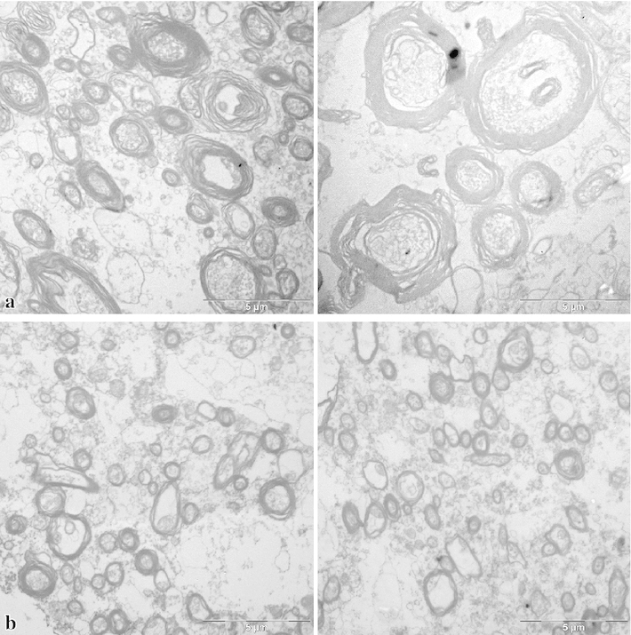
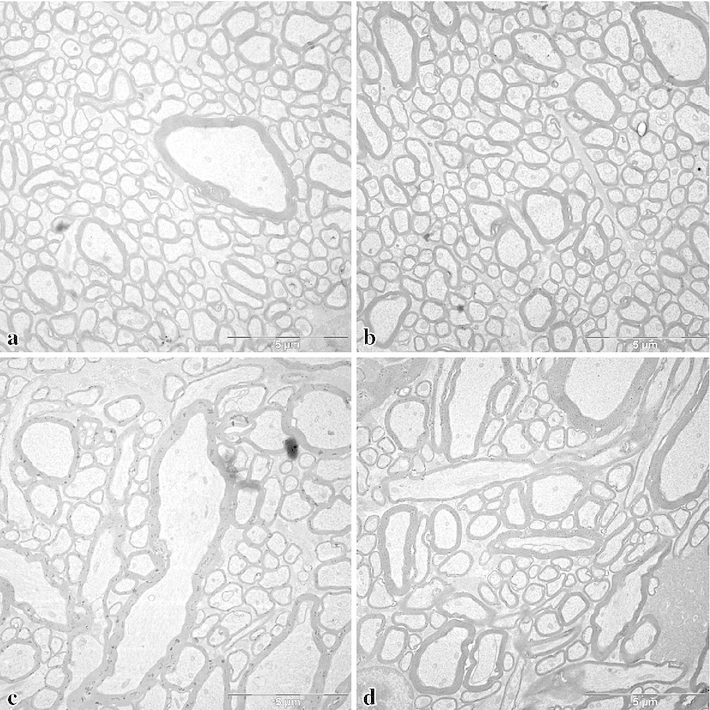
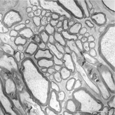
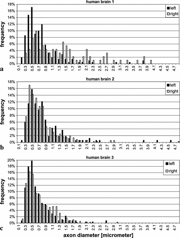
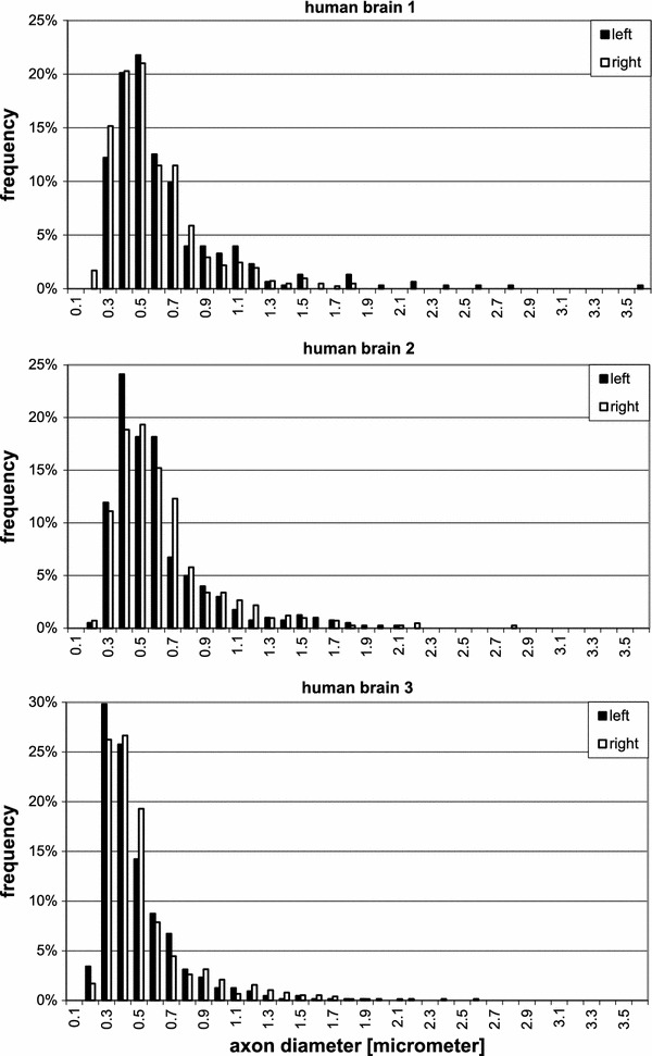
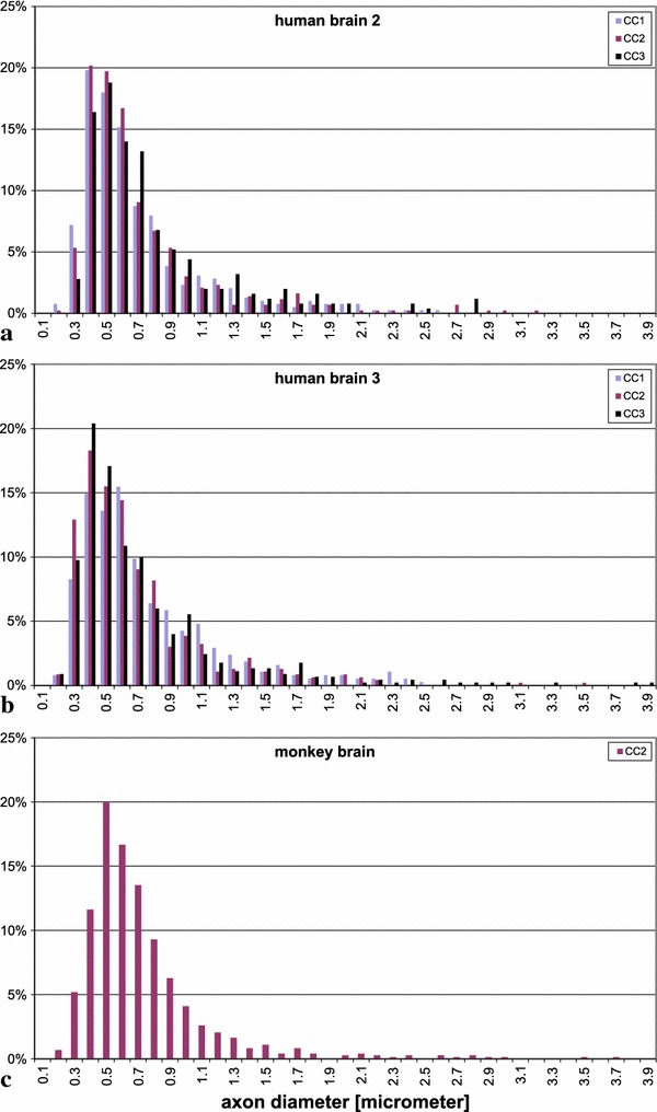
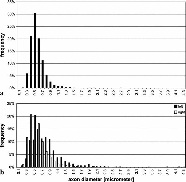
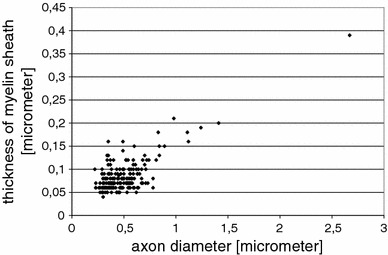
The aim of this study was to obtain information on the axonal diameters of cortico-cortical fibres in the human brain, connecting distant regions of the same hemisphere via the white matter. Samples for electron microscopy were taken from the region of the superior longitudinal fascicle and from the transitional white matter between temporal and frontal lobe where the uncinate and inferior occipitofrontal fascicle merge. We measured the inner diameter of cross sections of myelinated axons. For comparison with data from the literature on the human corpus callosum, we also took samples from that region. For comparison with well-fixed material, we also included samples from corresponding regions of a monkey brain (Macaca mulatta). Fibre diameters in human brains ranged from 0.16 to 9 μm. Distributions of diameters were similar in the three systems of cortico-cortical fibres investigated, both in humans and the monkey, with most of the average values below 1 μm diameter and a small population of much thicker fibres. Within individual human brains, the averages were larger in the superior longitudinal fascicle than in the transitional zone between temporal and frontal lobe. An asymmetry between left and right could be found in one of the human brains, as well as in the monkey brain. A correlation was also found between the thickness of the myelin sheath and the inner axon diameter for axons whose calibre was greater than about 0.6 μm. The results are compared to white matter data in other mammals and are discussed with respect to conduction velocity, brain size, cognition, as well as diffusion weighted imaging studies.
Overview
The claims and statistics of this paper haven’t been checked yet.
Run the check — free, no account needed. It reads the open-access text and checks the sources it cites.
Integrity signals
Registry
Cites
Statistics
Figures
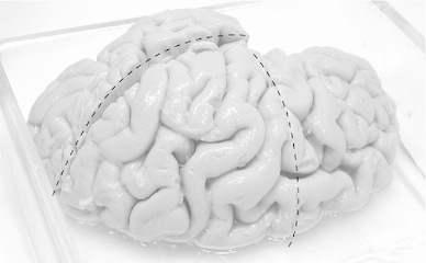
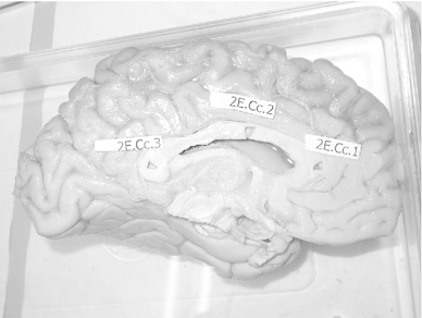
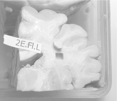
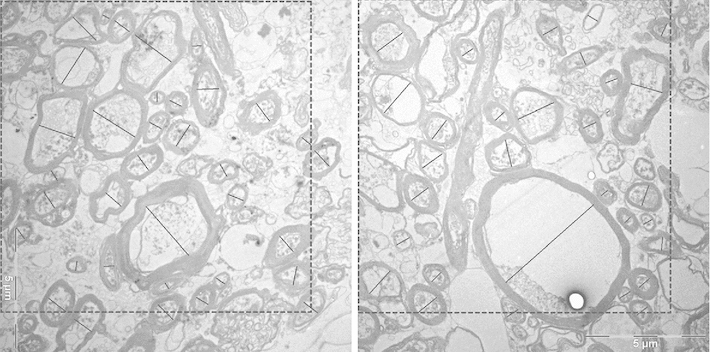
Verification
Claims the paper makes
The claims and statistics of this paper haven’t been checked yet.
Run the check — free, no account needed. It reads the open-access text and checks the sources it cites.
Claims other papers make about this one
Each is checked against this paper’s own text: does it say what the citing paper says it does?
The source backs part of the claim, not all of it — or backs it for a different population, condition or comparison.
In the citing paper
“For example, certain axonal and dendritic domains are less than 0.50 μm in diameter (Shepherd and Harris, 1998), and myelinated axons at three different sites of the corpus callosum in the human brain are estimated to have axon diameters of ≥0.50 μm, in 70–90% of cases (range 0.16–3.73 μm, mean 0.73 ± 0.55 μm) (Liewald et al., 2014).”
From Human Brain/Cloud Interface (2019) · cites this paper as Liewald et al., 2014
Experimental — accuracy not yet measured
Self-check
Percentages, sums and reported differences re-computed from the paper’s own numbers.
The self-check needs the paper’s full text.
Citations
Cites
The structure of the nervous system of the nematode Caenorhabditis elegans
White, J. G., Southgate, E., Thomson, J. N., & Brenner, S. (1986) · Philosophical Transactions of the Royal Society of London. B, Biological Sciences · doi:10.1098/rstb.1986.0056 · Cited by 5,589
Fiber Tract–based Atlas of Human White Matter Anatomy
Wakana, S., Jiang, H., Nagae‐Poetscher, L. M., van Zijl, P. C. M., & Mori, S. (2004) · Radiology · doi:10.1148/radiol.2301021640 · Cited by 2,023
The Fine Structure of the Nervous System.
Tennyson, V. M. (1970) · Archives of Neurology · doi:10.1001/archneur.1970.00480240094019 · Cited by 1,946
Lectu re Notes in Biomathematics
Levin, S. A. (1983) · Cited by 1,568
Fiber tract-based atlas of human white matter anatomy
Wakana, S. (2006) · Medical Entomology and Zoology · Cited by 1,531
Fiber composition of the human corpus callosum
Aboitiz, F., Scheibel, A. B., Fisher, R. S., & Eran Zaidel, B. (1992) · Brain Research · doi:10.1016/0006-8993(92)90178-c · Cited by 1,467
(2009) · Elsevier eBooks · doi:10.1016/c2009-1-03742-3 · Cited by 1,335
The Human Central Nervous System
Nieuwenhuys, R., Voogd, J., & van Huijzen, C. (2008) · doi:10.1007/978-3-540-34686-9 · Cited by 1,262
The Central Nervous System of Vertebrates
Nieuwenhuys, R., ten Donkelaar, H. J., & Nicholson, C. (1998) · doi:10.1007/978-3-642-18262-4 · Cited by 1,249
Cortex: Statistics and Geometry of Neuronal Connectivity
Braitenberg, V., & Schüz, A. (1998) · doi:10.1007/978-3-662-03733-1 · Cited by 1,178
Self-citation
Research works printed but not linked: 5
- Aboitiz F, Ide A, Olivares R (2003) Corpus callosum morphology in relation to cerebral asymmetries in the postmortem human. In: Zaidel E, Iacoboni M (eds) The parallel brain: the cognitive neuroscience of the corpus callosum. MIT Press, Cambridge, pp 33–46
- Allman J (1999) Evolving brains. Scientific American Library, New York
- Bürgel U (1999) Kartierung langer Faserbahnen im adulten menschlichen Gehirn. Geschlechtsunterschiede, Lateralisation und Variabilität. Dissertation at the Faculty of Medicine, Heinrich-Heine-Universität Düsseldorf
- Häggqvist G (1936) Analyse der Faserverteilung in einem Rückenmarkquerschnitt (Th 3). Z Mikrosk Anat Forsch 39:1–34
- Palay LS, Chan-Palay V (1974) Cereb Cortex. Springer, Berlin
Cited by
Sorted by citations
The Human Connectome Project's neuroimaging approach
Glasser, M. F., Smith, S. M., Marcus, D. S., Andersson, J. L. R., Auerbach, E. J., Behrens, T. E. J., Coalson, T. S., Harms, M. P., Jenkinson, M., Moeller, S., et al. (2016) · Nature Neuroscience · doi:10.1038/nn.4361 · Cited by 1,090
Investigating white matter fibre density and morphology using fixel-based analysis.
Raffelt, D. A., Tournier, J.-D., Smith, R. E., Vaughan, D. N., Jackson, G., Ridgway, G. R., & Connelly, A. (2017) · NeuroImage · doi:10.1016/j.neuroimage.2016.09.029 · Cited by 620
“However, most white matter voxels are known to contain crossing fibres (Jeurissen et al., 2012), and voxel-averaged measures such as FA are not fibre-specific (or even erroneous) in such regions, which confounds interpretation of apparent differences (Douaud et al., 2011; Pierpaoli et al., 2001;…”
BackgroundQuantifying brain microstructure with diffusion MRI: Theory and parameter estimation
Novikov, D. S., Fieremans, E., Jespersen, S. N., & Kiselev, V. G. (2019) · NMR in Biomedicine · doi:10.1002/nbm.3998 · Cited by 581
MRI in traumatic spinal cord injury: from clinical assessment to neuroimaging biomarkers
Freund, P., Seif, M., Weiskopf, N., Friston, K. J., Fehlings, M. G., Thompson, A. J., & Curt, A. E. P. (2019) · The Lancet Neurology · doi:10.1016/s1474-4422(19)30138-3 · Cited by 225
Noninvasive quantification of axon radii using diffusion MRI
Veraart, J., Nunes, D., Rudrapatna, U. S., Fieremans, E., Jones, D. K., Novikov, D. S., & Shemesh, N. (2020) · eLife · doi:10.7554/elife.49855 · Cited by 185
Metadata
- Authors
- Liewald, D., Miller, R., Logothetis, N., Wagner, H.-J., & Schüz, A.
- Affiliations
- Max Planck Institute for Biological Cybernetics (DE)
- University of Otago (NZ)
- University of Tübingen (DE)
- Corresponding author
- Schüz, Almut
- Venue
- Biological Cybernetics
- Published
- 2014-08-21
- Volume
- 108
- Issue
- 5
- Pages
- 541-557
- ISSN
- 0340-1200 (ISSN-L)
- 1432-0770
- PMC
- PMC4228120
- OpenAlex
- W2046529900
- Cited by
- 322
- References
- 98
- Citations by year
- 2026: 16 · 2025: 28 · 2024: 25 · 2023: 36 · 2022: 39 · 2021: 37 · 2020: 39 · 2019: 35 · 2018: 28 · 2017: 14 · 2016: 21 · 2015: 4
- FWCI
- 10.17
- Type
- journal article
- Language
- en
- Open access
- hybrid
- doi.org
- Locations
- Biological Cybernetics (publishedVersion, cc-by)
- Europe PMC (PubMed Central) (publishedVersion, cc-by)
- PubMed Central (publishedVersion, cc-by)
- PubMed (publishedVersion)
- MPG.PuRe (Max Planck Society) (submittedVersion, other-oa)
- Max Planck Digital Library (submittedVersion)
- Topics
- Health Sciences › Medicine › Radiology, Nuclear Medicine and Imaging › Advanced Neuroimaging Techniques and Applications
- Health Sciences › Medicine › Anatomy › Classical Anatomy and Histology
- Life Sciences › Neuroscience › Cognitive Neuroscience › Visual perception and processing mechanisms
- Keywords
- axon diameter, cortical white matter, superior longitudinal fasciculus, corpus callosum, myelinated axons, myelin thickness, hemispheric lateralization, human brain, macaque brain, conduction velocity, diffusion-weighted imaging
- MeSH terms
- Aged, Aged, 80 and over, Animals, Axons, Cerebral Cortex, Humans, Functional Laterality, Macaca mulatta, Male, Myelin Sheath, Electron Microscope Tomography, White Matter
About these checks
Registry, retraction, Cites and statistics results are computed, not judged, with one exception: a printed reference that no index lists is read once by a model before it counts as a citation error. Claims are extracted by a model and kept only when their quote appears verbatim in the text; each cited source is then checked, with a model’s help, for whether it says what the claim relies on. Those verdicts can be wrong — read the claim’s quote and the cited source before relying on one.
Statistics and arithmetic are recomputed, not judged.
Confidence bands are shown only for fields calibrated on our annotated test set. None is calibrated yet, so no band is shown.
What counts as a claim ›How we measure confidence ›How we check this ›
Are you an author, or do you see a problem with this page? Request a review
