

The DOI matches its Crossref record; 40 of the 40 references it cites were found in a registry or an index.
Fully Automated Deep Learning System for Bone Age Assessment
Open accessAbstract
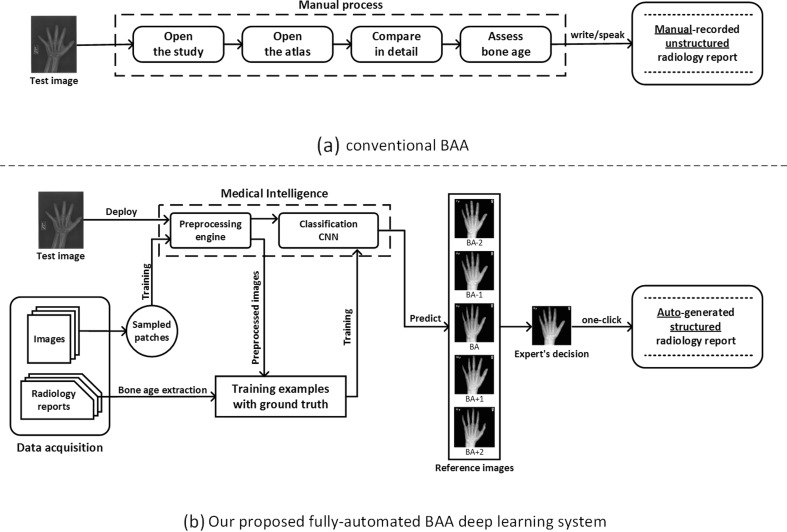
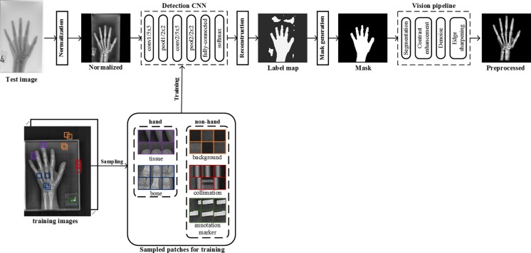
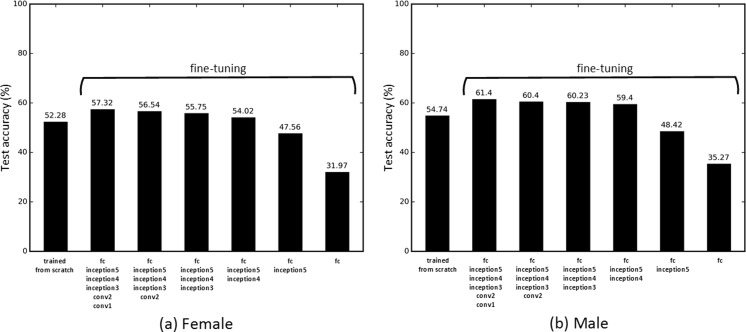
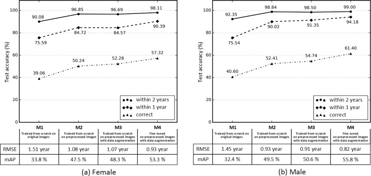
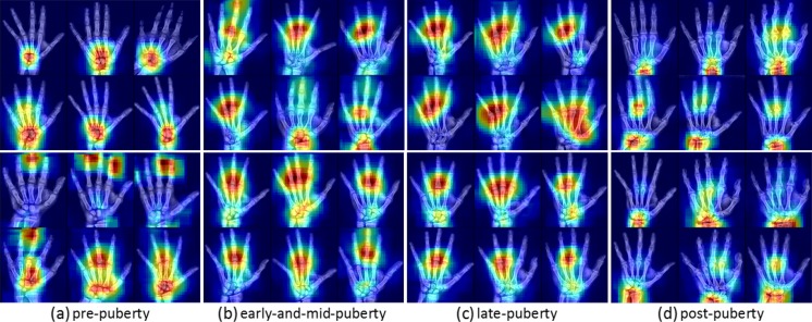
Skeletal maturity progresses through discrete phases, a fact that is used routinely in pediatrics where bone age assessments (BAAs) are compared to chronological age in the evaluation of endocrine and metabolic disorders. While central to many disease evaluations, little has changed to improve the tedious process since its introduction in 1950. In this study, we propose a fully automated deep learning pipeline to segment a region of interest, standardize and preprocess input radiographs, and perform BAA. Our models use an ImageNet pretrained, fine-tuned convolutional neural network (CNN) to achieve 57.32 and 61.40% accuracies for the female and male cohorts on our held-out test images. Female test radiographs were assigned a BAA within 1 year 90.39% and within 2 years 98.11% of the time. Male test radiographs were assigned 94.18% within 1 year and 99.00% within 2 years. Using the input occlusion method, attention maps were created which reveal what features the trained model uses to perform BAA. These correspond to what human experts look at when manually performing BAA. Finally, the fully automated BAA system was deployed in the clinical environment as a decision supporting system for more accurate and efficient BAAs at much faster interpretation time (<2 s) than the conventional method.
Overview
The claims and statistics of this paper haven’t been checked yet.
Run the check — free, no account needed. It reads the open-access text and checks the sources it cites.
Integrity signals
Registry
Cites
Statistics
Figures
Verification
Claims the paper makes
The claims and statistics of this paper haven’t been checked yet.
Run the check — free, no account needed. It reads the open-access text and checks the sources it cites.
Claims other papers make about this one
Each is checked against this paper’s own text: does it say what the citing paper says it does?
In the citing paper
“BoneXpert is one of the first commercial applications of AI in radiology [25] and appeared to be the only AI application in clinical use across several hospitals in The Netherlands.”
From Implementation of artificial intelligence (AI) applications in radiology: hindering and facilitating factors (2020) · cites this paper as [25]
Self-check
Percentages, sums and reported differences re-computed from the paper’s own numbers.
The self-check needs the paper’s full text.
Citations
Cites
Deep Residual Learning for Image Recognition
He, K., Zhang, X., Ren, S., & Sun, J. (2016) · 2016 IEEE Conference on Computer Vision and Pattern Recognition (CVPR) · doi:10.1109/cvpr.2016.90 · Cited by 235,474
Very Deep Convolutional Networks for Large-Scale Image Recognition
Simonyan, K., & Zisserman, A. (2014) · arXiv · doi:10.48550/arxiv.1409.1556 · Cited by 114,148
ImageNet classification with deep convolutional neural networks
Krizhevsky, A., Sutskever, I., & Hinton, G. E. (2017) · Communications of the ACM · doi:10.1145/3065386 · Cited by 112,155
LeCun, Y., Bengio, Y., & Hinton, G. (2015) · Nature · doi:10.1038/nature14539 · Cited by 84,126
ImageNet: A large-scale hierarchical image database
Deng, J., Dong, W., Socher, R., Li, L.-J., Li, K., & Fei-Fei, L. (2009) · 2009 IEEE Conference on Computer Vision and Pattern Recognition · doi:10.1109/cvpr.2009.5206848 · Cited by 65,333
Gradient-based learning applied to document recognition
Lecun, Y., Bottou, L., Bengio, Y., & Haffner, P. (1998) · Proceedings of the IEEE · doi:10.1109/5.726791 · Cited by 58,805
Going deeper with convolutions
Szegedy, C., Liu, W., Jia, Y., Sermanet, P., Reed, S., Anguelov, D., Erhan, D., Vanhoucke, V., & Rabinovich, A. (2015) · 2015 IEEE Conference on Computer Vision and Pattern Recognition (CVPR) · doi:10.1109/cvpr.2015.7298594 · Cited by 48,562
ImageNet Large Scale Visual Recognition Challenge
Russakovsky, O., Deng, J., Su, H., Krause, J., Satheesh, S., Ma, S., Huang, Z., Karpathy, A., Khosla, A., Bernstein, M., et al. (2015) · International Journal of Computer Vision · doi:10.1007/s11263-015-0816-y · Cited by 39,380
Rich Feature Hierarchies for Accurate Object Detection and Semantic Segmentation
Girshick, R., Donahue, J., Darrell, T., & Malik, J. (2014) · 2014 IEEE Conference on Computer Vision and Pattern Recognition · doi:10.1109/cvpr.2014.81 · Cited by 32,684
Visualizing and understanding convolutional networks
Zeiler, M. D., & Fergus, R. (2014) · Lecture Notes in Computer Science · doi:10.1007/978-3-319-10590-1_53 · Cited by 15,839
Research works printed but not linked: 1
- Greulich WW, Pyle SI. Radiographic atlas of skeletal development of the hand and wrist. Am J Med Sci. pdfs.journals. lww.com ; 1959; Available: http://pdfs.journals.lww.com/amjmedsci/1959/09000/Radiographic_Atlas_of_Skeletal_Development_of_the.30.pdf · DOI
Cited by
Sorted by citations
Items 401–452 of 452
Research on Key Technologies of synonym automatic extraction based on multithreaded web crawler
Junling, L., Chen, Y., Mingzhu, Z., Yang, H., Zuoping, W., Bao, Y., & Hong, O. (2020) · International Conference on Innovation in Artificial Intelligence (ICIAI) · doi:10.1145/3390557.3394293
Bone age assessment using metric learning on small dataset of hand radiographs
Madan, S., Gandhi, T. K., & Chaudhury, S. (2020) · Elsevier eBooks · doi:10.1016/b978-0-12-819295-5.00010-x
Valtchinov, V. I., Seo, J. B., Hida, T., & Hatabu, H. (2020) · Medical radiology · doi:10.1007/978-3-030-43539-4_15
Tıbbi Cihaz Sektöründe Kullanılan Derin Öğrenme Yöntemlerine Genel Bakış
GÜNDÜZ, O., Tepe, C., Şenyer, N., & Odabaş, M. S. (2021) · Black Sea Journal of Engineering and Science · doi:10.34248/bsengineering.858918
Computer-aided analysis system for bone age in x-ray image using deep neural network
Kuo, W.-C., Hsiao, W.-T., & Chen, C.-J. (2021) · International Forum on Medical Imaging in Asia (IFMIA) · doi:10.1117/12.2590718
Pediatric Skeletal Age Assessment Using Deep Learning Proceedings
Desai, S. D., & Kadiyavar, P. T. (2021) · Advances in intelligent systems and computing · doi:10.1007/978-981-16-0171-2_12
A Review of Transfer Learning for Medical Image Analysis
Hee, K. E., Alejandro, A., Maros, M. E., Santhanam, N., Jannesari, M., & Ganslandt, T. (2021)
Magnetic Resonance Imaging in the Diagnosis, Characterization and Management of Crohn’s Fistula
Vincent, M. L., Lung, P. F. C., & Sahnan, K. (2021) · Coloproctology · doi:10.1007/978-3-030-30902-2_15-1
Raine, J., MBChB, J. W. G., Vliet, G. V., MB, B. J. I. W., & Deladoëy, J. (2019) · Wiley eBooks · doi:10.1002/9781119386230.ch3
Deshmukh, S., & Khaparde, A. (2022) · Research on Biomedical Engineering · doi:10.1007/s42600-022-00225-z
Magnetic Resonance Imaging in the Diagnosis, Characterization, and Management of Crohn’s Fistula
Vincent, M. L., Lung, P. F. C., & Sahnan, K. (2022) · Coloproctology · doi:10.1007/978-3-030-76670-2_15
Human age prediction from bone X-ray Scan images by using advanced Deep CNN
Ashraf, M. U., Choudhary, J., & Singh, D. P. (2022) · International Conference on Advance Computing and Innovative Technologies in Engineering (ICACITE) · doi:10.1109/icacite53722.2022.9823550
Ding, W., Zong, Z., Ding, X., & Mao, K. (2022) · IET Image Processing · doi:10.1049/ipr2.12675
Data Imbalance in Immunity Bone Age Assessment System Using Independent Autoencoders
Peng, C.-T., Chan, Y., & Yu, S.-S. (2022) · IEEE Eurasia Conference on Biomedical Engineering, Healthcare and Sustainability (ECBIOS) · doi:10.1109/ecbios54627.2022.9944988
COVID-19 Detection by Using Handcrafted Features Extracted From Chest CT-Scan Images
Shinde, A. V., & Shinde, S. V. (2023) · International Conference on Information Systems and Computer Networks (ISCON) · doi:10.1109/iscon57294.2023.10112122
Convolutional Neural Network Model for Sex Determination Using Femur Bones
Nasien, D., Adiya, M. H., Afrianty, I., Farkhan, M., & Butar-Butar, R. J. H. (2023) · JOIV International Journal on Informatics Visualization · doi:10.30630/joiv.7.4.01711
Prediction of Bone Age in the Wrist Bone Region Based on the TW3-C Carpal Method
Lian, Z., Bai, G., Sun, K., & Zhao, M. (2024) · Frontiers in artificial intelligence and applications · doi:10.3233/faia231305
Mehrotra, S., Agarhari, A., Malik, S., Yuvaraj, K., Jeevananthan, P., & Mehta, P. S. (2023) · International Conference on Power Energy, Environment and Intelligent Control (PEEIC) · doi:10.1109/peeic59336.2023.10450701
Optimized Deep Learning for Human age Estimation
Patil, K. A. (2024) · Journal of Electrical Systems · doi:10.52783/jes.1525
Convolutional Neural Network Model for Sex Determination Using Femur Bones
Nasien, D., Adiya, M. H., Afrianty, I., Farkhan, M., & Butar-Butar, R. J. H. (2023) · JOIV International Journal on Informatics Visualization · doi:10.62527/joiv.7.4.1711
Bone Age Prediction Using Edge Detection and ResNet50
Rajendra, G., Satwika, G. L., Jahan, J., Krishna, P. S. V., & Rohith, N. (2024) · Lecture notes in networks and systems · doi:10.1007/978-981-97-6726-7_24
Predição de idade óssea a partir de imagens radiográficas do carpo usando aprendizado profundo
Malanga, R. G., Botelho, V. R., Pianoschi, T. A., de Andrade, J. R. M., Garcia, G. R., Lykawka, R., Bacelar, A., & Becker, C. D. L. (2024) · Journal of Health Informatics · doi:10.59681/2175-4411.v16.iespecial.2024.1361
Recent Orthodontic Advancements: A Systematic Review
Naureen, S., Kiani, H. G., & Asad, S. (2024) · Pakistan Journal of Medicine and Dentistry · doi:10.36283/ziun-pjmd13-4/021
Advancing Bone Age Estimation with Xception: A Deep Learning Model for Pediatric Radiograph Analysis
Jain, E., & Singh, A. (2024) · International Conference on Data Intelligence and Cognitive Informatics (ICDICI) · doi:10.1109/icdici62993.2024.10810764
Streamlining Kidney Stone Detection through Image Processing and Deep Learning
Raju, B., & Abhilash, G. (2025) · doi:10.38124/ijisrt/25mar1609
DisCLE-BAR: A Dynamic Region-of-Interest Guided Framework for Hand Bone Age Assessment
Qiu, J., Tan, T., & Shen, G. (2025) · IEEE Transactions on Instrumentation and Measurement · doi:10.1109/tim.2025.3582326
AI bone lesion classifier with sensitivity-driven optimization for radiographs
Rinott, B., Zeltser-Dekel, C., Ilivitzki, A., Militianu, D., & Bercovich, E. (2025) · Clinical Radiology · doi:10.1016/j.crad.2025.107075
Deep Learning Based Web Application for Prediction of Bone Age Range and Gender
Jena, R., Behera, D., Puhan, S., Mohanty, M., Mohapatra, S. K., Dora, C., & Chakravarty, S. (2025) · International Conference on Innovations in Intelligent Systems: Advancements in Computing, Communication, and Cybersecurity (ISAC3) · doi:10.1109/isac364032.2025.11156344
Current Landscape and Commercialization of AI Models in Musculoskeletal Imaging
Kang, C. H. (2025) · Journal of the Korean Society of Radiology · doi:10.3348/jksr.2025.0058
Research hotspots and trends of pediatric bone age: A bibliometric and visualization analysis
Tang, H., Ma, Z., & Gao, L. (2025) · Lasers in Medical Science · doi:10.1007/s10103-025-04643-0
Deep Learning-Based Sanders Classification
Koç, Ö., Hancı, N. B., Erdem, O., & Tuna, F. (2025) · Signal Processing: Algorithms, Architectures, Arrangements, and Applications · doi:10.23919/spa65537.2025.11215099
Pediatric Bone Age Assessment and Growth Analysis Through Deep Learning Techniques
Prajna, M., Alva, A. S., Menezes, J., Lasrado, R. J., Castellino, R., & Rao, N. (2025) · IEEE International Conference on Distributed Computing, VLSI, Electrical Circuits and Robotics (DISCOVER) · doi:10.1109/discover66922.2025.11258948
Additional file 3 of Transfer learning for medical image classification: a literature review
Kim, H. E., Cosa‐Linan, A., Santhanam, N., Jannesari, M., Maros, M. E., & Ganslandt, T. (2022) · Figshare · doi:10.6084/m9.figshare.19593962
Wang, X., Liu, Y., Miao, X., Chen, Y., Cao, X., Zhang, Y., Li, S., & Zhou, Q. (2022) · Figshare · doi:10.6084/m9.figshare.21341204
Orlova, I. A., Akopyan, Z. A., Plisyuk, A. G., Tarasova, E. V., Борисов, Е. Н., Dolgushin, G. O., Khvatova, E. I., Grigoryan, M. A., Адыгамовна, Г. Л., & Kamalov, A. A. (2024) · Figshare · doi:10.6084/m9.figshare.26603956
Additional file 1 of Transfer learning for medical image classification: a literature review
Kim, H. E., Cosa‐Linan, A., Santhanam, N., Jannesari, M., Maros, M. E., & Ganslandt, T. (2022) · Figshare · doi:10.6084/m9.figshare.19593956
Additional file 2 of Transfer learning for medical image classification: a literature review
Kim, H. E., Cosa‐Linan, A., Santhanam, N., Jannesari, M., Maros, M. E., & Ganslandt, T. (2022) · Figshare · doi:10.6084/m9.figshare.19593959
Ma, T., Ye, T., Deng, L., Tang, Y., TakMan, L. O., Tse, K., Wu, J., Huang, Y., Li, Z., & Lu, R. (2025) · IEEE International Conference on Industrial Informatics (INDIN) · doi:10.1109/indin64977.2025.11279617
Nomoto, D., Fukuda, M., Ariji, Y., Nakanishi, T., Tsuji, K., & Kubo, H. (2026) · Oral Surgery Oral Medicine Oral Pathology and Oral Radiology · doi:10.1016/j.oooo.2026.01.026
Yue, J., Bai, G., Huang, W., Sun, K., & Wang, H. (2025) · International Conference on Virtual Reality and Visualization · doi:10.1109/icvrv67992.2025.00092
Exploring the feasibility of self-supervised Learning for X-ray bone image analysis
Zhao, Y., Ye, B., Ding, Q., & Zhang, X. (2026) · Applied Mathematics for Modern Challenges · doi:10.3934/ammc.2026002
Erravelli, B., Bandi, L., Srujith, C. H., Kodamala, P. R., Ravulakollu, K. K., Sambangi, S. S. S., & Kuthadi, D. (2026) · Research Square · doi:10.21203/rs.3.rs-9190460/v1 · Cited by 1
Formes 2D et 3D des otolithes : de la structuration à la classification des poissons
Andrialovanirina, N. (2024) · theses.fr (ABES) · doi:10.70675/22629e0az63a4z435cz935az5e66ec2a508d
Jamila, M., & Mohd.Arfat (2026) · Intelligent Hospital · doi:10.1016/j.inhs.2026.100089
Waheed, S. M. (2026) · AI & Society · doi:10.1007/s00146-026-03145-z
Development of radiographic references for skeletal maturity in Indian children
Khadilkar, V. V., Khadilkar, A. V., Oza, C. M., Mondkar, S., & Shah, N. (2026) · Journal of Pediatric Endocrinology and Metabolism · doi:10.1515/jpem-2026-0164
Deng, Y., Qiang, L., Wang, T., Zheng, H., He, Y., Li, Y., Zhu, W., & Li, Y. (2026) · European Journal of Radiology · doi:10.1016/j.ejrad.2026.113135
Avinaash, A., Bhadresh, Pandey, P., & Jayaraman, U. (2026) · Proceedings - International Conference on Image Processing · doi:10.1109/icip61757.2026.11630337
Lozano-Lopez, M. A., Román-Rojas, D., Gálvez, J., & Espinoza-Valdez, A. (2026) · Technologies · doi:10.3390/technologies14090520
Kaya, Y., Çelik, B., Genç, M. Z., & Çelik, M. E. (2025) · The Korean Journal of Orthodontics · doi:10.4041/kjod25.157
Sannalingappa, A. K. K., Reddy, C. N., Sadda, B. R., & Angappan, S. (2026) · Biomedical Signal Processing and Control · doi:10.1016/j.bspc.2026.111488
Miao, S., Jiang, Y., Huang, W., Jiang, Y., Wang, Q., Sun, M., Liu, Z., & Wang, R. (2026) · Ultrasound in Medicine & Biology · doi:10.1016/j.ultrasmedbio.2026.09.018
Metadata
- Authors
- Lee, H., Tajmir, S., Lee, J., Zissen, M., Yeshiwas, B. A., Alkasab, T. K., Choy, G., & Do, S.
- Affiliations
- Harvard University (US)
- Corresponding author
- Do, Synho
- Venue
- Journal of Digital Imaging
- Published
- 2017-03-08
- Volume
- 30
- Issue
- 4
- Pages
- 427-441
- ISSN
- 0897-1889 (ISSN-L)
- 1618-727X
- PMC
- PMC5537090
- OpenAlex
- W2592765733
- Cited by
- 452
- References
- 40
- Citations by year
- 2026: 21 · 2025: 37 · 2024: 45 · 2023: 57 · 2022: 55 · 2021: 62 · 2020: 75 · 2019: 47 · 2018: 45 · 2017: 8
- FWCI
- 276.78
- Type
- journal article
- Language
- en
- Open access
- hybrid
- doi.org
- Locations
- Journal of Imaging Informatics in Medicine (publishedVersion, cc-by)
- PubMed Central (publishedVersion, cc-by)
- PubMed (publishedVersion)
- Digital Access to Scholarship at Harvard (DASH) (Harvard University) (submittedVersion, cc-by)
- Topics
- Social Sciences › Arts and Humanities › Archeology (arts and humanities) › Forensic Anthropology and Bioarchaeology Studies
- Physical Sciences › Engineering › Biomedical Engineering › Medical Imaging and Analysis
- Health Sciences › Medicine › Health Informatics › Artificial Intelligence in Healthcare and Education
- Keywords
- bone age assessment, skeletal maturity, convolutional neural networks, attention maps, decision support system, pediatric endocrine disorders, automated image analysis
- MeSH terms
- Machine Learning, Adolescent, Adult, Age Determination by Skeleton, Child, Female, Hand, Humans, Male, Software, Neural Networks, Computer, Decision Support Systems, Clinical
About these checks
Registry, retraction, Cites and statistics results are computed, not judged, with one exception: a printed reference that no index lists is read once by a model before it counts as a citation error. Claims are extracted by a model and kept only when their quote appears verbatim in the text; each cited source is then checked, with a model’s help, for whether it says what the claim relies on. Those verdicts can be wrong — read the claim’s quote and the cited source before relying on one.
Statistics and arithmetic are recomputed, not judged.
Confidence bands are shown only for fields calibrated on our annotated test set. None is calibrated yet, so no band is shown.
What counts as a claim ›How we measure confidence ›How we check this ›
Are you an author, or do you see a problem with this page? Request a review
