

The DOI matches its Crossref record; 40 of the 40 references it cites were found in a registry or an index.
Fully Automated Deep Learning System for Bone Age Assessment
Open accessAbstract
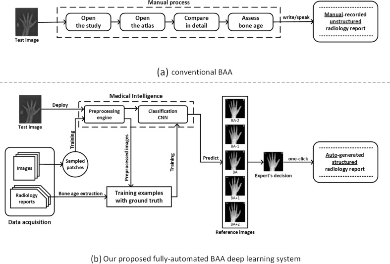
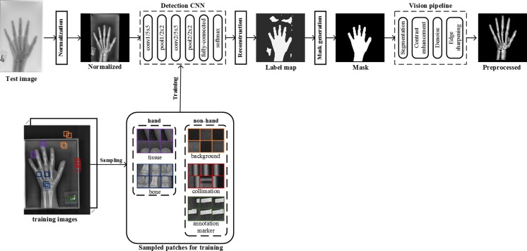
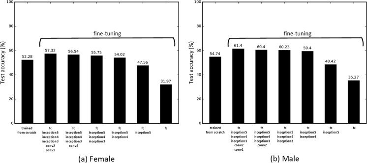
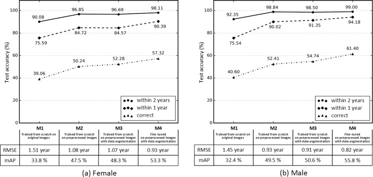
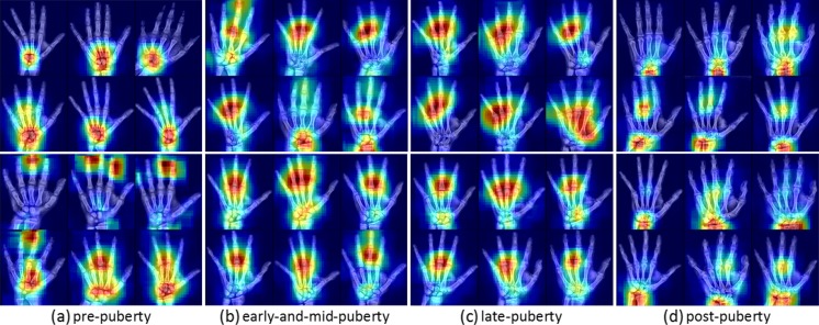
Skeletal maturity progresses through discrete phases, a fact that is used routinely in pediatrics where bone age assessments (BAAs) are compared to chronological age in the evaluation of endocrine and metabolic disorders. While central to many disease evaluations, little has changed to improve the tedious process since its introduction in 1950. In this study, we propose a fully automated deep learning pipeline to segment a region of interest, standardize and preprocess input radiographs, and perform BAA. Our models use an ImageNet pretrained, fine-tuned convolutional neural network (CNN) to achieve 57.32 and 61.40% accuracies for the female and male cohorts on our held-out test images. Female test radiographs were assigned a BAA within 1 year 90.39% and within 2 years 98.11% of the time. Male test radiographs were assigned 94.18% within 1 year and 99.00% within 2 years. Using the input occlusion method, attention maps were created which reveal what features the trained model uses to perform BAA. These correspond to what human experts look at when manually performing BAA. Finally, the fully automated BAA system was deployed in the clinical environment as a decision supporting system for more accurate and efficient BAAs at much faster interpretation time (<2 s) than the conventional method.
Overview
The claims and statistics of this paper haven’t been checked yet.
Run the check — free, no account needed. It reads the open-access text and checks the sources it cites.
Integrity signals
Registry
Cites
Statistics
Figures
Verification
Claims the paper makes
The claims and statistics of this paper haven’t been checked yet.
Run the check — free, no account needed. It reads the open-access text and checks the sources it cites.
Claims other papers make about this one
Each is checked against this paper’s own text: does it say what the citing paper says it does?
In the citing paper
“BoneXpert is one of the first commercial applications of AI in radiology [25] and appeared to be the only AI application in clinical use across several hospitals in The Netherlands.”
From Implementation of artificial intelligence (AI) applications in radiology: hindering and facilitating factors (2020) · cites this paper as [25]
Self-check
Percentages, sums and reported differences re-computed from the paper’s own numbers.
The self-check needs the paper’s full text.
Citations
Cites
Deep Residual Learning for Image Recognition
He, K., Zhang, X., Ren, S., & Sun, J. (2016) · 2016 IEEE Conference on Computer Vision and Pattern Recognition (CVPR) · doi:10.1109/cvpr.2016.90 · Cited by 235,474
Very Deep Convolutional Networks for Large-Scale Image Recognition
Simonyan, K., & Zisserman, A. (2014) · arXiv · doi:10.48550/arxiv.1409.1556 · Cited by 114,148
ImageNet classification with deep convolutional neural networks
Krizhevsky, A., Sutskever, I., & Hinton, G. E. (2017) · Communications of the ACM · doi:10.1145/3065386 · Cited by 112,155
LeCun, Y., Bengio, Y., & Hinton, G. (2015) · Nature · doi:10.1038/nature14539 · Cited by 84,126
ImageNet: A large-scale hierarchical image database
Deng, J., Dong, W., Socher, R., Li, L.-J., Li, K., & Fei-Fei, L. (2009) · 2009 IEEE Conference on Computer Vision and Pattern Recognition · doi:10.1109/cvpr.2009.5206848 · Cited by 65,333
Gradient-based learning applied to document recognition
Lecun, Y., Bottou, L., Bengio, Y., & Haffner, P. (1998) · Proceedings of the IEEE · doi:10.1109/5.726791 · Cited by 58,805
Going deeper with convolutions
Szegedy, C., Liu, W., Jia, Y., Sermanet, P., Reed, S., Anguelov, D., Erhan, D., Vanhoucke, V., & Rabinovich, A. (2015) · 2015 IEEE Conference on Computer Vision and Pattern Recognition (CVPR) · doi:10.1109/cvpr.2015.7298594 · Cited by 48,562
ImageNet Large Scale Visual Recognition Challenge
Russakovsky, O., Deng, J., Su, H., Krause, J., Satheesh, S., Ma, S., Huang, Z., Karpathy, A., Khosla, A., Bernstein, M., et al. (2015) · International Journal of Computer Vision · doi:10.1007/s11263-015-0816-y · Cited by 39,380
Rich Feature Hierarchies for Accurate Object Detection and Semantic Segmentation
Girshick, R., Donahue, J., Darrell, T., & Malik, J. (2014) · 2014 IEEE Conference on Computer Vision and Pattern Recognition · doi:10.1109/cvpr.2014.81 · Cited by 32,684
Visualizing and understanding convolutional networks
Zeiler, M. D., & Fergus, R. (2014) · Lecture Notes in Computer Science · doi:10.1007/978-3-319-10590-1_53 · Cited by 15,839
Caffe: Convolutional Architecture for Fast Feature Embedding
Jia, Y., Shelhamer, E., Donahue, J., Karayev, S., Long, J., Girshick, R., Guadarrama, S., & Darrell, T. (2014) · Proceedings of the 22nd ACM international conference on Multimedia · doi:10.1145/2647868.2654889 · Cited by 11,077
Shin, H.-C., Roth, H. R., Gao, M., Lu, L., Xu, Z., Nogues, I., Yao, J., Mollura, D., & Summers, R. M. (2016) · IEEE Transactions on Medical Imaging · doi:10.1109/tmi.2016.2528162 · Cited by 5,759
RADIOGRAPHIC ATLAS OF SKELETAL DEVELOPMENT OF THE HAND AND WRIST
GREULICH, W. W., & Pyle, S. I. (1959) · The American Journal of the Medical Sciences · doi:10.1097/00000441-195909000-00030 · Cited by 5,562
Deep Inside Convolutional Networks: Visualising Image Classification Models and Saliency Maps
Simonyan, K., Vedaldi, A., & Zisserman, A. (2013) · arXiv · doi:10.48550/arxiv.1312.6034 · Cited by 5,370
Deep Residual Learning for Image Recognition
He, K., Zhang, X., Ren, S., & Sun, J. (2015) · arXiv · doi:10.48550/arxiv.1512.03385 · Cited by 4,196
Caltech-UCSD Birds-200-2011 Dataset
Wah, C. (2024) · CaltechAUTHORS (California Institute of Technology) · doi:10.57702/i9oeewf6 · Cited by 3,620
Automated Flower Classification over a Large Number of Classes
Nilsback, M.-E., & Zisserman, A. (2008) · 2008 Sixth Indian Conference on Computer Vision, Graphics & Image Processing · doi:10.1109/icvgip.2008.47 · Cited by 3,442
Convolutional Neural Networks for Medical Image Analysis: Full Training or Fine Tuning?
Tajbakhsh, N., Shin, J. Y., Gurudu, S. R., Hurst, R. T., Kendall, C. B., Gotway, M. B., & Liang, J. (2016) · IEEE Transactions on Medical Imaging · doi:10.1109/tmi.2016.2535302 · Cited by 3,277
Greenspan, H. K., van Ginneken, B., & Summers, R. M. (2016) · IEEE Transactions on Medical Imaging · doi:10.1109/tmi.2016.2553401 · Cited by 1,725
Lung pattern classification for interstitial lung diseases using a deep convolutional neural network
Anthimopoulos, M., Christodoulidis, S., Ebner, L., Christe, A., & Mougiakakou, S. (2016) · IEEE Transactions on Medical Imaging · doi:10.1109/tmi.2016.2535865 · Cited by 1,343
An Analysis of Deep Neural Network Models for Practical Applications
Canziani, A., Paszke, A., & Culurciello, E. (2016) · arXiv · doi:10.48550/arxiv.1605.07678 · Cited by 969
Segmenting Retinal Blood Vessels With Deep Neural Networks
Liskowski, P., & Krawiec, K. (2016) · IEEE Transactions on Medical Imaging · doi:10.1109/tmi.2016.2546227 · Cited by 904
ImageNet Large Scale Visual Recognition Challenge
Russakovsky, O., Deng, J., Su, H., Krause, J., Satheesh, S., Ma, S., Huang, Z., Karpathy, A., Khosla, A., Bernstein, M., et al. (2014) · arXiv · doi:10.48550/arxiv.1409.0575 · Cited by 567
The BoneXpert Method for Automated Determination of Skeletal Maturity
Thodberg, H. H., Kreiborg, S., Juul, A., & Pedersen, K. D. (2009) · IEEE Transactions on Medical Imaging · doi:10.1109/tmi.2008.926067 · Cited by 438
van Grinsven, M. J. J. P., van Ginneken, B., Hoyng, C. B., Theelen, T., & Sanchez, C. I. (2016) · IEEE Transactions on Medical Imaging · doi:10.1109/tmi.2016.2526689 · Cited by 426
Assessment of Skeletal Maturity and Prediction of Adult Height (TW2 Method)
Garn, S. M., & Tanner, J. M. (1986) · Man · doi:10.2307/2802657 · Cited by 314
Multi-Instance Deep Learning: Discover Discriminative Local Anatomies for Bodypart Recognition
Yan, Z., Zhan, Y., Peng, Z., Liao, S., Shinagawa, Y., Zhang, S., Metaxas, D., & Zhou, X. S. (2016) · IEEE Transactions on Medical Imaging · doi:10.1109/tmi.2016.2524985 · Cited by 227
King, D. G., Steventon, D. M., O'Sullivan, M. P., Cook, A., Hornsby, V. P. L., Jefferson, I. G., & King, P. R. (1994) · British Journal of Radiology · doi:10.1259/0007-1285-67-801-848 · Cited by 169
Hand Bone Age: A Digital Atlas of Skeletal Maturity
Gilsanz, V., & Ratib, O. (2005) · Digital Access to Libraries (Université catholique de Louvain (UCL), l'Université de Namur (UNamur) and the Université Saint-Louis (USL-B)) · doi:10.1007/978-3-642-23762-1 · Cited by 132
Automatic bone age assessment for young children from newborn to 7-year-old using carpal bones
Zhang, A., Gertych, A., & Liu, B. J. (2007) · Computerized Medical Imaging and Graphics · doi:10.1016/j.compmedimag.2007.02.008 · Cited by 125
L??EXPLORATION CHIRURGICALE ST??R??OTAXIQUE DU LOBE TEMPORAL DANS L??EPILEPSIE TEMPORALE
Talairach, J., Tournoux, P., & DAVID, M. (1959) · The American Journal of the Medical Sciences · doi:10.1097/00000441-195909000-00032 · Cited by 79
Digital hand atlas and web-based bone age assessment: system design and implementation
Cao, F. F., Huang, H. K., Piętka, E., & Gilsanz, V. (2000) · Computerized Medical Imaging and Graphics · doi:10.1016/s0895-6111(00)00026-4 · Cited by 75
Somkantha, K., Theera‐Umpon, N., & Auephanwiriyakul, S. (2011) · Journal of Imaging Informatics in Medicine · doi:10.1007/s10278-011-9372-3 · Cited by 57
Automatic Bone Age Measurement Using Computerized Image Analysis
Tanner, J. M., & Gibbons, R. D. (1994) · Journal of Pediatric Endocrinology and Metabolism · doi:10.1515/jpem.1994.7.2.141 · Cited by 56
Leveraging the Wisdom of the Crowd for Fine-Grained Recognition
Deng, J., Krause, J., Stark, M. M., & Fei-Fei, L. (2015) · IEEE Transactions on Pattern Analysis and Machine Intelligence · doi:10.1109/tpami.2015.2439285 · Cited by 52
Tanner, J. M., Oshman, D., Lindgren, G., Grunbaum, J. A., Elouki, R., & Labarthe, D. R. (2008) · Hormone Research · doi:10.1159/000184211 · Cited by 49
Automated classification system for Bone Age X-ray images
Seok, J., Hyun, B., Kasa-Vubu, J. Z., & Girard, A. (2012) · Conference proceedings/Conference proceedings - IEEE International Conference on Systems, Man, and Cybernetics · doi:10.1109/icsmc.2012.6377701 · Cited by 22
Zhang, J., Lin, F., & Ding, X. (2016) · Korean Journal of Radiology · doi:10.3348/kjr.2016.17.3.435 · Cited by 20
Cameron, N. (1984) · Annals of Human Biology · doi:10.1080/03014468400007151 · Cited by 13
Comparison of the Greulich-Pyle and Tanner Whitehouse (TW3) Methods in Bone age Assessment
Kim, S., 오연정, Shin, J. Y., 이영준, & Lee, K. (2008) · Annals of Pediatric Endocrinology & Metabolism · Cited by 7
Research works printed but not linked: 1
- Greulich WW, Pyle SI. Radiographic atlas of skeletal development of the hand and wrist. Am J Med Sci. pdfs.journals. lww.com ; 1959; Available: http://pdfs.journals.lww.com/amjmedsci/1959/09000/Radiographic_Atlas_of_Skeletal_Development_of_the.30.pdf · DOI
Cited by
Sorted by citations
Transfer learning for medical image classification: a literature review
Kim, H. E., Cosa‐Linan, A., Santhanam, N., Jannesari, M., Maros, M. E., & Ganslandt, T. (2022) · BMC Medical Imaging · doi:10.1186/s12880-022-00793-7 · Cited by 1,117
Current Applications and Future Impact of Machine Learning in Radiology
Choy, G., Khalilzadeh, O., Michalski, M., Do, S., Samir, A. E., Pianykh, O. S., Geis, J. R., Pandharipande, P. V., Brink, J. A., & Dreyer, K. J. (2018) · Radiology · doi:10.1148/radiol.2018171820 · Cited by 800
Self-citationDeep learning in medical imaging and radiation therapy
Sahiner, B., Pezeshk, A. X., Hadjiiski, L. M., Wang, X., Drukker, K., Cha, K. H., Summers, R. M., & Giger, M. L. (2018) · Medical Physics · doi:10.1002/mp.13264 · Cited by 763
Thrall, J. H., Li, X., Li, Q., Cruz, C., Do, S., Dreyer, K., & Brink, J. (2018) · Journal of the American College of Radiology · doi:10.1016/j.jacr.2017.12.026 · Cited by 710
Self-citationArtificial Intelligence: The Ambiguous Labor Market Impact of Automating Prediction
Agrawal, A., Gans, J. S., & Goldfarb, A. (2019) · Journal of Economic Perspectives · doi:10.1257/jep.33.2.31 · Cited by 685
Metadata
- Authors
- Lee, H., Tajmir, S., Lee, J., Zissen, M., Yeshiwas, B. A., Alkasab, T. K., Choy, G., & Do, S.
- Affiliations
- Harvard University (US)
- Corresponding author
- Do, Synho
- Venue
- Journal of Digital Imaging
- Published
- 2017-03-08
- Volume
- 30
- Issue
- 4
- Pages
- 427-441
- ISSN
- 0897-1889 (ISSN-L)
- 1618-727X
- PMC
- PMC5537090
- OpenAlex
- W2592765733
- Cited by
- 452
- References
- 40
- Citations by year
- 2026: 21 · 2025: 37 · 2024: 45 · 2023: 57 · 2022: 55 · 2021: 62 · 2020: 75 · 2019: 47 · 2018: 45 · 2017: 8
- FWCI
- 276.78
- Type
- journal article
- Language
- en
- Open access
- hybrid
- doi.org
- Locations
- Journal of Imaging Informatics in Medicine (publishedVersion, cc-by)
- PubMed Central (publishedVersion, cc-by)
- PubMed (publishedVersion)
- Digital Access to Scholarship at Harvard (DASH) (Harvard University) (submittedVersion, cc-by)
- Topics
- Social Sciences › Arts and Humanities › Archeology (arts and humanities) › Forensic Anthropology and Bioarchaeology Studies
- Physical Sciences › Engineering › Biomedical Engineering › Medical Imaging and Analysis
- Health Sciences › Medicine › Health Informatics › Artificial Intelligence in Healthcare and Education
- Keywords
- bone age assessment, skeletal maturity, convolutional neural networks, attention maps, decision support system, pediatric endocrine disorders, automated image analysis
- MeSH terms
- Machine Learning, Adolescent, Adult, Age Determination by Skeleton, Child, Female, Hand, Humans, Male, Software, Neural Networks, Computer, Decision Support Systems, Clinical
About these checks
Registry, retraction, Cites and statistics results are computed, not judged, with one exception: a printed reference that no index lists is read once by a model before it counts as a citation error. Claims are extracted by a model and kept only when their quote appears verbatim in the text; each cited source is then checked, with a model’s help, for whether it says what the claim relies on. Those verdicts can be wrong — read the claim’s quote and the cited source before relying on one.
Statistics and arithmetic are recomputed, not judged.
Confidence bands are shown only for fields calibrated on our annotated test set. None is calibrated yet, so no band is shown.
What counts as a claim ›How we measure confidence ›How we check this ›
Are you an author, or do you see a problem with this page? Request a review
