

The DOI matches its Crossref record; 40 of the 40 references it cites were found in a registry or an index.
Fully Automated Deep Learning System for Bone Age Assessment
Open accessAbstract
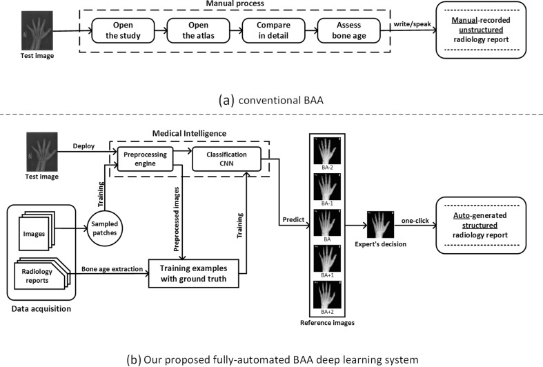
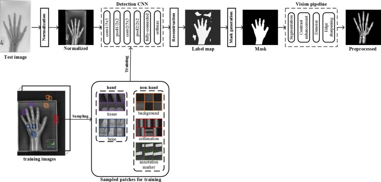
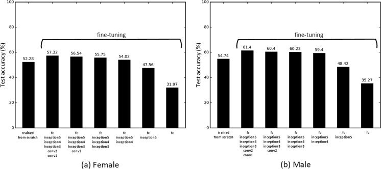
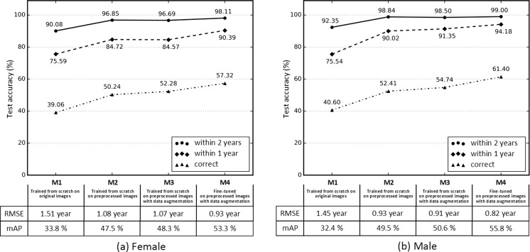
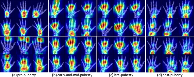
Skeletal maturity progresses through discrete phases, a fact that is used routinely in pediatrics where bone age assessments (BAAs) are compared to chronological age in the evaluation of endocrine and metabolic disorders. While central to many disease evaluations, little has changed to improve the tedious process since its introduction in 1950. In this study, we propose a fully automated deep learning pipeline to segment a region of interest, standardize and preprocess input radiographs, and perform BAA. Our models use an ImageNet pretrained, fine-tuned convolutional neural network (CNN) to achieve 57.32 and 61.40% accuracies for the female and male cohorts on our held-out test images. Female test radiographs were assigned a BAA within 1 year 90.39% and within 2 years 98.11% of the time. Male test radiographs were assigned 94.18% within 1 year and 99.00% within 2 years. Using the input occlusion method, attention maps were created which reveal what features the trained model uses to perform BAA. These correspond to what human experts look at when manually performing BAA. Finally, the fully automated BAA system was deployed in the clinical environment as a decision supporting system for more accurate and efficient BAAs at much faster interpretation time (<2 s) than the conventional method.
Overview
The claims and statistics of this paper haven’t been checked yet.
Run the check — free, no account needed. It reads the open-access text and checks the sources it cites.
Integrity signals
Registry
Cites
Statistics
Figures
Verification
Claims the paper makes
The claims and statistics of this paper haven’t been checked yet.
Run the check — free, no account needed. It reads the open-access text and checks the sources it cites.
Claims other papers make about this one
Each is checked against this paper’s own text: does it say what the citing paper says it does?
In the citing paper
“BoneXpert is one of the first commercial applications of AI in radiology [25] and appeared to be the only AI application in clinical use across several hospitals in The Netherlands.”
From Implementation of artificial intelligence (AI) applications in radiology: hindering and facilitating factors (2020) · cites this paper as [25]
Self-check
Percentages, sums and reported differences re-computed from the paper’s own numbers.
The self-check needs the paper’s full text.
Citations
Cites
Deep Residual Learning for Image Recognition
He, K., Zhang, X., Ren, S., & Sun, J. (2016) · 2016 IEEE Conference on Computer Vision and Pattern Recognition (CVPR) · doi:10.1109/cvpr.2016.90 · Cited by 235,474
Very Deep Convolutional Networks for Large-Scale Image Recognition
Simonyan, K., & Zisserman, A. (2014) · arXiv · doi:10.48550/arxiv.1409.1556 · Cited by 114,148
ImageNet classification with deep convolutional neural networks
Krizhevsky, A., Sutskever, I., & Hinton, G. E. (2017) · Communications of the ACM · doi:10.1145/3065386 · Cited by 112,155
LeCun, Y., Bengio, Y., & Hinton, G. (2015) · Nature · doi:10.1038/nature14539 · Cited by 84,126
ImageNet: A large-scale hierarchical image database
Deng, J., Dong, W., Socher, R., Li, L.-J., Li, K., & Fei-Fei, L. (2009) · 2009 IEEE Conference on Computer Vision and Pattern Recognition · doi:10.1109/cvpr.2009.5206848 · Cited by 65,333
Gradient-based learning applied to document recognition
Lecun, Y., Bottou, L., Bengio, Y., & Haffner, P. (1998) · Proceedings of the IEEE · doi:10.1109/5.726791 · Cited by 58,805
Going deeper with convolutions
Szegedy, C., Liu, W., Jia, Y., Sermanet, P., Reed, S., Anguelov, D., Erhan, D., Vanhoucke, V., & Rabinovich, A. (2015) · 2015 IEEE Conference on Computer Vision and Pattern Recognition (CVPR) · doi:10.1109/cvpr.2015.7298594 · Cited by 48,562
ImageNet Large Scale Visual Recognition Challenge
Russakovsky, O., Deng, J., Su, H., Krause, J., Satheesh, S., Ma, S., Huang, Z., Karpathy, A., Khosla, A., Bernstein, M., et al. (2015) · International Journal of Computer Vision · doi:10.1007/s11263-015-0816-y · Cited by 39,380
Rich Feature Hierarchies for Accurate Object Detection and Semantic Segmentation
Girshick, R., Donahue, J., Darrell, T., & Malik, J. (2014) · 2014 IEEE Conference on Computer Vision and Pattern Recognition · doi:10.1109/cvpr.2014.81 · Cited by 32,684
Visualizing and understanding convolutional networks
Zeiler, M. D., & Fergus, R. (2014) · Lecture Notes in Computer Science · doi:10.1007/978-3-319-10590-1_53 · Cited by 15,839
Research works printed but not linked: 1
- Greulich WW, Pyle SI. Radiographic atlas of skeletal development of the hand and wrist. Am J Med Sci. pdfs.journals. lww.com ; 1959; Available: http://pdfs.journals.lww.com/amjmedsci/1959/09000/Radiographic_Atlas_of_Skeletal_Development_of_the.30.pdf · DOI
Cited by
Sorted by citations
Transfer learning for medical image classification: a literature review
Kim, H. E., Cosa‐Linan, A., Santhanam, N., Jannesari, M., Maros, M. E., & Ganslandt, T. (2022) · BMC Medical Imaging · doi:10.1186/s12880-022-00793-7 · Cited by 1,117
Current Applications and Future Impact of Machine Learning in Radiology
Choy, G., Khalilzadeh, O., Michalski, M., Do, S., Samir, A. E., Pianykh, O. S., Geis, J. R., Pandharipande, P. V., Brink, J. A., & Dreyer, K. J. (2018) · Radiology · doi:10.1148/radiol.2018171820 · Cited by 800
Self-citationDeep learning in medical imaging and radiation therapy
Sahiner, B., Pezeshk, A. X., Hadjiiski, L. M., Wang, X., Drukker, K., Cha, K. H., Summers, R. M., & Giger, M. L. (2018) · Medical Physics · doi:10.1002/mp.13264 · Cited by 763
Thrall, J. H., Li, X., Li, Q., Cruz, C., Do, S., Dreyer, K., & Brink, J. (2018) · Journal of the American College of Radiology · doi:10.1016/j.jacr.2017.12.026 · Cited by 710
Self-citationArtificial Intelligence: The Ambiguous Labor Market Impact of Automating Prediction
Agrawal, A., Gans, J. S., & Goldfarb, A. (2019) · Journal of Economic Perspectives · doi:10.1257/jep.33.2.31 · Cited by 685
Artificial Intelligence in Anesthesiology
Hashimoto, D. A., Witkowski, E. R., Gao, L., Meireles, O. R., & Rosman, G. (2019) · Anesthesiology · doi:10.1097/aln.0000000000002960 · Cited by 591
Convolutional Neural Networks for Radiologic Images: A Radiologist’s Guide
Soffer, S., Ben-Cohen, A., Shimon, O., Amitai, M. M., Greenspan, H. K., & Klang, E. (2019) · Radiology · doi:10.1148/radiol.2018180547 · Cited by 528
McBee, M. P., Awan, O. A. R., Colucci, A. T., Ghobadi, C. W., Kadom, N., Kansagra, A. P., Tridandapani, S., & Auffermann, W. F. (2018) · Academic Radiology · doi:10.1016/j.acra.2018.02.018 · Cited by 512
The RSNA Pediatric Bone Age Machine Learning Challenge
Halabi, S. S., Prevedello, L. M. S., Kalpathy–Cramer, J., Mamonov, A. B., Bilbily, A., Cicero, M. D., Pan, I., Pereira, L. A., Sousa, R. T., Abdala, N., et al. (2018) · Radiology · doi:10.1148/radiol.2018180736 · Cited by 465
Hung, K. F., Montalvao, C., Tanaka, R., Kawai, T., & Bornstein, M. M. (2019) · Dentomaxillofacial Radiology · doi:10.1259/dmfr.20190107 · Cited by 417
Physician Confidence in Artificial Intelligence: An Online Mobile Survey
Oh, S., Kim, J. H., Choi, S.-W., Lee, H. J., Hong, J., & Kwon, S. H. (2019) · Journal of Medical Internet Research · doi:10.2196/12422 · Cited by 387
Self-citationOrhan, K., Bayrakdar, İ. Ş., Ezhov, M., Kravtsov, A., & Özyürek, T. (2020) · International Endodontic Journal · doi:10.1111/iej.13265 · Cited by 369
Strohm, L., Hehakaya, C., Ranschaert, E. R., Boon, W. P. C., & Moors, E. H. M. (2020) · European Radiology · doi:10.1007/s00330-020-06946-y · Cited by 329
Hiraiwa, T., Ariji, Y., Fukuda, M., Kise, Y., Nakata, K., Katsumata, A., Fujita, H., & Ariji, E. (2018) · Dentomaxillofacial Radiology · doi:10.1259/dmfr.20180218 · Cited by 339
Mall, P. K., Singh, P. K., Srivastav, S., Narayan, V., Paprzycki, M., Jaworska, T., & Ganzha, M. (2023) · Healthcare Analytics · doi:10.1016/j.health.2023.100216 · Cited by 310
Urakawa, T., Tanaka, Y., Goto, S., Matsuzawa, H., Watanabe, K., & Endo, N. (2018) · Skeletal Radiology · doi:10.1007/s00256-018-3016-3 · Cited by 271
Mousavi Baigi, S. F., Sarbaz, M., Ghaddaripouri, K., Ghaddaripouri, M., Mousavi, A. S., & Kimiafar, K. (2023) · Health Science Reports · doi:10.1002/hsr2.1138 · Cited by 255
Explanation Methods in Deep Learning: Users, Values, Concerns and Challenges
Ras, G., van Gerven, M., & Haselager, P. (2018) · The Springer Series on Challenges in Machine Learning · doi:10.1007/978-3-319-98131-4_2 · Cited by 255
Li, F., Ruijs, N., & Lü, Y. (2022) · AI · doi:10.3390/ai4010003 · Cited by 284
A deep learning approach for dental implant planning in cone-beam computed tomography images
Bayrakdar, S. K., Orhan, K., Bayrakdar, İ. Ş., Bilgir, E., Ezhov, M., Gusarev, M., & Shumilov, E. (2021) · BMC Medical Imaging · doi:10.1186/s12880-021-00618-z · Cited by 228
Deep Learning to Classify Radiology Free-Text Reports
Chen, M. C., Ball, R. L., Yang, L., Moradzadeh, N., Chapman, B. E., Larson, D. B., Langlotz, C. P., Amrhein, T. J., & Lungren, M. P. (2018) · Radiology · doi:10.1148/radiol.2017171115 · Cited by 217
Murata, M., Ariji, Y., Ohashi, Y., Kawai, T., Fukuda, M., Funakoshi, T., Kise, Y., Nozawa, M., Katsumata, A., Fujita, H., et al. (2018) · Oral Radiology · doi:10.1007/s11282-018-0363-7 · Cited by 212
Management of Thyroid Nodules Seen on US Images: Deep Learning May Match Performance of Radiologists
Buda, M., Wildman‐Tobriner, B., Hoang, J. K., Thayer, D., Tessler, F. N., Middleton, W. D., & Mazurowski, M. A. (2019) · Radiology · doi:10.1148/radiol.2019181343 · Cited by 215
Assessment of Convolutional Neural Networks for Automated Classification of Chest Radiographs
Dunnmon, J. A., Yi, D., Langlotz, C. P., Ré, C., Rubin, D. L., & Lungren, M. P. (2019) · Radiology · doi:10.1148/radiol.2018181422 · Cited by 206
Paediatric Bone Age Assessment Using Deep Convolutional Neural Networks
Iglovikov, V., Rakhlin, A., Kalinin, A. A., & Shvets, A. A. (2018) · Lecture notes in computer science · doi:10.1007/978-3-030-00889-5_34 · Cited by 195
When Machines Think: Radiology’s Next Frontier
Dreyer, K. J., & Geis, J. R. (2017) · Radiology · doi:10.1148/radiol.2017171183 · Cited by 183
Wang, X., Yang, W. H., Weinreb, J. C., Han, J., Li, Q., Kong, X., Yan, Y., Ke, Z., Luo, B., Liu, T., et al. (2017) · Scientific Reports · doi:10.1038/s41598-017-15720-y · Cited by 200
Attitudes and perceptions of dental students towards artificial intelligence
Yüzbaşıoğlu, E. (2020) · Journal of Dental Education · doi:10.1002/jdd.12385 · Cited by 225
Ariji, Y., Fukuda, M., Kise, Y., Nozawa, M., Yanashita, Y., Fujita, H., Katsumata, A., & Ariji, E. (2018) · Oral Surgery Oral Medicine Oral Pathology and Oral Radiology · doi:10.1016/j.oooo.2018.10.002 · Cited by 180
Kim, J. R., Shim, W. H., Yoon, H. M., Hong, S. H., Lee, J. S., Cho, Y. A., & Kim, S. (2017) · American Journal of Roentgenology · doi:10.2214/ajr.17.18224 · Cited by 170
Self-citationArtificial Intelligence in Musculoskeletal Imaging: Current Status and Future Directions
Gyftopoulos, S., Lin, D. J., Knöll, F., Doshi, A. M., Rodrigues, T. C., & Recht, M. P. (2019) · American Journal of Roentgenology · doi:10.2214/ajr.19.21117 · Cited by 170
Lee, H., Troschel, F. M., Tajmir, S., Fuchs, G., Mario, J., Fintelmann, F. J., & Do, S. (2017) · Journal of Imaging Informatics in Medicine · doi:10.1007/s10278-017-9988-z · Cited by 164
Self-citationPedoia, V., Norman, B., Mehany, S. N., Bucknor, M. D., Link, T. M., & Majumdar, S. (2018) · Journal of Magnetic Resonance Imaging · doi:10.1002/jmri.26246 · Cited by 165
Artificial Intelligence in Radiology: Current Technology and Future Directions
Syed, A., & Zoga, A. (2018) · Seminars in Musculoskeletal Radiology · doi:10.1055/s-0038-1673383 · Cited by 140
Cardiovascular Stents: A Review of Past, Current, and Emerging Devices
Udriște, A. S. S., Niculescu, A.-G., Grumezescu, A., & Bădilă, E. (2021) · Materials · doi:10.3390/ma14102498 · Cited by 125
Dallora, A. L., Anderberg, P., Kvist, O., Mendes, E., Ruiz, S. D., & Berglund, J. S. (2019) · PLoS ONE · doi:10.1371/journal.pone.0220242 · Cited by 152
Artificial intelligence in medicine: what is it doing for us today?
Becker, A. (2019) · Health Policy and Technology · doi:10.1016/j.hlpt.2019.03.004 · Cited by 137
Parakh, A., Lee, H., Lee, J. H., Eisner, B. H., Sahani, D. V., & Do, S. (2019) · Radiology Artificial Intelligence · doi:10.1148/ryai.2019180066 · Cited by 146
Self-citationHussain, L., Nguyen, T., Li, H., Abbasi, A. A., Lone, K. J., Zhao, Z., Zaib, M., Chen, A. L., & Duong, T. Q. (2020) · BioMedical Engineering OnLine · doi:10.1186/s12938-020-00831-x · Cited by 135
Current applications and future directions of deep learning in musculoskeletal radiology
Chea, P., & Mandell, J. C. (2019) · Skeletal Radiology · doi:10.1007/s00256-019-03284-z · Cited by 134
Artificial Intelligence in Clinical Decision Support: a Focused Literature Survey
Stefania Montani, B., & Striani, M. (2019) · Yearbook of Medical Informatics · doi:10.1055/s-0039-1677911 · Cited by 138
Tajmir, S., Lee, H., Shailam, R., Gale, H. I., Nguyen, J. C., Westra, S. J., Lim, R., Yune, S., Gee, M. S., & Do, S. (2018) · Skeletal Radiology · doi:10.1007/s00256-018-3033-2 · Cited by 128
Self-citationDeep Learning in Radiology: Does One Size Fit All?
Erickson, B. J., Korfiatis, P. D., Kline, T. L., Akkus, Z., Philbrick, K. A., & Weston, A. D. (2018) · Journal of the American College of Radiology · doi:10.1016/j.jacr.2017.12.027 · Cited by 120
Pedoia, V., Lee, J., Norman, B., Link, T. M., & Majumdar, S. (2019) · Osteoarthritis and Cartilage · doi:10.1016/j.joca.2019.02.800 · Cited by 125
Self-citationApplication of Artificial Intelligence in Orthodontics: Current State and Future Perspectives
Liu, J., Zhang, C., & Shan, Z. (2023) · Healthcare · doi:10.3390/healthcare11202760 · Cited by 149
Artificial Intelligence and Clinical Decision Support for Radiologists and Referring Providers
Bizzo, B. C., de Almeida, R. R., Michalski, M. H., & Alkasab, T. K. (2019) · Journal of the American College of Radiology · doi:10.1016/j.jacr.2019.06.010 · Cited by 100
Self-citationA modified YOLOv3 detection method for vision-based water surface garbage capture robot
Li, X., Tian, M., Kong, S., Wu, L., & Yu, J. (2020) · International Journal of Advanced Robotic Systems · doi:10.1177/1729881420932715 · Cited by 98
Tajmir, S., & Alkasab, T. K. (2018) · Academic Radiology · doi:10.1016/j.acra.2018.03.007 · Cited by 99
Self-citationDetection of rheumatoid arthritis from hand radiographs using a convolutional neural network
Üreten, K., Erbay, H., & Maraş, H. H. (2019) · Clinical Rheumatology · doi:10.1007/s10067-019-04487-4 · Cited by 91
MABAL: a Novel Deep-Learning Architecture for Machine-Assisted Bone Age Labeling
Mutasa, S., Chang, P., Ruzal‐Shapiro, C., & Ayyala, R. S. (2018) · Journal of Imaging Informatics in Medicine · doi:10.1007/s10278-018-0053-3 · Cited by 92
Seo, H., Hwang, J. J., Jeong, T.-S., & Shin, J. (2021) · Journal of Clinical Medicine · doi:10.3390/jcm10163591 · Cited by 98
Xiao, B., Xu, Y., Bi, X., Li, W., Ma, Z., Zhang, J., & Ma, X. (2019) · IEEE Internet of Things Journal · doi:10.1109/jiot.2019.2961132 · Cited by 87
Bui, T. D., Lee, J.-J., & Shin, J. (2019) · Artificial Intelligence in Medicine · doi:10.1016/j.artmed.2019.04.005 · Cited by 86
Self-citationForensic age estimation for pelvic X-ray images using deep learning
Li, Y., Huang, Z., Dong, X., Liang, W., Xue, H., Zhang, L., Zhang, Y., & Deng, Z. (2018) · European Radiology · doi:10.1007/s00330-018-5791-6 · Cited by 90
Nadeem, M. W., Goh, H. G., Ali, A., Hussain, M., Khan, M. A., & Ponnusamy, V. (2020) · Diagnostics · doi:10.3390/diagnostics10100781 · Cited by 85
Eng, D. K.-M., Khandwala, N., Long, J., Fefferman, N. R., Lala, S. V., Strubel, N. A., Milla, S. S., Filice, R. W., Sharp, S. E., Towbin, A. J., et al. (2021) · Radiology · doi:10.1148/radiol.2021204021 · Cited by 88
Fritz, B., Marbach, G., Civardi, F., Fucentese, S. F., & Pfirrmann, C. W. A. (2020) · Skeletal Radiology · doi:10.1007/s00256-020-03410-2 · Cited by 83
Towards fully automated third molar development staging in panoramic radiographs
Banar, N., Bertels, J., Laurent, F., Boedi, R. M., Tobel, J. D., Thevissen, P. W., & Vandermeulen, D. (2020) · International Journal of Legal Medicine · doi:10.1007/s00414-020-02283-3 · Cited by 89
Li, D., Xiao, C., Liu, Y., Chen, Z., Hassan, H., Su, L., Liu, J., Li, H., Xie, W., Zhong, W., et al. (2022) · Diagnostics · doi:10.3390/diagnostics12081788 · Cited by 94
Effects of Hypertension, Diabetes, and Smoking on Age and Sex Prediction from Retinal Fundus Images
Kim, Y. D., Noh, K. J., Byun, S. J., Lee, S., Kim, T., Sunwoo, L., Lee, K. J., Kang, S., Park, K. H., & Park, S. J. (2020) · Scientific Reports · doi:10.1038/s41598-020-61519-9 · Cited by 86
Traditional and New Methods of Bone Age Assessment-An Overview
Prokop-Piotrkowska, M., Marszałek-Dziuba, K., Moszczyńska, E., Szalecki, M., & Jurkiewicz, E. (2020) · Journal of Clinical Research in Pediatric Endocrinology · doi:10.4274/jcrpe.galenos.2020.2020.0091 · Cited by 81
TW3-Based Fully Automated Bone Age Assessment System Using Deep Neural Networks
Son, S. J., Song, Y.-M., Kim, N., Do, Y., Kwak, N., Lee, M. S., & Lee, B.-D. (2019) · IEEE Access · doi:10.1109/access.2019.2903131 · Cited by 80
Mesejo, P., Martos, R., Ibáñez, O. C., Novo, J., & Ortega, M. (2020) · Applied Sciences · doi:10.3390/app10144703 · Cited by 84
An Ensemble-Based Densely-Connected Deep Learning System for Assessment of Skeletal Maturity
Wang, S., Wang, X., Shen, Y., He, B., Zhao, X., Cheung, P. W. H., Cheung, J. P. Y., Luk, K. D. K., & Hu, Y. S. (2020) · IEEE Transactions on Systems Man and Cybernetics Systems · doi:10.1109/tsmc.2020.2997852 · Cited by 73
Basic of machine learning and deep learning in imaging for medical physicists
Manco, L., Maffei, N., Strolin, S., Vichi, S., Bottazzi, L., & Strigari, L. (2021) · Physica Medica · doi:10.1016/j.ejmp.2021.03.026 · Cited by 75
Booz, C., Yel, İ., Wichmann, J. L., Boettger, S., Kamali, A. A., Albrecht, M. H., Martin, S. S., Lenga, L., Huizinga, N. A., D’Angelo, T., et al. (2020) · European Radiology Experimental · doi:10.1186/s41747-019-0139-9 · Cited by 72
Alzubaidi, L., Al-Dulaimi, K., Salhi, A., Alammar, Z., Fadhel, M. A., Albahri, A. S., Alamoodi, A. H., Albahri, O. S., Hasan, A. F., Bai, J., et al. (2024) · Artificial Intelligence in Medicine · doi:10.1016/j.artmed.2024.102935 · Cited by 78
Pediatric Bone Age Assessment Using Deep Convolutional Neural NetworksPreprint
Iglovikov, V., Rakhlin, A., Kalinin, A. A., & Shvets, A. A. (2017) · bioRxiv (Cold Spring Harbor Laboratory) · doi:10.1101/234120 · Cited by 71
Application and Construction of Deep Learning Networks in Medical Imaging
Torres-Velázquez, M., Chen, W., Li, X., & McMillan, A. B. (2020) · IEEE Transactions on Radiation and Plasma Medical Sciences · doi:10.1109/trpms.2020.3030611 · Cited by 74
Automated age estimation of young individuals based on 3D knee MRI using deep learning
der Mauer, M. A., Well, E. J.-V., Herrmann, J., Groth, M., Morlock, M. M., Maas, R., & Säring, D. (2020) · International Journal of Legal Medicine · doi:10.1007/s00414-020-02465-z · Cited by 68
Workflow Applications of Artificial Intelligence in Radiology and an Overview of Available Tools
Kapoor, N., Lacson, R. C., & Khorasani, R. (2020) · Journal of the American College of Radiology · doi:10.1016/j.jacr.2020.08.016 · Cited by 66
Automated Bone Age Assessment Using Artificial Intelligence: The Future of Bone Age Assessment
Lee, B.-D., & Lee, M. S. (2021) · Korean Journal of Radiology · doi:10.3348/kjr.2020.0941 · Cited by 70
Fan, F., Ke, W., Wu, W., Tian, X., Lyu, T., Liu, Y., Liao, P., Dai, X., Chen, H., & Deng, Z. (2020) · Forensic Science International · doi:10.1016/j.forsciint.2020.110416 · Cited by 70
Pan, I., Agarwal, S., & Merck, D. L. (2019) · Journal of Imaging Informatics in Medicine · doi:10.1007/s10278-019-00180-9 · Cited by 64
Technology Can Augment, but Not Replace, Critical Human Skills Needed for Patient Care
Alrassi, J., Katsufrakis, P. J., & Chandran, L. (2021) · Academic Medicine · doi:10.1097/acm.0000000000003733 · Cited by 60
A deep learning-based computer-aided diagnosis method of X-ray images for bone age assessment
Li, S., Liu, B., Li, S., Zhu, X., Yan, Y., & Zhang, D. (2021) · Complex & Intelligent Systems · doi:10.1007/s40747-021-00376-z · Cited by 61
Deep learning for lithological classification of carbonate rock micro-CT images
dos Anjos, C. E. M., Avila, M. R. V., Vasconcelos, A. G. P., Neta, A. P. M., Medeiros, L. C., Evsukoff, A. G., Surmas, R., & Landau, L. (2021) · Computational Geosciences · doi:10.1007/s10596-021-10033-6 · Cited by 63
Orhan, K., Belgın, C. A., Manulis, D., Golitsyna, M., Bayrak, S., Aksoy, S., Sanders, A., Önder, M., Ezhov, M., Shamshiev, M., et al. (2023) · Imaging Science in Dentistry · doi:10.5624/isd.20230109 · Cited by 59
Kim, E., Oh, I.-S., So, J.-E., Kang, J., Le, V. N. T., Tak, M.-K., & Lee, D. (2021) · Journal of Clinical Medicine · doi:10.3390/jcm10225400 · Cited by 62
Lee, H., Mansouri, M., Tajmir, S., Lev, M. H., & Do, S. (2017) · Journal of Imaging Informatics in Medicine · doi:10.1007/s10278-017-0025-z · Cited by 56
Self-citationLearning osteoarthritis imaging biomarkers from bone surface spherical encoding
Martinez, A. M., Calivá, F., Flament, I., Liu, F., Lee, J., Cao, P., Shah, R., Majumdar, S., & Pedoia, V. (2020) · Magnetic Resonance in Medicine · doi:10.1002/mrm.28251 · Cited by 60
Self-citationD’Angelo, T., Caudo, D., Blandino, A., Albrecht, M. H., Vogl, T. J., Gruenewald, L. D., Gaeta, M., Yel, İ., Koch, V., Martin, S. S., et al. (2022) · Journal of Clinical Ultrasound · doi:10.1002/jcu.23321 · Cited by 59
Skeletal Maturity Recognition Using a Fully Automated System With Convolutional Neural Networks
Wang, S., Shen, Y., Shi, C., Yin, P., Wang, Z., Cheung, P. W. H., Cheung, J. P. Y., Luk, K. D. K., & Hu, Y. S. (2018) · IEEE Access · doi:10.1109/access.2018.2843392 · Cited by 56
Atici, S. F., Ansari, R., Allareddy, V., Suhaym, O., Çetin, A. E., & Elnagar, M. H. (2022) · PLoS ONE · doi:10.1371/journal.pone.0269198 · Cited by 59
Koitka, S., Kim, M., Qu, M., Fischer, A., Friedrich, C. M., & Nensa, F. (2020) · Medical Image Analysis · doi:10.1016/j.media.2020.101743 · Cited by 54
Ryu, J., Lee, Y.-S., Mo, S.-P., Lim, K., Jung, S., & Kim, T. (2022) · BMC Oral Health · doi:10.1186/s12903-022-02466-x · Cited by 59
Residual Attention Based Network for Hand Bone Age Assessment
Wu, E., Kong, B., Wang, X., Bai, J., Lu, Y., Gao, F., Zhang, S., Cao, K., Song, Q., Lyu, S., et al. (2019) · IEEE International Symposium on Biomedical Imaging (ISBI) · doi:10.1109/isbi.2019.8759332 · Cited by 53
Fully Automated Bone Age Assessment on Large-Scale Hand X-Ray Dataset
Pan, X., Zhao, Y., Chen, H., Wei, D., Zhao, C., & Wei, Z. (2020) · International Journal of Biomedical Imaging · doi:10.1155/2020/8460493 · Cited by 53
A multi-scale data fusion framework for bone age assessment with convolutional neural networks
Liu, Y., Zhang, C., Cheng, J., Chen, X., & Wang, Z. J. (2019) · Computers in Biology and Medicine · doi:10.1016/j.compbiomed.2019.03.015 · Cited by 52
Chen, C., Chen, Z., Jin, X., Li, L., Speier, W. F., & Arnold, C. W. (2021) · IEEE Journal of Biomedical and Health Informatics · doi:10.1109/jbhi.2021.3095128 · Cited by 52
Applications of Artificial Intelligence in Musculoskeletal Imaging: From the Request to the Report
Gorelik, N., & Gyftopoulos, S. (2020) · Canadian Association of Radiologists Journal · doi:10.1177/0846537120947148 · Cited by 50
Fully Automatic Model Based on SE-ResNet for Bone Age Assessment
He, J., & Jiang, D. (2021) · IEEE Access · doi:10.1109/access.2021.3074713 · Cited by 50
Artificial intelligence to diagnosis distal radius fracture using biplane plain X-rays
Oka, K., Shiode, R., Yoshii, Y., Tanaka, H., Iwahashi, T., & Murase, T. (2021) · Journal of Orthopaedic Surgery and Research · doi:10.1186/s13018-021-02845-0 · Cited by 49
Data Augmentation in Training Deep Learning Models for Medical Image Analysis
Abdollahi, B., Tomita, N., & Hassanpour, S. (2020) · Intelligent systems reference library · doi:10.1007/978-3-030-42750-4_6 · Cited by 50
Aboalshamat, K. T. (2022) · The Open Dentistry Journal · doi:10.2174/18742106-v16-e2208110 · Cited by 60
eXplainable Artificial Intelligence (XAI) in aging clock models
Kalyakulina, A., Yusipov, I., Moskalev, A., Franceschi, C., & Ivanchenko, M. (2023) · Ageing Research Reviews · doi:10.1016/j.arr.2023.102144 · Cited by 46
Innovation in the imaging of perianal fistula: a step towards personalised medicine
Sahnan, K., Adegbola, S. O., Tozer, P. J., Patel, U. B., Ilangovan, R., Warusavitarne, J. H., Faiz, O. D., Hart, A. L., Phillips, R. K. S., & Lung, P. F. C. (2018) · Therapeutic Advances in Gastroenterology · doi:10.1177/1756284818775060 · Cited by 47
Bone age assessment based on deep convolution neural network incorporated with segmentation
Gao, Y., Zhu, T., & Xu, X. (2020) · International Journal of Computer Assisted Radiology and Surgery · doi:10.1007/s11548-020-02266-0 · Cited by 49
Yune, S., Lee, H., Kim, M., Tajmir, S., Gee, M. S., & Do, S. (2018) · Journal of Imaging Informatics in Medicine · doi:10.1007/s10278-018-0148-x · Cited by 50
Self-citationDeep learning recognition of diseased and normal cell representation
Iqbal, M. S., Ahmad, I. A., Bin, L., Khan, S., & Rodrigues, J. J. P. C. (2020) · Transactions on Emerging Telecommunications Technologies · doi:10.1002/ett.4017 · Cited by 46
Metadata
- Authors
- Lee, H., Tajmir, S., Lee, J., Zissen, M., Yeshiwas, B. A., Alkasab, T. K., Choy, G., & Do, S.
- Affiliations
- Harvard University (US)
- Corresponding author
- Do, Synho
- Venue
- Journal of Digital Imaging
- Published
- 2017-03-08
- Volume
- 30
- Issue
- 4
- Pages
- 427-441
- ISSN
- 0897-1889 (ISSN-L)
- 1618-727X
- PMC
- PMC5537090
- OpenAlex
- W2592765733
- Cited by
- 452
- References
- 40
- Citations by year
- 2026: 21 · 2025: 37 · 2024: 45 · 2023: 57 · 2022: 55 · 2021: 62 · 2020: 75 · 2019: 47 · 2018: 45 · 2017: 8
- FWCI
- 276.78
- Type
- journal article
- Language
- en
- Open access
- hybrid
- doi.org
- Locations
- Journal of Imaging Informatics in Medicine (publishedVersion, cc-by)
- PubMed Central (publishedVersion, cc-by)
- PubMed (publishedVersion)
- Digital Access to Scholarship at Harvard (DASH) (Harvard University) (submittedVersion, cc-by)
- Topics
- Social Sciences › Arts and Humanities › Archeology (arts and humanities) › Forensic Anthropology and Bioarchaeology Studies
- Physical Sciences › Engineering › Biomedical Engineering › Medical Imaging and Analysis
- Health Sciences › Medicine › Health Informatics › Artificial Intelligence in Healthcare and Education
- Keywords
- bone age assessment, skeletal maturity, convolutional neural networks, attention maps, decision support system, pediatric endocrine disorders, automated image analysis
- MeSH terms
- Machine Learning, Adolescent, Adult, Age Determination by Skeleton, Child, Female, Hand, Humans, Male, Software, Neural Networks, Computer, Decision Support Systems, Clinical
About these checks
Registry, retraction, Cites and statistics results are computed, not judged, with one exception: a printed reference that no index lists is read once by a model before it counts as a citation error. Claims are extracted by a model and kept only when their quote appears verbatim in the text; each cited source is then checked, with a model’s help, for whether it says what the claim relies on. Those verdicts can be wrong — read the claim’s quote and the cited source before relying on one.
Statistics and arithmetic are recomputed, not judged.
Confidence bands are shown only for fields calibrated on our annotated test set. None is calibrated yet, so no band is shown.
What counts as a claim ›How we measure confidence ›How we check this ›
Are you an author, or do you see a problem with this page? Request a review
