

The DOI matches its Crossref record.
LncRNA NEAT1 Enhances Glioma Progression via Regulating the miR-128-3p/ITGA5 Axis
Abstract
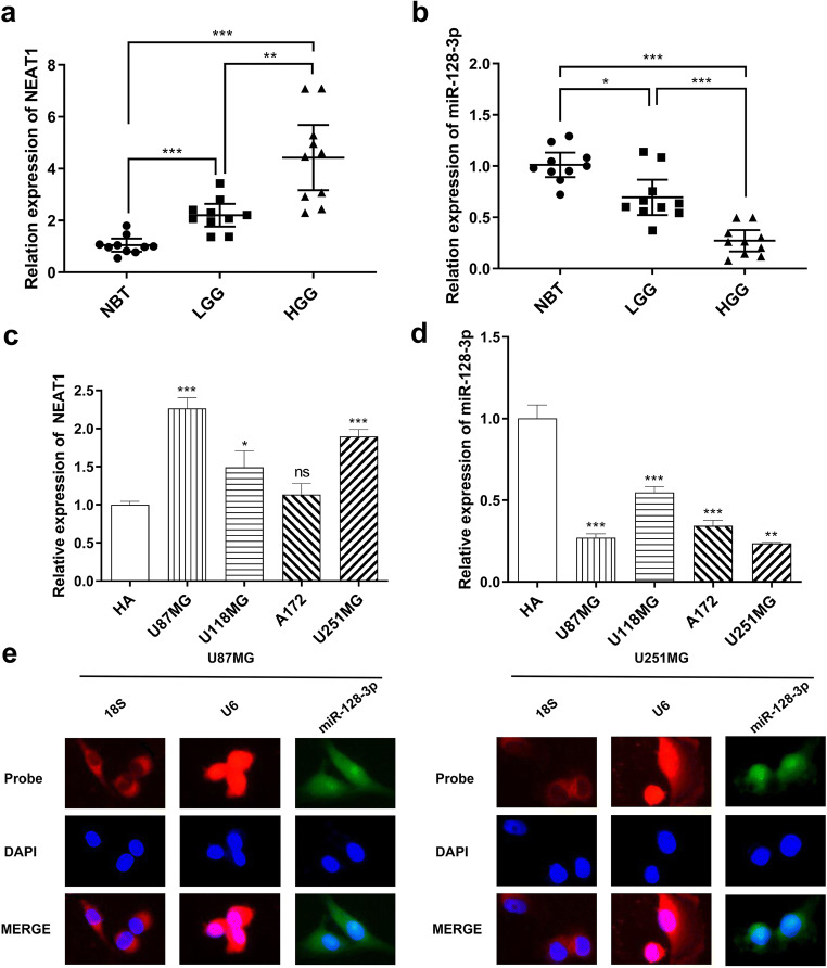
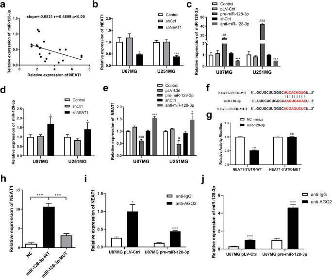
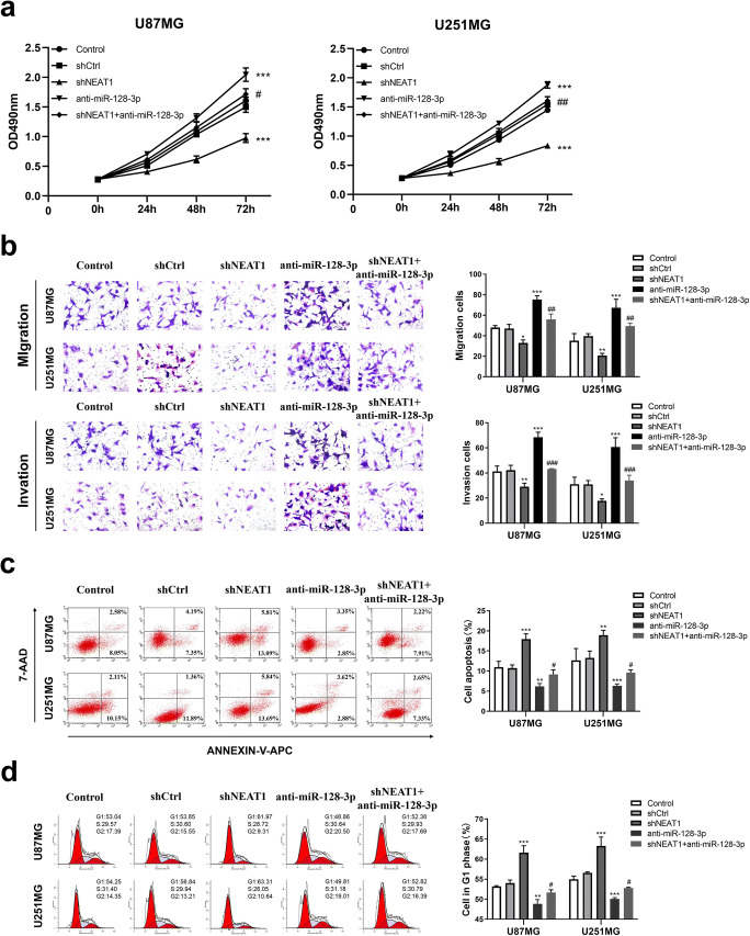
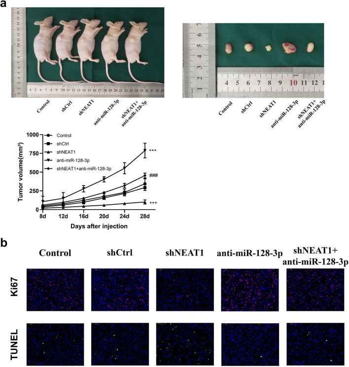
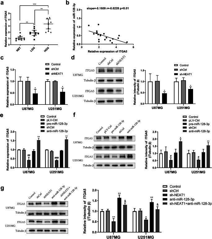
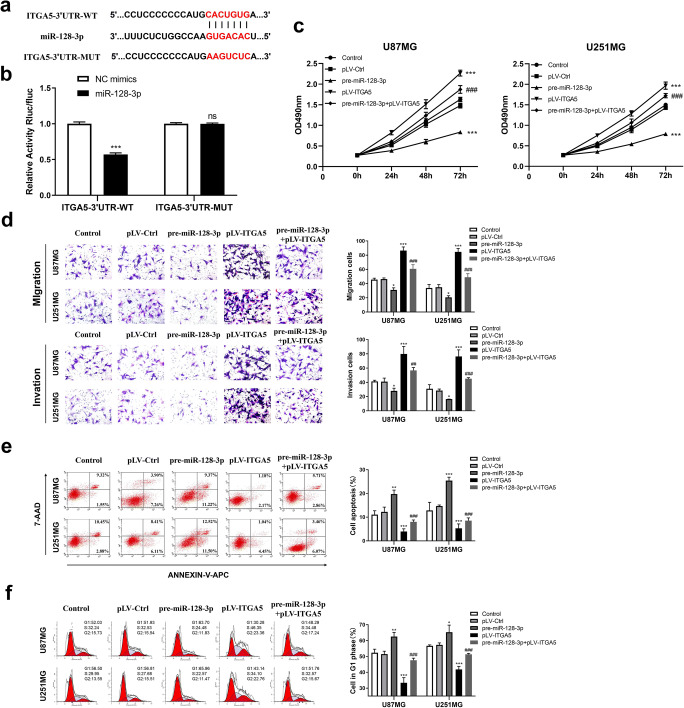
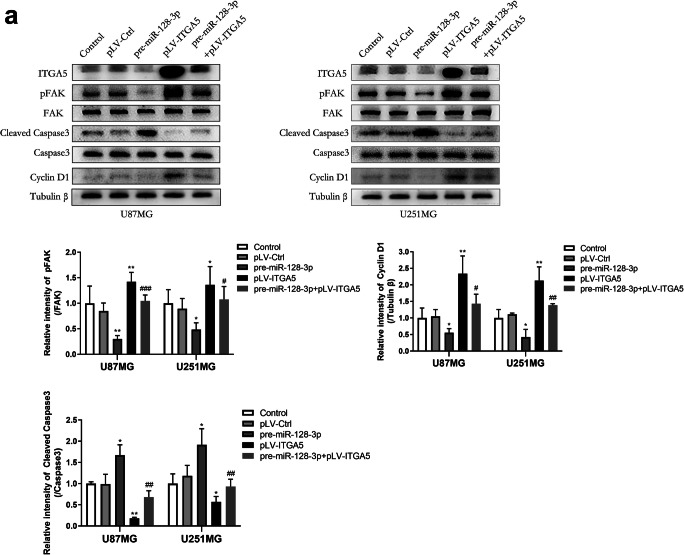
Accumulating evidences indicate that long non-coding RNA nuclear paraspeckle assembly transcript 1 (NEAT1) promotes the progression of glioma. In this study, we postulated that NEAT1 may act as a miR-128-3p sponge. Relative levels of NEAT1 and miR-128-3p expression in human glioma samples and GBM cells were detected using quantitative real-time PCR. By means of CCK-8 assays, transwell assays, and flow cytometric analysis, the biological functions of miR-128-3p and NEAT1 were investigated in U87MG and U251MG human GBM cell lines with stable miR-128-3p and NEAT1 knockdown or overexpression. The luciferase reports, RNA pull-down assay, and RNA immunoprecipitation assay were conducted to determine the relevance of NEAT1 and miR-128-3p in glioma. As a result, high expression of NEAT1 and lack of miR-128-3p were observed in glioma specimens and cells. By binding to anti-oncogene miR-128-3p in the nucleus, NEAT1 enhanced tumorigenesis and glioma development. Further experiments suggested that ITGA5 expression was increased in glioma tissues and was found to be connected with miR-128-3p. Additionally, NEAT1 facilitated ITGA5 expression via competitively binding to miR-128-3p. For this reason, ITGA5 would not be decomposed by miR-128-3p and could activate FAK signaling pathway, thereby promoting cell growth. Collectively, these results indicated that the NEAT1/miR-128-3p/ITGA5 axis was involved in glioma initiation and progression, and might offer a potential novel strategy for treatment of glioma.
Overview
The claims and statistics of this paper haven’t been checked yet.
Run the check — free, no account needed. It reads the open-access text and checks the sources it cites.
Integrity signals
Registry
Cites
Statistics
Figures
Verification
Claims the paper makes
The claims and statistics of this paper haven’t been checked yet.
Run the check — free, no account needed. It reads the open-access text and checks the sources it cites.
Claims other papers make about this one
Each is checked against this paper’s own text: does it say what the citing paper says it does?
The cited source says what the claim attributes to it.
In the citing paper
“The interaction between lncRNA NEAT1 (Nuclear paraspeckle assembly transcript 1) and miR-128-3p that resulted in the sponging of this microRNA contributed not only to glioma pathogenesis,172 but also to the progression of spinal cord injury-mediated NPP.173”
From MiR-128-3p – a gray eminence of the human central nervous system (2024) · cites this paper as [172]
Self-check
Percentages, sums and reported differences re-computed from the paper’s own numbers.
The self-check needs the paper’s full text.
Citations
Cites
No references could be resolved yet.
Research works printed but not linked: 48
- Louis D, Perry A, Reifenberger G, von Deimling A, Figarella-Branger D, Cavenee W, Ohgaki H, Wiestler O et al (2016) The 2016 World Health Organization classification of tumors of the central nervous system: a summary. Acta Neuropathol 131(6):803–820. https://doi.org/10.1007/s00401-016-1545-1 · DOI
- Van Meir E, Hadjipanayis C, Norden A, Shu H, Wen P, Olson J (2010) Exciting new advances in neuro-oncology: the avenue to a cure for malignant glioma. CA Cancer J Clin 60(3):166–193. https://doi.org/10.3322/caac.20069 · DOI · Paper page
- Ostrom Q, Bauchet L, Davis F, Deltour I, Fisher J, Langer C, Pekmezci M, Schwartzbaum J et al (2014) The epidemiology of glioma in adults: a "state of the science" review. Neuro-oncology 16(7):896–913. https://doi.org/10.1093/neuonc/nou087 · DOI
- Fan Z, Zheng J, Xue Y, Liu X, Wang D, Yang C, Ma J, Liu L et al (2018) NR2C2-uORF targeting UCA1-miR-627-5p-NR2C2 feedback loop to regulate the malignant behaviors of glioma cells. Cell Death Dis 9(12):1165. https://doi.org/10.1038/s41419-018-1149-x · DOI
- Wu P, Cai J, Chen Q, Han B, Meng X, Li Y, Li Z, Wang R et al (2019) Lnc-TALC promotes O-methylguanine-DNA methyltransferase expression via regulating the c-Met pathway by competitively binding with miR-20b-3p. Nat Commun 10(1):2045. https://doi.org/10.1038/s41467-019-10025-2 · DOI
- Gao X, Xia X, Li F, Zhang M, Zhou H, Wu X, Zhong J, Zhao Z et al (2021) Circular RNA-encoded oncogenic E-cadherin variant promotes glioblastoma tumorigenicity through activation of EGFR-STAT3 signalling. Nat Cell Biol 23(3):278–291. https://doi.org/10.1038/s41556-021-00639-4 · DOI
- Gutschner T, Diederichs S (2012) The hallmarks of cancer: a long non-coding RNA point of view. RNA Biol 9(6):703–719 · DOI
- Zhang X, Leung G (2014) Long non-coding RNAs in glioma: functional roles and clinical perspectives. Neurochem Int 77:78–85. https://doi.org/10.1016/j.neuint.2014.05.008 · DOI
- Akkipeddi S, Velleca AJ, Carone DM (2020) Probing the function of long noncoding RNAs in the nucleus. Chromosom Res 28(1):87–110 · DOI
- Cheng Z, Liang X, Zhang C, Wang R, Wei T, Ning B, Poreba E, Li L et al (2021) SOX9-transactived long non-coding RNA NEAT1 promotes the self-renewal of liver cancer stem cells through PKA/Hippo signaling. Signal Transduct Target Ther 6(1):87. https://doi.org/10.1038/s41392-021-00466-x · DOI
- Wen S, Wei Y, Zen C, Xiong W, Niu Y, Zhao Y (2020) Long non-coding RNA NEAT1 promotes bone metastasis of prostate cancer through N6-methyladenosine. Mol Cancer 19(1):171. https://doi.org/10.1186/s12943-020-01293-4 · DOI
- Ma P, Pan Y, Yang F, Fang Y, Liu W, Zhao C, Yu T, Xie M et al (2020) KLF5-modulated lncRNA NEAT1 contributes to tumorigenesis by acting as a scaffold for BRG1 to silence GADD45A in gastric cancer. Molec Ther Nucleic Acids 22:382–395. https://doi.org/10.1016/j.omtn.2020.09.003 · DOI
- Caliò A, Harada S, Brunelli M, Pedron S, Segala D, Portillo S, Magi-Galluzzi C, Netto G et al (2021) TFEB rearranged renal cell carcinoma. A clinicopathologic and molecular study of 13 cases. Tumors harboring MALAT1-TFEB, ACTB-TFEB, and the novel NEAT1-TFEB translocations constantly express PDL1. Mod Pathol 34(4):842–850. https://doi.org/10.1038/s41379-020-00713-6 · DOI
- Wang C, Chen Y, Wang Y, Liu X, Liu Y, Li Y, Chen H, Fan C et al (2019) Inhibition of COX-2, mPGES-1 and CYP4A by isoliquiritigenin blocks the angiogenic Akt signaling in glioma through ceRNA effect of miR-194-5p and lncRNA NEAT1. J Experiment Clin Cancer Res 38(1):371. https://doi.org/10.1186/s13046-019-1361-2 · DOI
- Yu H, Xu A, Wu B, Wang M, Chen Z (2021) Long noncoding RNA NEAT1 promotes progression of glioma as a ceRNA by sponging miR-185-5p to stimulate DNMT1/mTOR signaling. J Cell Physiol 236(1):121–130. https://doi.org/10.1002/jcp.29644 · DOI
- Zhou K, Zhang C, Yao H, Zhang X, Zhou Y, Che Y, Huang Y (2018) Knockdown of long non-coding RNA NEAT1 inhibits glioma cell migration and invasion via modulation of SOX2 targeted by miR-132. Mol Cancer 17(1):105. https://doi.org/10.1186/s12943-018-0849-2 · DOI
- Gao X, Zang J, Zheng M, Zhang Y, Yue K, Cao X, Cao Y, Li X et al (2021) Temozolomide treatment induces HMGB1 to promote the formation of glioma stem cells via the TLR2/NEAT1/Wnt pathway in glioblastoma. Front Cell Develop Biol 9:620883. https://doi.org/10.3389/fcell.2021.620883 · DOI
- He L, Hannon GJ (2004) MicroRNAs : small RNAs with a big role in gene regulation. Nat Rev Genet 5(8):631–631 · DOI
- Liu T, Zhang X, Du L, Wang Y, Liu X, Tian H, Wang L, Li P et al (2019) Exosome-transmitted miR-128-3p increase chemosensitivity of oxaliplatin-resistant colorectal cancer. Mol Cancer 18(1):43. https://doi.org/10.1186/s12943-019-0981-7 · DOI
- Breunig C, Erdem N, Bott A, Greiwe J, Reinz E, Bernhardt S, Giacomelli C, Wachter A et al (2018) TGFβ1 regulates HGF-induced cell migration and hepatocyte growth factor receptor MET expression via C-ets-1 and miR-128-3p in basal-like breast cancer. Mol Oncol 12(9):1447–1463. https://doi.org/10.1002/1878-0261.12355 · DOI
- Qu C, Yan C, Cao W, Li F, Qu Y, Guan K, Si C, Yu Z et al (2020) miR-128-3p contributes to mitochondrial dysfunction and induces apoptosis in glioma cells via targeting pyruvate dehydrogenase kinase 1. IUBMB Life 72(3):465–475. https://doi.org/10.1002/iub.2212 · DOI
- Jay SD, David AC (2010) Integrins in cancer: biological implications and therapeutic opportunities. Nat Rev Cancer 10 · DOI
- Seguin L, Desgrosellier JS, Weis SM, Cheresh DA (2015) Integrins and cancer: regulators of cancer stemness, metastasis, and drug resistance. Trends Cell Biol 25(4):234–240 · DOI · Paper page
- Hynes RO (2002) Integrins: bidirectional, allosteric signaling machines. Cell 110(6):673–687 · DOI
- Deng Y, Wan Q, Yan W (2019) Integrin α5/ITGA5 promotes the proliferation, migration, invasion and progression of oral squamous carcinoma by epithelial–mesenchymal transition. Cancer Manag Res 11:9609–9620 · DOI
- Kuninty PR, Bansal R, Geus S, Mardhian DF, Prakash J (2019) ITGA5 inhibition in pancreatic stellate cells attenuates desmoplasia and potentiates efficacy of chemotherapy in pancreatic cancer. Sci Adv 5(9):eaax2770 · DOI
- Pantano F, Croset M, Driouch K, Bednarz-Knoll N, Iuliani M, Ribelli G, Bonnelye E, Wikman H et al (2021) Integrin alpha5 in human breast cancer is a mediator of bone metastasis and a therapeutic target for the treatment of osteolytic lesions. Oncogene 40(7):1284–1299. https://doi.org/10.1038/s41388-020-01603-6 · DOI
- Wang X, Che X, Yu Y, Cheng Y, Bai M, Yang Z, Guo Q, Xie X et al (2020) Hypoxia-autophagy axis induces VEGFA by peritoneal mesothelial cells to promote gastric cancer peritoneal metastasis through an integrin α5-fibronectin pathway. J Experiment Clin Cancer Res 39(1):221. https://doi.org/10.1186/s13046-020-01703-x · DOI
- Blandin A, Cruz Da Silva E, Mercier M, Glushonkov O, Didier P, Dedieu S, Schneider C, Devy J et al (2021) Gefitinib induces EGFR and α5β1 integrin co-endocytosis in glioblastoma cells. Cell Mol Life Sci 78(6):2949–2962. https://doi.org/10.1007/s00018-020-03686-6 · DOI
- Cosset EC, Godet E-W, Guérin G, Froelich B, Pinel P (2012) Involvement of the TGFβ pathway in the regulation of α 5β 1 integrins by caveolin-1 in human glioblastoma. Int J Cancer 131(3):601–611 · DOI
- Janouskova H, Maglott A, Leger DY, Bossert C, Noulet F, Guerin E, Guenot D, Pinel S et al (2012) Integrin α5β1 plays a critical role in resistance to temozolomide by interfering with the p53 pathway in high-grade glioma. Eur J Cancer 48(14):S67–S67 · DOI
- Tay Y, Rinn J, Pandolfi P (2014) The multilayered complexity of ceRNA crosstalk and competition. Nature 505(7483):344–352. https://doi.org/10.1038/nature12986 · DOI · Paper page
- Liang H, Zhang J, Zen K, Zhang C, Chen X (2013) Nuclear microRNAs and their unconventional role in regulating non-coding RNAs. Protein Cell 4(5):325–330. https://doi.org/10.1007/s13238-013-3001-5 · DOI
- Leucci E, Patella F, Waage J, Holmstr M K, Lindow M, Porse B, Kauppinen S, Lund AH (2013) microRNA-9 targets the long non-coding RNA MALAT1 for degradation in the nucleus. Scientific Reports 3 · DOI
- Izaurralde E (2012) Elucidating the temporal order of silencing. EMBO Rep 13(8):662–663. https://doi.org/10.1038/embor.2012.91 · DOI
- Calalb M, Polte T, Hanks S (1995) Tyrosine phosphorylation of focal adhesion kinase at sites in the catalytic domain regulates kinase activity: a role for Src family kinases. Mol Cell Biol 15(2):954–963. https://doi.org/10.1128/mcb.15.2.954 · DOI
- Devi S, Yadav R, Arya R (2021) Altered actin dynamics in cell migration of GNE mutant cells. Front Cell Develop Biol 9:603742. https://doi.org/10.3389/fcell.2021.603742 · DOI
- Sanai N, Berger MS (2018) Surgical oncology for gliomas: the state of the art. Nature Reviews Clinical Oncology · DOI
- Bush N, Chang SM, Berger MS (2016) Current and future strategies for treatment of glioma. Neurosurg Rev 40(1):1–14 · DOI
- Miao S, Lee KW (2015) From discovery to function: the expanding roles of long non-coding RNAs in physiology and disease. Endocr Rev 1:25–64
- Wu D, Wang S, Wen X, Han X, Wang Y, Fan S, Zhang Z, Shan Q et al (2019) Long noncoding RNA nuclear enriched abundant transcript 1 impacts cell proliferation, invasion, and migration of glioma through regulating miR-139-5p/ CDK6. J Cell Physiol 234(5):5972–5987. https://doi.org/10.1002/jcp.27093 · DOI
- Zhen Y, Nan Y, Guo S, Zhang L, Li G, Yue S, Liu X (2019) Knockdown of NEAT1 repressed the malignant progression of glioma through sponging miR-107 and inhibiting CDK14. J Cell Physiol 234(7):10671–10679. https://doi.org/10.1002/jcp.27727 · DOI
- Liu X, Liang Y, Song R, Yang G, Han J, Lan Y, Pan S, Zhu M et al (2018) Long non-coding RNA NEAT1-modulated abnormal lipolysis via ATGL drives hepatocellular carcinoma proliferation. Mol Cancer 17(1):90 · DOI
- Chen Y, Wu N, Liu L, Dong H, Liu X (2020) microRNA-128-3p overexpression inhibits breast cancer stem cell characteristics through suppression of Wnt signalling pathway by down-regulating NEK2. J Cell Mol Med 24(13):7353–7369. https://doi.org/10.1111/jcmm.15317 · DOI
- Fu C, Li D, Zhang X, Liu N, Chi G, Jin X (2018) LncRNA PVT1 Facilitates tumorigenesis and progression of glioma via regulation of MiR-128-3p/GREM1 axis and BMP signaling pathway. Neurotherapeutics 15(4):1139–1157. https://doi.org/10.1007/s13311-018-0649-9 · DOI · Paper page
- Feng L, Ma J, Ji J, Ji H, Liu Y, Hu W (2017) miR-330-5p suppresses glioblastoma cell proliferation and invasiveness through targeting ITGA5. Bioscience Reports:BSR20170019 · DOI
- Orso F, Quirico L, Virga F, Penna E, Dettori D, Cimino D, Coppo R, Grassi E, Elia AR, Brusa D (2016) miR-214 and miR-148b targeting inhibits dissemination of melanoma and breast cancer. Cancer Research 5151-5162 · DOI
- Dingemans A, Boogaart V, Vosse BA, Suylen R, Griffioen AW, Thijssen VL (2010) Integrin expression profiling identifies integrin alpha5 and beta1 as prognostic factors in early stage non-small cell lung cancer. Molecular Cancer 9(1):152 · DOI
Cited by
Sorted by citations
MiR-128-3p – a gray eminence of the human central nervous system
Kiel, K., Król, S. K., Bronisz, A., & Godlewski, J. (2024) · Molecular Therapy Nucleic Acids · doi:10.1016/j.omtn.2024.102141 · Cited by 15
Metadata
- Authors
- Chen, J., Wang, H., Wang, J., Niu, W., Deng, C., & Zhou, M.
- Venue
- Molecular Neurobiology
- Published
- 2021-07-15
- Volume
- 58
- Issue
- 10
- Pages
- 5163-5177
- ISSN
- 0893-7648
- 1559-1182
- PMC
- PMC8497354
- Cited by
- 26
- References
- 0
- Language
- en
- Funders
- National Natural Science Foundation of China (No.81672503 and No.81702484)
About these checks
Registry, retraction, Cites and statistics results are computed, not judged, with one exception: a printed reference that no index lists is read once by a model before it counts as a citation error. Claims are extracted by a model and kept only when their quote appears verbatim in the text; each cited source is then checked, with a model’s help, for whether it says what the claim relies on. Those verdicts can be wrong — read the claim’s quote and the cited source before relying on one.
Statistics and arithmetic are recomputed, not judged.
Confidence bands are shown only for fields calibrated on our annotated test set. None is calibrated yet, so no band is shown.
What counts as a claim ›How we measure confidence ›How we check this ›
Are you an author, or do you see a problem with this page? Request a review
