

The DOI matches its Crossref record; 2 of the 2 references it cites were found in a registry or an index.
Osteoblastic and anti-osteoclastic activities of strontium-substituted silicocarnotite ceramics: In vitro and in vivo studies
Overview
The claims and statistics of this paper haven’t been checked yet.
Run the check — free, no account needed. It reads the open-access text and checks the sources it cites.
Integrity signals
Registry
Cites
Statistics
Figures
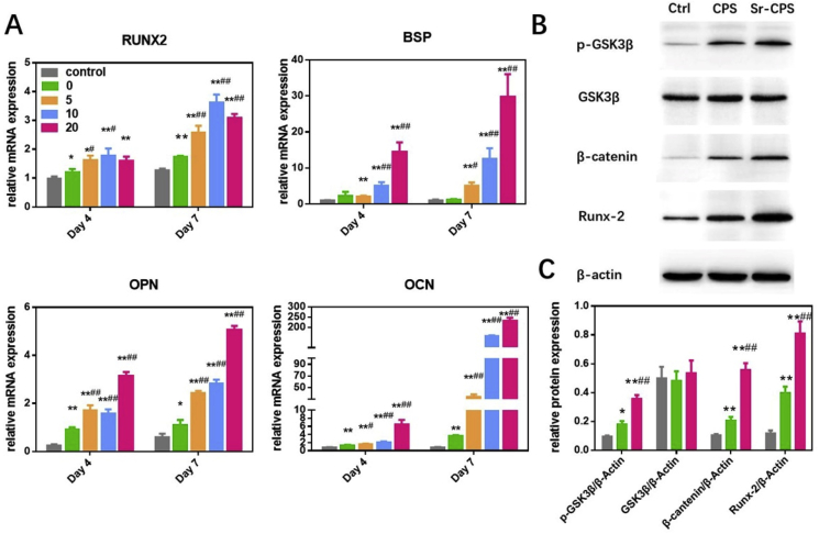
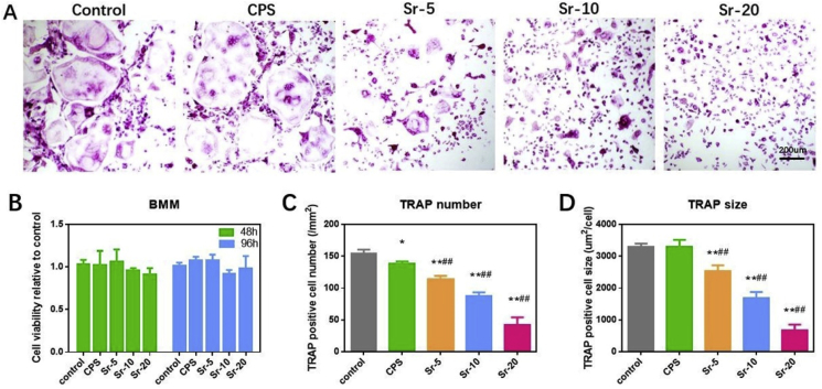
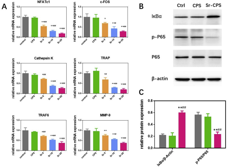
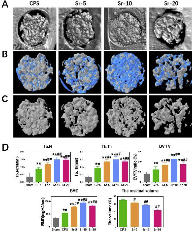
Verification
Claims the paper makes
The claims and statistics of this paper haven’t been checked yet.
Run the check — free, no account needed. It reads the open-access text and checks the sources it cites.
Claims other papers make about this one
Each is checked against this paper’s own text: does it say what the citing paper says it does?
The source backs part of the claim, not all of it — or backs it for a different population, condition or comparison.
In the citing paper
“The imbalance between osteoblast and osteoclast activity further complicates bone defect repair in DM9,10.”
From Microenvironment-responsive multifunctional enzyme-linked hydrogel for diabetic bone defect regeneration. (2025) · cites this paper as [9]
Experimental — accuracy not yet measured
Self-check
Percentages, sums and reported differences re-computed from the paper’s own numbers.
The self-check needs the paper’s full text.
Citations
Cites
Osteoclast differentiation and activation
Boyle, W. J., Simonet, W. S., & Lacey, D. L. (2003) · Nature · doi:10.1038/nature01658 · Cited by 6,670 · cited 1×
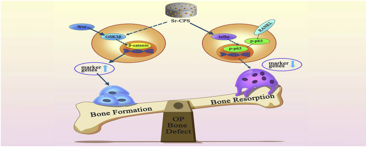
“When RANKL binds to its receptor RANK, tumor-necrosis factor (TNF) receptor-associated factor 6 (TRAF6) will be activated thus stimulating the NF-kB pathway [31,32].”
BackgroundCalcium phosphate cements for bone substitution: Chemistry, handling and mechanical properties
Zhang, J., Liu, W., Schnitzler, V., Tancret, F., & Bouler, J.-M. (2014) · Acta Biomaterialia · doi:10.1016/j.actbio.2013.11.001 · Cited by 667
Research works printed but not linked: 43
- Rachner, 2011, Osteoporosis: now and the future, Lancet, 377, 1276, 10.1016/S0140-6736(10)62349-5 · DOI · Paper page
- Compston, 2019, Osteoporosis, Lancet, 393, 364, 10.1016/S0140-6736(18)32112-3 · DOI · Paper page
- Akesson, 2013, Capture the Fracture: a Best Practice Framework and global campaign to break the fragility fracture cycle, Osteoporos. Int., 24, 2135, 10.1007/s00198-013-2348-z · DOI
- Lane, 2006, Epidemiology, etiology, and diagnosis of osteoporosis, Am. J. Obstet. Gynecol., 194, S3, 10.1016/j.ajog.2005.08.047 · DOI
- Chen, 2017, Matrine prevents bone loss in ovariectomized mice by inhibiting RANKL-induced osteoclastogenesis, Faseb. J. : Off. Publ. Feder. Am. Soc. Exp. Biol., 31, 4855, 10.1096/fj.201700316R · DOI
- Chow, 2011, Low-magnitude high-frequency vibration (LMHFV) enhances bone remodeling in osteoporotic rat femoral fracture healing, J. Orthop. Res. : Off. Publ. Orthop. Res. Soc., 29, 746, 10.1002/jor.21303 · DOI
- Cheng, 2016, Comparison of unilateral versus bilateral percutaneous kyphoplasty for the treatment of patients with osteoporosis vertebral compression fracture (OVCF): a systematic review and meta-analysis, Eur. Spine J. : official publication of the European Spine Society, the European Spinal Deformity Society, and the European Section of the Cervical Spine Research Society, 25, 3439, 10.1007/s00586-016-4395-6 · DOI
- Balasubramanian, 2019, Risk of subsequent fracture after prior fracture among older women, Osteoporos. Int., 30, 79, 10.1007/s00198-018-4732-1 · DOI
- Egol, 2015, Bone grafting: sourcing, timing, strategies, and alternatives, J. Orthop. Trauma, 29, S10, 10.1097/BOT.0000000000000460 · DOI
- Duan, 2013, Cytocompatibility and osteogenic activity of a novel calcium phosphate silicate bioceramic: Silicocarnotite, J. Biomed. Mater. Res., 101, 1955, 10.1002/jbm.a.34497 · DOI
- Yuan, 2010, Osteoinductive ceramics as a synthetic alternative to autologous bone grafting, Proc. Natl. Acad. Sci. U.S.A., 107, 13614, 10.1073/pnas.1003600107 · DOI · Paper page
- Motisuke, 2017, Influence of Si substitution on the reactivity of alpha-tricalcium phosphate, Mater. Sci. Eng. C Mater. Biol. Appl., 75, 816, 10.1016/j.msec.2017.02.099 · DOI
- Khan, 2014, Bioactive behavior of silicon substituted calcium phosphate based bioceramics for bone regeneration, Mater. Sci. Eng. C Mater. Biol. Appl., 35, 245, 10.1016/j.msec.2013.11.013 · DOI
- Lu, 2012, Mechanical properties and in vitro bioactivity of Ca5(PO4)2SiO4 bioceramic, J. Biomater. Appl., 26, 637, 10.1177/0885328210383599 · DOI
- Pavone, 2017, Pharmacological therapy of osteoporosis: a systematic current review of literature, Front. Pharmacol., 8, 803, 10.3389/fphar.2017.00803 · DOI
- Hodges, 1950, The strontium content of human bones, J. Biol. Chem., 185, 519, 10.1016/S0021-9258(18)56336-5 · DOI
- Kargozar, 2019, Multiple and promising applications of strontium (Sr)-Containing bioactive glasses in bone tissue engineering, Front. Bioeng. Biotechnol., 7, 161, 10.3389/fbioe.2019.00161 · DOI
- El-Hajj Fuleihan, 2004, Strontium ranelate--a novel therapy for osteoporosis or a permutation of the same?, N. Engl. J. Med., 350, 504, 10.1056/NEJMe038229 · DOI
- Sips, 1995, Intestinal strontium absorption: from bioavailability to validation of a simple test representative for intestinal calcium absorption, Clin. Chem., 41, 1446, 10.1093/clinchem/41.10.1446 · DOI
- Yang, 2011, Strontium enhances osteogenic differentiation of mesenchymal stem cells and in vivo bone formation by activating Wnt/catenin signaling, Stem Cell., 29, 981, 10.1002/stem.646 · DOI
- Zhu, 2016, Strontium inhibits titanium particle-induced osteoclast activation and chronic inflammation via suppression of NF-kappaB pathway, Sci. Rep., 6
- Chen, 2019, Inhibitory effects of N-[2-(4-acetyl-1-piperazinyl) phenyl]-2-(2-chlorophenoxy) acetamide on osteoclast differentiation in vitro via the downregulation of TRAF6, Int. J. Mol. Sci., 20, 10.3390/ijms20205196 · DOI
- An, 2016, Natural products for treatment of bone erosive diseases: the effects and mechanisms on inhibiting osteoclastogenesis and bone resorption, Int. Immunopharm., 36, 118, 10.1016/j.intimp.2016.04.024 · DOI
- O'Donnell, 2010, Materials characterisation and cytotoxic assessment of strontium-substituted bioactive glasses for bone regeneration, J. Mater. Chem., vol. 20, 8934, 10.1039/c0jm01139h · DOI
- O'Donnell, 2008, Structural analysis of a series of strontium-substituted apatites, Acta Biomater., 4, 1455, 10.1016/j.actbio.2008.04.018 · DOI
- Mcmillan, 1983, Raman-spectroscopic studies of silicate and related glass structure—a review, 106, 57
- Minear, 2010, Wnt proteins promote bone regeneration, Sci. Transl. Med., 2, 10.1126/scitranslmed.3000231 · DOI
- Ling, 2009, Wnt signaling controls the fate of mesenchymal stem cells, Gene, 433, 1, 10.1016/j.gene.2008.12.008 · DOI
- Holmen, 2005, Essential role of beta-catenin in postnatal bone acquisition, J. Biol. Chem., 280, 21162, 10.1074/jbc.M501900200 · DOI · Paper page
- Zhi, 2020, l-tetrahydropalmatine suppresses osteoclastogenesis in vivo and in vitro via blocking RANK-TRAF6 interactions and inhibiting NF-kappaB and MAPK pathways, J. Cell Mol. Med., 24, 785, 10.1111/jcmm.14790 · DOI
- Yamaguchi, 2012, The intact strontium ranelate complex stimulates osteoblastogenesis and suppresses osteoclastogenesis by antagonizing NF-kappaB activation, Mol. Cell. Biochem., 359, 399, 10.1007/s11010-011-1034-8 · DOI
- Bai, 2017, Dual crosslinked chondroitin sulfate injectable hydrogel formed via continuous Diels-Alder (DA) click chemistry for bone repair, Carbohydr. Polym., 166, 123, 10.1016/j.carbpol.2017.02.062 · DOI
- Morales, 2004, Effects of 1alpha,25-dihydroxyvitamin D3 and growth hormone on apoptosis and proliferation in UMR 106 osteoblast-like cells, Endocrinology, 145, 87, 10.1210/en.2003-0718 · DOI
- Zuo, 2012, Osteoblastogenesis regulation signals in bone remodeling, Osteoporos. Int., 23, 1653, 10.1007/s00198-012-1909-x · DOI
- Holstein, 2019, A patent review of bisphosphonates in treating bone disease, Expert Opin. Ther. Pat., 29, 315, 10.1080/13543776.2019.1608180 · DOI
- Wojda, 2018, Parathyroid hormone for bone regeneration, J. Orthop. Res. : Off. Publ. Orthop. Res. Soc., 36, 2586, 10.1002/jor.24075 · DOI
- Bonnelye, 2008, Dual effect of strontium ranelate: stimulation of osteoblast differentiation and inhibition of osteoclast formation and resorption in vitro, Bone, 42, 129, 10.1016/j.bone.2007.08.043 · DOI
- Lin, 2013, Enhanced osteoporotic bone regeneration by strontium-substituted calcium silicate bioactive ceramics, Biomaterials, 34, 10028, 10.1016/j.biomaterials.2013.09.056 · DOI
- Naruphontjirakul, 2018, In vitro osteogenesis by intracellular uptake of strontium containing bioactive glass nanoparticles, Acta Biomater., 66, 67, 10.1016/j.actbio.2017.11.008 · DOI
- Gentleman, 2010, The effects of strontium-substituted bioactive glasses on osteoblasts and osteoclasts in vitro, Biomaterials, 31, 3949, 10.1016/j.biomaterials.2010.01.121 · DOI
- Hutmacher, 2000, Scaffolds in tissue engineering bone and cartilage, Biomaterials, 21, 2529, 10.1016/S0142-9612(00)00121-6 · DOI
- Hill, 2005, Canonical Wnt/beta-catenin signaling prevents osteoblasts from differentiating into chondrocytes, Dev. Cell, 8, 727, 10.1016/j.devcel.2005.02.013 · DOI
- Abu-Amer, 2013, NF-kappaB signaling and bone resorption, Osteoporos. Int., 24, 2377, 10.1007/s00198-013-2313-x · DOI
Cited by
Sorted by citations
Niu, Y., Wang, Z., Shi, Y., Dong, L., & Wang, C. (2021) · Bioactive Materials · doi:10.1016/j.bioactmat.2020.08.012 · Cited by 213
Zhou, H., He, Z., Cao, Y., Chu, L., Liang, B., Yu, K., & Deng, Z. (2024) · Theranostics · doi:10.7150/thno.97412 · Cited by 115
Fu, X., Luo, Z., Guo, Y., Meng, W., Jin, S., Chen, J., Cai, Y., Luo, Z., Huang, C., Chen, A., et al. (2025) · Nature Communications · doi:10.1038/s41467-025-65165-5 · Cited by 44
Metadata
- Authors
- Zeng, J., Guo, J., Sun, Z., Deng, F., Ning, C., & Xie, Y.
- ORCID
- Xie, Youzhuan
- Venue
- Bioactive Materials
- Publisher
- Elsevier BV
- Published
- 2020-09
- Volume
- 5
- Issue
- 3
- Pages
- 435-446
- ISSN
- 2452-199X
- PMC
- PMC7138933
- Cited by
- 46
- References
- 2
- Language
- en
- Licence
- Funders
- Ministry of Science and Technology of the People's Republic of China
About these checks
Registry, retraction, Cites and statistics results are computed, not judged, with one exception: a printed reference that no index lists is read once by a model before it counts as a citation error. Claims are extracted by a model and kept only when their quote appears verbatim in the text; each cited source is then checked, with a model’s help, for whether it says what the claim relies on. Those verdicts can be wrong — read the claim’s quote and the cited source before relying on one.
Statistics and arithmetic are recomputed, not judged.
Confidence bands are shown only for fields calibrated on our annotated test set. None is calibrated yet, so no band is shown.
What counts as a claim ›How we measure confidence ›How we check this ›
Are you an author, or do you see a problem with this page? Request a review
