

The DOI matches its Crossref record; 52 of the 52 references it cites were found in a registry or an index.
Marginal sealing around integral bilayer scaffolds for repairing osteochondral defects based on photocurable silk hydrogels
Open accessAbstract
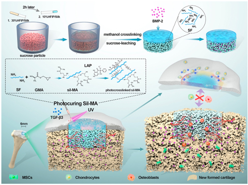
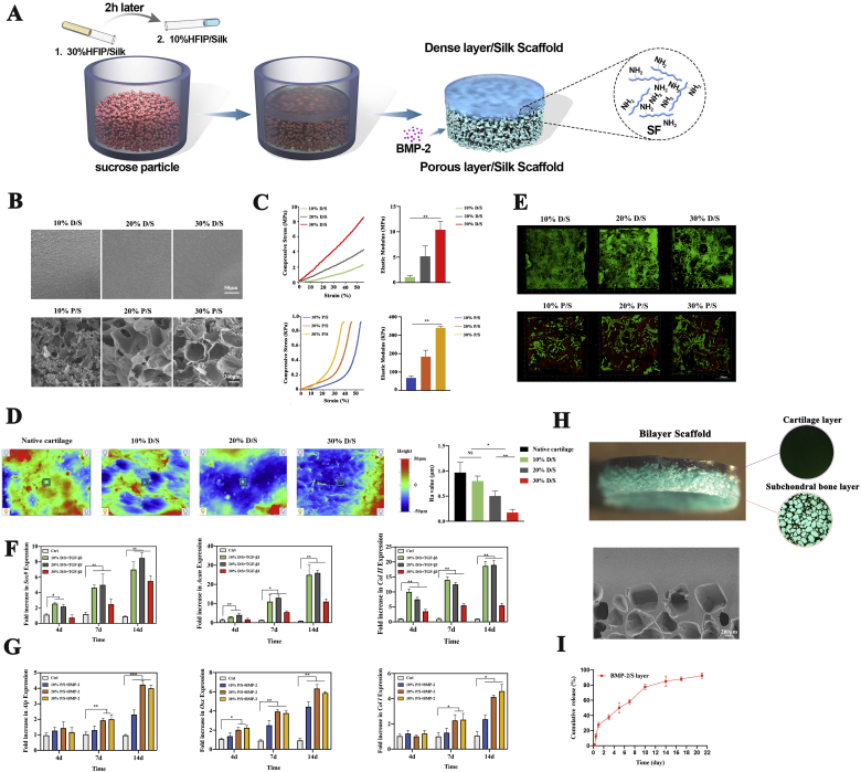
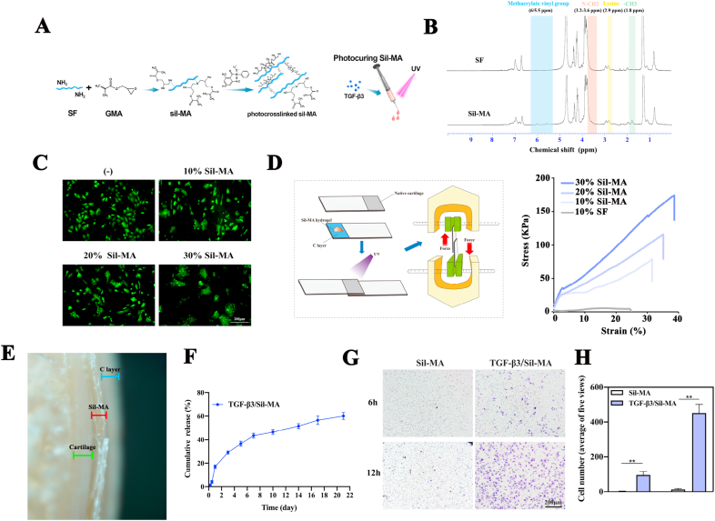
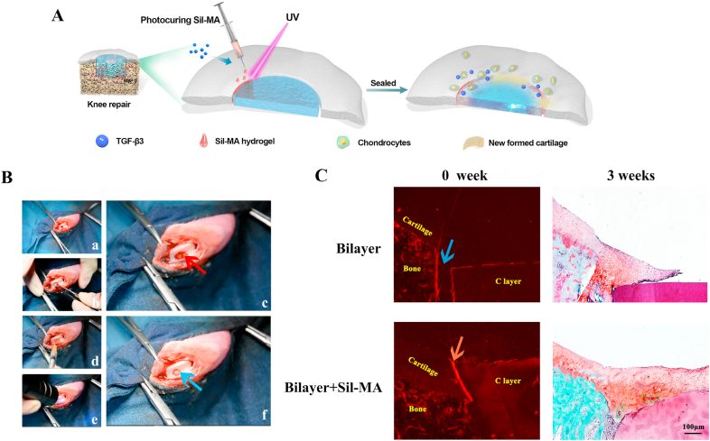
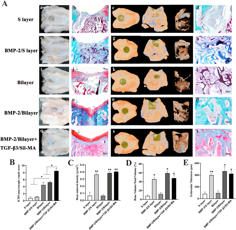
Osteochondral repair remains a major challenge in current clinical practice despite significant advances in tissue engineering. In particular, the lateral integration of neocartilage into surrounding native cartilage is a difficult and inadequately addressed problem that determines the success of tissue repair. Here, a novel design of an integral bilayer scaffold combined with a photocurable silk sealant for osteochondral repair is reported. First, we fabricated a bilayer silk scaffold with a cartilage layer resembling native cartilage in surface morphology and mechanical strength and a BMP-2-loaded porous subchondral bone layer that facilitated the osteogenic differentiation of BMSCs. Second, a TGF-β3-loaded methacrylated silk fibroin sealant (Sil-MA) exhibiting biocompatibility and good adhesive properties was developed and confirmed to promote chondrocyte migration and differentiation. Importantly, this TGF-β3-loaded Sil-MA hydrogel provided a bridge between the cartilage layer of the scaffold and the surrounding cartilage and then guided new cartilage to grow towards and replace the degraded cartilage layer from the surrounding native cartilage in the early stage of knee repair. Thus, osteochondral regeneration and superior lateral integration were achieved in vivo by using this composite. These results demonstrate that the new approach of marginal sealing around the cartilage layer of bilayer scaffolds with Sil-MA hydrogel has tremendous potential for clinical use in osteochondral regeneration.
Overview
The claims and statistics of this paper haven’t been checked yet.
Run the check — free, no account needed. It reads the open-access text and checks the sources it cites.
Integrity signals
Registry
Cites
Statistics
Figures
Verification
Claims the paper makes
The claims and statistics of this paper haven’t been checked yet.
Run the check — free, no account needed. It reads the open-access text and checks the sources it cites.
Claims other papers make about this one
Each is checked against this paper’s own text: does it say what the citing paper says it does?
The source backs part of the claim, not all of it — or backs it for a different population, condition or comparison.
In the citing paper
“This design solves the bone–cartilage connection problem”
From Recent Advances in Silk Fibroin-Based Composites for Bone Repair Applications: A Review (2025) · cites this paper as [80]
Experimental — accuracy not yet measuredThe cited source says what the claim attributes to it.
In the citing paper
“can effectively promote the regeneration of osteochondral cartilage”
From Recent Advances in Silk Fibroin-Based Composites for Bone Repair Applications: A Review (2025) · cites this paper as [80]
The cited source says what the claim attributes to it.
In the citing paper
“which is a potential strategy for osteochondral repair”
From Recent Advances in Silk Fibroin-Based Composites for Bone Repair Applications: A Review (2025) · cites this paper as [80]
The cited source says what the claim attributes to it.
In the citing paper
“achieves horizontal integration of the newborn cartilage with the surrounding natural cartilage through a light-cured hydrogel”
From Recent Advances in Silk Fibroin-Based Composites for Bone Repair Applications: A Review (2025) · cites this paper as [80]
Self-check
Percentages, sums and reported differences re-computed from the paper’s own numbers.
The self-check needs the paper’s full text.
Citations
Cites
Extracellular-matrix tethering regulates stem-cell fate
Trappmann, B., Gautrot, J. E., Connelly, J. T., Strange, D. G. T., Li, Y., Oyen, M. L., Stuart, M. A. C., Boehm, H., Li, B., Viola Vogel, B., et al. (2012) · Nature Materials · doi:10.1038/nmat3339 · Cited by 1,570
Silk fibroin biomaterials for tissue regenerations
Kundu, B., Rajkhowa, R., Kundu, S. C., & Wang, X. (2013) · Advanced Drug Delivery Reviews · doi:10.1016/j.addr.2012.09.043 · Cited by 1,391
Role of material surfaces in regulating bone and cartilage cell response
Boyan, B. D., Hummert, T. W., Dean, D. D., & Schwartz, Z. (1996) · Biomaterials · doi:10.1016/0142-9612(96)85758-9 · Cited by 1,331
Unlike Bone, Cartilage Regeneration Remains Elusive
Huey, D. J., Hu, J. C. Y., & Athanasiou, K. A. (2012) · Science · doi:10.1126/science.1222454 · Cited by 1,209
Fairbanks, B. D., Schwartz, M. P., Bowman, C. N., & Anseth, K. S. (2009) · Biomaterials · doi:10.1016/j.biomaterials.2009.08.055 · Cited by 1,206
Review: tissue engineering for regeneration of articular cartilage
Temenoff, J. S., & Mikos, A. G. (2000) · Biomaterials · doi:10.1016/s0142-9612(99)00213-6 · Cited by 1,056
Precisely printable and biocompatible silk fibroin bioink for digital light processing 3D printing
Kim, S. H., Yeon, Y. K., Lee, J. M., Chao, J. R., Lee, Y. J., Seo, Y. B., Sultan, M. T., Lee, O. J., Lee, J. S., Yoon, S., et al. (2018) · Nature Communications · doi:10.1038/s41467-018-03759-y · Cited by 998
Interplay of matrix stiffness and protein tethering in stem cell differentiation
Wen, J. H., Vincent, L. G., Fuhrmann, A., Choi, Y. S., Hribar, K. C., Taylor‐Weiner, H., Chen, S., & Engler, A. J. (2014) · Nature Materials · doi:10.1038/nmat4051 · Cited by 939
The chemistry and engineering of polymeric hydrogel adhesives for wound closure: a tutorial
Ghobril, C., & Grinstaff, M. W. (2015) · Chemical Society Reviews · doi:10.1039/c4cs00332b · Cited by 926
Three-dimensional (3D) printed scaffold and material selection for bone repair
Zhang, L., Yang, G., Johnson, B. N., & Jia, X. (2018) · Acta Biomaterialia · doi:10.1016/j.actbio.2018.11.039 · Cited by 889
Self-citationPorous 3-D Scaffolds from Regenerated Silk Fibroin
Nazarov, R., Jin, H., & Kaplan, D. L. (2004) · Biomacromolecules · doi:10.1021/bm034327e · Cited by 888
Engineering vascularized tissue
Jain, R. K., Au, P., Tam, J., Duda, D. G., & Fukumura, D. (2005) · Nature Biotechnology · doi:10.1038/nbt0705-821 · Cited by 808
Multifunctional chondroitin sulphate for cartilage tissue–biomaterial integration
Wang, D., Varghese, S., Sharma, B., Strehin, I. A., Fermanian, S., Gorham, J. M., Fairbrother, D. H., Cascio, B. M., & Elisseeff, J. H. (2007) · Nature Materials · doi:10.1038/nmat1890 · Cited by 695
Lee, C. H., Cook, J. L., Mendelson, A., Moioli, E. K., Yao, H., & Mao, J. J. (2010) · The Lancet · doi:10.1016/s0140-6736(10)60668-x · Cited by 614
Hydrogels for the Repair of Articular Cartilage Defects
Spiller, K. L., Maher, S. A., & Lowman, A. M. (2011) · Tissue Engineering Part B Reviews · doi:10.1089/ten.teb.2011.0077 · Cited by 484
Digital light processing 3D printed silk fibroin hydrogel for cartilage tissue engineering
Hong, H., Seo, Y. B., Kim, D. Y., Lee, J. S., Lee, Y. J., Lee, H., Ajiteru, O., Sultan, M. T., Lee, O. J., Kim, S. H., et al. (2019) · Biomaterials · doi:10.1016/j.biomaterials.2019.119679 · Cited by 473
4D-bioprinted silk hydrogels for tissue engineering
Kim, S. H., Seo, Y. B., Yeon, Y. K., Lee, Y. J., Park, H. S., Sultan, M. T., Lee, J. M., Lee, J. S., Lee, O. J., Hong, H., et al. (2020) · Biomaterials · doi:10.1016/j.biomaterials.2020.120281 · Cited by 333
Mechanical Properties of Natural Cartilage and Tissue-Engineered Constructs
Little, C. J., Bawolin, N. K., & Chen, D. (2011) · Tissue Engineering Part B Reviews · doi:10.1089/ten.teb.2010.0572 · Cited by 325
A Blood-Resistant Surgical Glue for Minimally Invasive Repair of Vessels and Heart Defects
Lang, N., Pereira, M. J. N., Lee, Y., Friehs, I., Vasilyev, N. V., Feins, E. N., Ablasser, K., O’Cearbhaill, E. D., Xu, C., Fabozzo, A., et al. (2014) · Science Translational Medicine · doi:10.1126/scitranslmed.3006557 · Cited by 319
Polymer Fiber Scaffolds for Bone and Cartilage Tissue Engineering
Zhang, Y., Liu, X., Zeng, L., Zhang, J. C., Zuo, J., Zou, J., Ding, J., & Chen, X. (2019) · Advanced Functional Materials · doi:10.1002/adfm.201903279 · Cited by 295
Evaluation of the reasons for failure of integration during cartilage repair. A review
Khan, I. M., Gilbert, S. J., Singhrao, S. K., Duance, V. C., & Archer, C. W. (2008) · European Cells and Materials · doi:10.22203/ecm.v016a04 · Cited by 279
Qiao, Z., Lian, M., Yu, H., Sun, B., Zhang, X. Y., Jiang, W., Li, H., Hao, Y., & Dai, K. (2020) · Biomaterials · doi:10.1016/j.biomaterials.2020.120385 · Cited by 231
An Adhesive Hydrogel with “Load‐Sharing” Effect as Tissue Bandages for Drug and Cell Delivery
Chen, J., Wang, D., Wang, L., Liu, W. H., Chiu, A., Shariati, K., Liu, Q., Wang, X., Zhong, Z., Webb, J., et al. (2020) · Advanced Materials · doi:10.1002/adma.202001628 · Cited by 215
Zhang, H., Huang, H., Hao, G., Zhang, Y., Ding, H., Fan, Z., & Sun, L. (2020) · Advanced Functional Materials · doi:10.1002/adfm.202006697 · Cited by 153
Zhou, L., van Osch, G. J. V. M., Malda, J., Stoddart, M. J., Lai, Y., Richards, R. G., Ho, K. K., & Qin, L. (2020) · Advanced Healthcare Materials · doi:10.1002/adhm.202001008 · Cited by 149
Bravenboer, J. V. D. B., der Maur, C. D. I., Bos, P. K., Feenstra, L., Verhaar, J. A. N., Weinans, H., & van Osch, G. J. V. M. (2004) · Arthritis Research & Therapy · doi:10.1186/ar1216 · Cited by 127
Luo, Z., Jiang, L., Xu, Y., Li, H., Xu, W., Wu, S., Wang, Y., Tang, Z., Lv, Y., & Yang, L. (2015) · Biomaterials · doi:10.1016/j.biomaterials.2015.01.001 · Cited by 123
3D Molecularly Functionalized Cell‐Free Biomimetic Scaffolds for Osteochondral Regeneration
Li, L., Li, J., Guo, J., Zhang, H., Zhang, X., Yin, C., Wang, L., Zhu, Y., & Yao, Q. (2018) · Advanced Functional Materials · doi:10.1002/adfm.201807356 · Cited by 111
Chen, Y., Wu, T., Huang, S., Suen, C.-W. W., Cheng, X., Li, J., Hou, H., She, G., Zhang, H., Wang, H., et al. (2019) · ACS Applied Materials & Interfaces · doi:10.1021/acsami.9b01532 · Cited by 110
Enzymatically Cross-Linked Silk Fibroin-Based Hierarchical Scaffolds for Osteochondral Regeneration
Ribeiro, V. P., Pina, S., Costa, J. B., Cengiz, I. F., García‐Fernández, L., Fernández‐Gutiérrez, M., Paiva, O. C., Oliveira, A. L., San‐Román, J., Oliveira, J. M., et al. (2019) · ACS Applied Materials & Interfaces · doi:10.1021/acsami.8b21259 · Cited by 109
Dahlin, R. L., Ni, M., Meretoja, V. V., Kasper, F. K., & Mikos, A. G. (2013) · Biomaterials · doi:10.1016/j.biomaterials.2013.09.086 · Cited by 108
Zhou, F., Hong, Y., Zhang, X., Yang, L., Li, ., Jiang, D., Bunpetch, V., Hu, Y., Ouyang, H., & Zhang, S. (2018) · Applied Materials Today · doi:10.1016/j.apmt.2018.08.005 · Cited by 106
Bioactive Pore‐Forming Bone Adhesives Facilitating Cell Ingrowth for Fracture Healing
Xu, L., Gao, S., Zhou, R., Zhou, F., Qiao, Y., & Qiu, D. (2020) · Advanced Materials · doi:10.1002/adma.201907491 · Cited by 100
Inhibition of integrative cartilage repair by proteoglycan 4 in synovial fluid
Englert, C., McGowan, K. B., Klein, T. J., Giurea, A., Schumacher, B. L., & Sah, R. L. (2005) · Arthritis & Rheumatism · doi:10.1002/art.20986 · Cited by 99
Mohanraj, B., Duan, G., Peredo, A. P., Kim, M., Tu, F., Lee, D., Dodge, G. R., & Mauck, R. L. (2019) · Advanced Functional Materials · doi:10.1002/adfm.201807909 · Cited by 86
Rapidly photocurable silk fibroin sealant for clinical applications
Kim, S. H., Lee, Y. J., Chao, J. R., Kim, D. Y., Sultan, M. T., Lee, H. J., Lee, J. M., Lee, J. S., Lee, O. J., Hong, H., et al. (2020) · NPG Asia Materials · doi:10.1038/s41427-020-0227-6 · Cited by 77
Armstrong, J. P. K., Shakur, R., Horne, J. P., Dickinson, S. C., Armstrong, C. T., Lau, K., Kadiwala, J., Lowe, R., Seddon, A. M., Mann, S., et al. (2015) · Nature Communications · doi:10.1038/ncomms8405 · Cited by 73
Wang, X., Song, X., Li, T., Chen, J., Cheng, G., Yang, L., & Chen, C. (2019) · The American Journal of Sports Medicine · doi:10.1177/0363546519856355 · Cited by 70
Zhang, W., Chen, L., Zhang, A., Liu, H., Jiang, Y., Li, X., Sheng, R., Yao, Q., & Chen, J. (2020) · Bioactive Materials · doi:10.1016/j.bioactmat.2020.05.003 · Cited by 70
Self-citationMechanical improvements to reinforced porous silk scaffolds
Gil, E. S., Kluge, J. A., Rockwood, D. N., Rajkhowa, R., Wang, L., Wang, X., & Kaplan, D. L. (2011) · Journal of Biomedical Materials Research Part A · doi:10.1002/jbm.a.33158 · Cited by 69
Lee, J., Jeon, O., Kong, M., Abdeen, A. A., Shin, J., Lee, H. N., Lee, Y. B., Sun, W., Bandaru, P., Alt, D. S., et al. (2020) · Science Advances · doi:10.1126/sciadv.aaz5913 · Cited by 67
Liu, X., Wei, Y., Xuan, C., Liu, L., Chen, L., Chai, M., Zhang, Z., Wang, L., & Shi, X. (2020) · Advanced Healthcare Materials · doi:10.1002/adhm.202000076 · Cited by 66
Li, J. J., Kim, K.-S., Roohani‐Esfahani, S., Guo, J. T., Kaplan, D. L., & Zreiqat, H. (2015) · Journal of Materials Chemistry B · doi:10.1039/c5tb00353a · Cited by 63
Electric field-driven building blocks for introducing multiple gradients to hydrogels
Xu, G., Ding, Z., Lü, Q., Zhang, X., Zhou, X., Xiao, L., Lu, G., & Kaplan, D. L. (2020) · Protein & Cell · doi:10.1007/s13238-020-00692-z · Cited by 62
Hong, H., Lee, O. J., Lee, Y. J., Lee, J. S., Ajiteru, O., Lee, H., Suh, Y. J., Sultan, M. T., Kim, S. H., & Park, C. H. (2020) · Biomolecules · doi:10.3390/biom11010035 · Cited by 58
Gilbert, S. J., Singhrao, S. K., Khan, I. M., Gonzalez, L. G., Thomson, B. M., Burdon, D., Duance, V. C., & Archer, C. W. (2009) · Tissue Engineering Part A · doi:10.1089/ten.tea.2008.0361 · Cited by 56
Wu, T., Chen, Y., Liu, W., Tong, K. L., Suen, C.-W. W., Huang, S., Hou, H., She, G., Zhang, H., Zheng, X., et al. (2020) · Materials Science and Engineering C · doi:10.1016/j.msec.2020.110757 · Cited by 46
Boyan, B. D., Lincks, J., Lohmann, C. H., Sylvia, V. L., Cochran, D. L., Blanchard, C. R., Dean, D. D., & Schwartz, Z. (1999) · Journal of Orthopaedic Research® · doi:10.1002/jor.1100170322 · Cited by 38
Allon, A. A., Ng, K., Hammoud, S., Russell, B. H., Jones, C. M., Rivera, J. J., Schwartz, J., Höök, M., & Maher, S. A. (2012) · Journal of Biomedical Materials Research Part A · doi:10.1002/jbm.a.34144 · Cited by 24
He, D., Genecov, D. G., Herbert, M. A., Barceló, C. R., Elsalanty, M. E., Weprin, B. E., & Opperman, L. A. (2009) · Journal of neurosurgery · doi:10.3171/2009.1.jns08976 · Cited by 23
Kuang, B., Yang, Y., & Lin, H. (2019) · ACS Omega · doi:10.1021/acsomega.9b02270 · Cited by 15
Kosorn, W., Sakulsumbat, M., Lertwimol, T., Thavornyutikarn, B., Uppanan, P., Chantaweroad, S., & Janvikul, W. (2019) · Journal of Materials Science Materials in Medicine · doi:10.1007/s10856-019-6335-6 · Cited by 13
Cited by
Sorted by citations
Silk fibroin-based biomaterials for cartilage/osteochondral repair
Zhou, Z., Cui, J., Wu, S., Geng, Z., & Su, J. (2022) · Theranostics · doi:10.7150/thno.74548 · Cited by 226
Yang, Y., Zhao, X., Wang, S., Zhang, Y., Yang, A., Cheng, Y., & Chen, X. (2023) · Nature Communications · doi:10.1038/s41467-023-43334-8 · Cited by 182
Hu, Z., Lu, J., Zhang, T., Liang, H., Yuan, H., Su, D., Ding, W., Lian, R., Ge, Y., Liang, B., et al. (2022) · Bioactive Materials · doi:10.1016/j.bioactmat.2022.08.025 · Cited by 165
Multifunctional Hydrogel Based on Silk Fibroin Promotes Tissue Repair and Regeneration
Lin, D., Li, M., Wang, L., Cheng, J., Yang, Y., Wang, H., Ye, J., & Liu, Y. (2024) · Advanced Functional Materials · doi:10.1002/adfm.202405255 · Cited by 147
Silk Fibroin-Based Biomaterials for Tissue Engineering Applications
Li, G., & Sun, S. (2022) · Molecules · doi:10.3390/molecules27092757 · Cited by 144
Metadata
- Authors
- Wu, X., Zhou, M., Jiang, F., Yin, S., Lin, S., Yang, G., Lu, Y., Zhang, W., & Jiang, X.
- Affiliations
- Shanghai Jiao Tong University (CN)
- Corresponding author
- Jiang, Fei
- Zhang, Wenjie
- Jiang, Xinquan
- Venue
- Bioactive Materials
- Publisher
- Elsevier BV
- Published
- 2021-11
- Volume
- 6
- Issue
- 11
- Pages
- 3976-3986
- ISSN
- 2097-1192 (ISSN-L)
- 2452-199X
- PMC
- PMC8081879
- OpenAlex
- W3158673470
- Cited by
- 113
- References
- 52
- Citations by year
- 2026: 10 · 2025: 26 · 2024: 20 · 2023: 24 · 2022: 30 · 2021: 3
- FWCI
- 5.03
- Type
- journal article
- Language
- en
- Open access
- gold
- doi.org
- Licence
- Locations
- Bioactive Materials (publishedVersion, cc-by-nc-nd)
- PubMed Central (publishedVersion, cc-by-nc-nd)
- PubMed (publishedVersion)
- DOAJ (DOAJ: Directory of Open Access Journals) (submittedVersion, cc-by-sa)
- Funders
- National Natural Science Foundation of China (31700848, 81620108006, 81921002, 81991505)
- National Key Research and Development Program of China (2016YFC1102900)
- Topics
- Physical Sciences › Materials Science › Biomaterials › Silk-based biomaterials and applications
- Physical Sciences › Engineering › Biomedical Engineering › Bone Tissue Engineering Materials
- Health Sciences › Medicine › Rheumatology › Osteoarthritis Treatment and Mechanisms
- Keywords
- osteochondral regeneration, marginal sealing, bilayer scaffold, silk fibroin hydrogel, TGF-beta3, BMP-2, mesenchymal stem cells, chondrogenic differentiation, cartilage integration, knee cartilage repair, subchondral bone regeneration
About these checks
Registry, retraction, Cites and statistics results are computed, not judged, with one exception: a printed reference that no index lists is read once by a model before it counts as a citation error. Claims are extracted by a model and kept only when their quote appears verbatim in the text; each cited source is then checked, with a model’s help, for whether it says what the claim relies on. Those verdicts can be wrong — read the claim’s quote and the cited source before relying on one.
Statistics and arithmetic are recomputed, not judged.
Confidence bands are shown only for fields calibrated on our annotated test set. None is calibrated yet, so no band is shown.
What counts as a claim ›How we measure confidence ›How we check this ›
Are you an author, or do you see a problem with this page? Request a review
