Cited by 134Tissue Engineering and Regenerative Medicine3D Printing in Biomedical ResearchPluripotent Stem Cells Research

The DOI matches its Crossref record; 30 of the 31 references it cites were found in a registry or an index.

Human iPS derived progenitors bioengineered into liver organoids using an inverted colloidal crystal poly (ethylene glycol) scaffold

Open access

Abstract

Generation of human organoids from induced pluripotent stem cells (iPSCs) offers exciting possibilities for developmental biology, disease modelling and cell therapy. Significant advances towards those goals have been hampered by dependence on animal derived matrices (e.g. Matrigel), immortalized cell lines and resultant structures that are difficult to control or scale. To address these challenges, we aimed to develop a fully defined liver organoid platform using inverted colloid crystal (ICC) whose 3-dimensional mechanical properties could be engineered to recapitulate the extracellular niche sensed by hepatic progenitors during human development. iPSC derived hepatic progenitors (IH) formed organoids most optimally in ICC scaffolds constructed with 140 μm diameter pores coated with type I collagen in a two-step process mimicking liver bud formation. The resultant organoids were closer to adult tissue, compared to 2D and 3D controls, with respect to morphology, gene expression, protein secretion, drug metabolism and viral infection and could integrate, vascularise and function following implantation into livers of immune-deficient mice. Preliminary interrogation of the underpinning mechanisms highlighted the importance of TGFβ and hedgehog signalling pathways. The combination of functional relevance with tuneable mechanical properties leads us to propose this bioengineered platform to be ideally suited for a range of future mechanistic and clinical organoid related applications.

Overview

The claims and statistics of this paper haven’t been checked yet.

Run the check — free, no account needed. It reads the open-access text and checks the sources it cites.

A few free checks a day · papers already verified open instantly · results are public

Integrity signals

Registry

Crossref · 10.1016/j.biomaterials.2018.07.043

Cites

30 of 30 references verified0 citation errors · 0 not checkable

Statistics

Figures

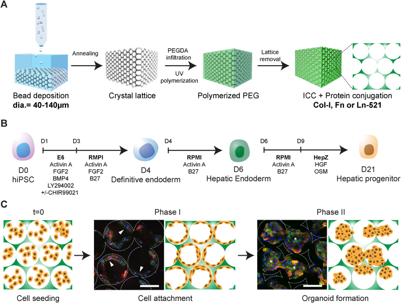
Fig. 1 Bioengineering liver organoids using ICC scaffold and iPSC-derived hepatic progenitor (IH). (A) Schematic illustration of ICC fabrication using a range of sacrificial monodispersed beads and ECM proteins for stem cell niche modulation. (B) Schematic of human iPSC-derived hepatic progenitors differentiation protocol. (C) Schematic of bioengineering liver organoids using IH and ICC in three main steps, cell seeding, cell attachment (Phase I) and organoid formation (Phase II). Confocal micrographs of human fetal liver cells (Fetal; CTNNB green; CK19 red) demonstrating the two-phase organoid formation in Col-I coated ICC with 140 μm pore size. Arrowheads indicate cells lining surface of ICC; asterisks represent cells forming clusters. Scale bar, 100 μm. (For interpretation of the references to colour in this figure legend, the reader is referred to the Web version of this article.)
Fig. 1 Bioengineering liver organoids using ICC scaffold and iPSC-derived hepatic progenitor (IH). (A) Schematic illustration of ICC fabrication using a range of sacrificial monodispersed beads and ECM proteins for stem cell niche modulation. (B) Schematic of human iPSC-derived hepatic progenitors differentiation protocol. (C) Schematic of bioengineering liver organoids using IH and ICC in three main steps, cell seeding, cell attachment (Phase I) and organoid formation (Phase II). Confocal micrographs of human fetal liver cells (Fetal; CTNNB green; CK19 red) demonstrating the two-phase organoid formation in Col-I coated ICC with 140 μm pore size. Arrowheads indicate cells lining surface of ICC; asterisks represent cells forming clusters. Scale bar, 100 μm. (For interpretation of the references to colour in this figure legend, the reader is referred to the Web version of this article.)CC BY · PubMed Central
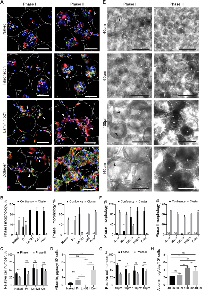
Fig. 2 Characterizing the effects of pore size and ECM proteins on liver organoid formation in ICC scaffold. (A) Confocal micrographs of IH (CTNNB, green; CK19, red) following seeding into non-coated (Naked), Fibronectin (Fn), Laminin 521 (Ln-521), and Collagen I (Col-I) coated ICC's. (B) Quantification of cellular attachment (confluency) and cluster formation (cluster) ability seen across different ECM's during Phase I and II. (C) Cell number relative to initial cell seeding number in Phase I and II. (D) Albumin secretion rate of IH seeded in different ECM coated ICC in Phase II (E) Bright field images revealing the morphologies of IH in ICC with different collagen coated pore sizes (40 μm, 60 μm, 100 μm and 140 μm) and the respective (F) morphological quantitation and (G) relative cell number in Phase I and II. (H) Albumin secretion rate of IH seeded in ICC with different pore sizes in Phase II. Arrowheads indicate cells lining surface of ICC; asterisks represent cells forming clusters. Scale bar, 100 μm. Mean ± sd, N = 4. *p < 0.05; **p < 0.005; ***p < 0.0005; ****p < 0.0001; ns non-significant. (For interpretation of the references to colour in this figure legend, the reader is referred to the Web version of this article.)
Fig. 2 Characterizing the effects of pore size and ECM proteins on liver organoid formation in ICC scaffold. (A) Confocal micrographs of IH (CTNNB, green; CK19, red) following seeding into non-coated (Naked), Fibronectin (Fn), Laminin 521 (Ln-521), and Collagen I (Col-I) coated ICC's. (B) Quantification of cellular attachment (confluency) and cluster formation (cluster) ability seen across different ECM's during Phase I and II. (C) Cell number relative to initial cell seeding number in Phase I and II. (D) Albumin secretion rate of IH seeded in different ECM coated ICC in Phase II (E) Bright field images revealing the morphologies of IH in ICC with different collagen coated pore sizes (40 μm, 60 μm, 100 μm and 140 μm) and the respective (F) morphological quantitation and (G) relative cell number in Phase I and II. (H) Albumin secretion rate of IH seeded in ICC with different pore sizes in Phase II. Arrowheads indicate cells lining surface of ICC; asterisks represent cells forming clusters. Scale bar, 100 μm. Mean ± sd, N = 4. *p < 0.05; **p < 0.005; ***p < 0.0005; ****p < 0.0001; ns non-significant. (For interpretation of the references to colour in this figure legend, the reader is referred to the Web version of this article.)CC BY · PubMed Central
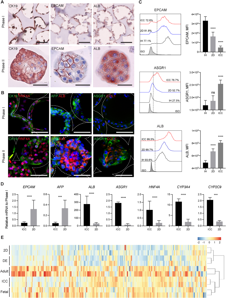
Fig. 3 Morphological and transcriptomic characterization of IH-ICC organoids. (A) Histochemical images demonstrating morphogenesis of IH from a single cell layer (Phase I top panel; scale bar, 100 μm) to organoids (Phase II bottom panel; scale bar, 50 μm) occurs in conjunction with differential protein expression of developmental markers CK19 (left), EPCAM (middle) and ALB (right). (B) Confocal micrographs highlighting upregulated protein expression of mature (ALB, ASGPR1, COL1) hepatic markers occurs in conjunction with down regulation of immature (CK19 and AFP) markers during transition of IH from Phase I (top panel; scale bar, 100 μm) to Phase II (bottom panel; scale bar, 100 μm) organoids. (C) FACS histogram and mean fluorescence intensity (MFI) analysis demonstrating hepatic maturation kinetics of IH in ICC vs 2D culture (N = 4). (D) Differential gene expression (by RT-PCR) of selected genes reveals a more mature hepatic signature of IH in ICC vs. 2D culture (N = 8). (e) Bi-clustering heatmap of 296 liver-specific genes across different primary (adult & fetal liver) and IH (DE, 2D & ICC) samples. Samples are linked by the dendrogram above to show the similarity of their gene expression patterns. Mean ± sd, *p < 0.05; **p < 0.005; ***p < 0.0005; ****p < 0.0001; ns nonsignificant.
Fig. 3 Morphological and transcriptomic characterization of IH-ICC organoids. (A) Histochemical images demonstrating morphogenesis of IH from a single cell layer (Phase I top panel; scale bar, 100 μm) to organoids (Phase II bottom panel; scale bar, 50 μm) occurs in conjunction with differential protein expression of developmental markers CK19 (left), EPCAM (middle) and ALB (right). (B) Confocal micrographs highlighting upregulated protein expression of mature (ALB, ASGPR1, COL1) hepatic markers occurs in conjunction with down regulation of immature (CK19 and AFP) markers during transition of IH from Phase I (top panel; scale bar, 100 μm) to Phase II (bottom panel; scale bar, 100 μm) organoids. (C) FACS histogram and mean fluorescence intensity (MFI) analysis demonstrating hepatic maturation kinetics of IH in ICC vs 2D culture (N = 4). (D) Differential gene expression (by RT-PCR) of selected genes reveals a more mature hepatic signature of IH in ICC vs. 2D culture (N = 8). (e) Bi-clustering heatmap of 296 liver-specific genes across different primary (adult & fetal liver) and IH (DE, 2D & ICC) samples. Samples are linked by the dendrogram above to show the similarity of their gene expression patterns. Mean ± sd, *p < 0.05; **p < 0.005; ***p < 0.0005; ****p < 0.0001; ns nonsignificant.CC BY · PubMed Central
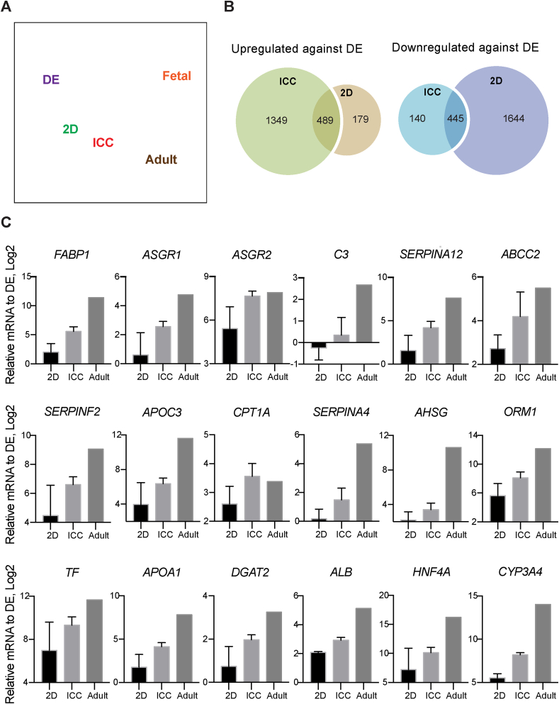
Fig. 4 The transcriptomic analysis of liver organoid in ICC. (A) Principle component analysis of RNA-seq data demonstrating the proximity of gene expression variance in 2D plot. (B) Venn diagrams showing the number of up and downregulated gene in ICC and 2D with respect to DE. (C) RT-PCR validation on top 18 liver-specific genes identified by RNA-seq analysis (N = 3) Mean ± sd.
Fig. 4 The transcriptomic analysis of liver organoid in ICC. (A) Principle component analysis of RNA-seq data demonstrating the proximity of gene expression variance in 2D plot. (B) Venn diagrams showing the number of up and downregulated gene in ICC and 2D with respect to DE. (C) RT-PCR validation on top 18 liver-specific genes identified by RNA-seq analysis (N = 3) Mean ± sd.CC BY · PubMed Central
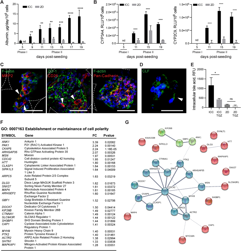
Fig. 5 Functional validation of organoids. (A) Albumin secretion rate of IH-ICC vs. 2D (N = 8). (B) Basal metabolic activity of Cytochrome P450 isoforms CYP3A4 and CYP2C9 in IH-ICC vs 2D (N = 8). RLU, relative luminescence unit. (C) Confocal micrographs showing expression of hepatocyte polarity markers (MRP2, ZO-1, CD26 and Pan-Cadherin) in IH-ICC organoids. Scale bar, 50 μm. White arrowhead points to apical region. (D) Accumulation of Cholyl-L-lysyl-fluorescein (CLS) in IH-ICC organoids after 40 min of CLF incubation followed by 40 min of washing. White arrowhead points to the CLF accumulation. (E) Effect of adding Troglitazone (TGZ) to CLS retention in IH-ICC (N = 4). (F) A list of uniquely upregulated genes in IH-ICC vs 2D that involved in establishment and maintenance of cell polarity. FC, fold change. (G) The STRING functional network predicted the associations between proteins (nodes) from regulated genes involved in cell polarity in IH-ICC. The cluster analysis was performed using KMEANS clustering algorithms. Mean ± sd, *p < 0.05; **p < 0.005; ***p < 0.0005; ****p < 0.0001; ns nonsignificant.
Fig. 5 Functional validation of organoids. (A) Albumin secretion rate of IH-ICC vs. 2D (N = 8). (B) Basal metabolic activity of Cytochrome P450 isoforms CYP3A4 and CYP2C9 in IH-ICC vs 2D (N = 8). RLU, relative luminescence unit. (C) Confocal micrographs showing expression of hepatocyte polarity markers (MRP2, ZO-1, CD26 and Pan-Cadherin) in IH-ICC organoids. Scale bar, 50 μm. White arrowhead points to apical region. (D) Accumulation of Cholyl-L-lysyl-fluorescein (CLS) in IH-ICC organoids after 40 min of CLF incubation followed by 40 min of washing. White arrowhead points to the CLF accumulation. (E) Effect of adding Troglitazone (TGZ) to CLS retention in IH-ICC (N = 4). (F) A list of uniquely upregulated genes in IH-ICC vs 2D that involved in establishment and maintenance of cell polarity. FC, fold change. (G) The STRING functional network predicted the associations between proteins (nodes) from regulated genes involved in cell polarity in IH-ICC. The cluster analysis was performed using KMEANS clustering algorithms. Mean ± sd, *p < 0.05; **p < 0.005; ***p < 0.0005; ****p < 0.0001; ns nonsignificant.CC BY · PubMed Central
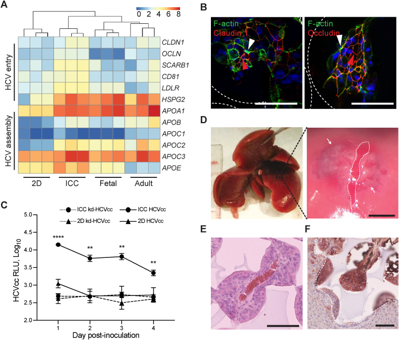
Fig. 6 Disease modelling and in vivo transplantation. (A) Heatmap and hierarchal clustering comparing expression of 12 genes involved in encoding HCV entry and assembly in IH-ICC vs 2D vs primary (adult, fetal) liver. (B) Confocal imaging showing expression of claudin 1 and occludin in IH-ICC organoids. Scale bar, 100 μm. White and red arrowheads point to apical and lateral regions respectively. (C) HCV expression of IH-ICC vs 2D following infection with HCV reporter virus expressing secreted GLuc (HCVcc, N = 4) or mock infected with knock down HCVcc (kd-HCVcc, N = 3) and subsequently were sampled and washed daily. RLU, relative luminescence unit. (D) Photograph showing location of surgical pocket formation on murine left lateral lobe (left) and appearance following IH-ICC transplantation (right). The white dashed line depicts the capsular incision and the limits of the sub-capsular scaffold implant are shown by the white arrows. Scale bar 1.5 mm (E) H&E staining of explant reveals neo-vasculature of IH-ICC. Scale bar, 100 μm. (F) Immuno-histochemical staining of explant for human albumin. Dashed white line indicates the boundary between implant and host liver. Scale bar, 100 μm. Mean ± sd; **p < 0.005, ****p < 0.0001, nd not detected. (For interpretation of the references to colour in this figure legend, the reader is referred to the Web version of this article.)
Fig. 6 Disease modelling and in vivo transplantation. (A) Heatmap and hierarchal clustering comparing expression of 12 genes involved in encoding HCV entry and assembly in IH-ICC vs 2D vs primary (adult, fetal) liver. (B) Confocal imaging showing expression of claudin 1 and occludin in IH-ICC organoids. Scale bar, 100 μm. White and red arrowheads point to apical and lateral regions respectively. (C) HCV expression of IH-ICC vs 2D following infection with HCV reporter virus expressing secreted GLuc (HCVcc, N = 4) or mock infected with knock down HCVcc (kd-HCVcc, N = 3) and subsequently were sampled and washed daily. RLU, relative luminescence unit. (D) Photograph showing location of surgical pocket formation on murine left lateral lobe (left) and appearance following IH-ICC transplantation (right). The white dashed line depicts the capsular incision and the limits of the sub-capsular scaffold implant are shown by the white arrows. Scale bar 1.5 mm (E) H&E staining of explant reveals neo-vasculature of IH-ICC. Scale bar, 100 μm. (F) Immuno-histochemical staining of explant for human albumin. Dashed white line indicates the boundary between implant and host liver. Scale bar, 100 μm. Mean ± sd; **p < 0.005, ****p < 0.0001, nd not detected. (For interpretation of the references to colour in this figure legend, the reader is referred to the Web version of this article.)CC BY · PubMed Central
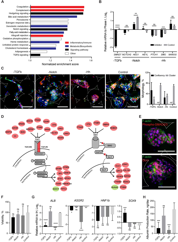
Fig. 7 Mechanisms of organoid formation. (A) Bar chart detailing gene sets uniquely enriched in IH-ICC over 2D. (B) Gene expression (by RT-PCR) in IH-ICC of selected pathway transcriptional targets, TGFb, Notch and Hedgehog (Hh) following addition of respective inhibitors (N = 4). (C) Confocal micrographs of IH in Phase II (CTNNB, green; CK19, red) demonstrating effects of inhibitors on IH-ICC morphogenesis. Morphological quantification of observations provided on right. (D) Regulatory networks of TGFb (left) and hedgehog (right). Genes labelled in red and green were identified as significantly up- and down regulated in IH-ICC over 2D. (E) Confocal micrographs showing translocation of phosphor-SMAD2/3 and GLI1 in response to TGFb and hedgehog pathway activation in IH-ICC organoids. (F) Cell viability in IH-ICC as a consequence of adding each inhibitor as determined by cell activity (N = 8). (G) Gene expression (by RT-PCR) of selected hepatic and biliary markers in IH-ICC following addition of each inhibitor above (N = 4). (H) Effect of inhibitors on IH-ICC hepatic function by albumin production rate (N = 4). Mean ± sd, *p < 0.05, **p < 0.005, ***p < 0.0005, ****p < 0.0001, ns nonsignificant. (For interpretation of the references to colour in this figure legend, the reader is referred to the Web version of this article.)
Fig. 7 Mechanisms of organoid formation. (A) Bar chart detailing gene sets uniquely enriched in IH-ICC over 2D. (B) Gene expression (by RT-PCR) in IH-ICC of selected pathway transcriptional targets, TGFb, Notch and Hedgehog (Hh) following addition of respective inhibitors (N = 4). (C) Confocal micrographs of IH in Phase II (CTNNB, green; CK19, red) demonstrating effects of inhibitors on IH-ICC morphogenesis. Morphological quantification of observations provided on right. (D) Regulatory networks of TGFb (left) and hedgehog (right). Genes labelled in red and green were identified as significantly up- and down regulated in IH-ICC over 2D. (E) Confocal micrographs showing translocation of phosphor-SMAD2/3 and GLI1 in response to TGFb and hedgehog pathway activation in IH-ICC organoids. (F) Cell viability in IH-ICC as a consequence of adding each inhibitor as determined by cell activity (N = 8). (G) Gene expression (by RT-PCR) of selected hepatic and biliary markers in IH-ICC following addition of each inhibitor above (N = 4). (H) Effect of inhibitors on IH-ICC hepatic function by albumin production rate (N = 4). Mean ± sd, *p < 0.05, **p < 0.005, ***p < 0.0005, ****p < 0.0001, ns nonsignificant. (For interpretation of the references to colour in this figure legend, the reader is referred to the Web version of this article.)CC BY · PubMed Central

Verification

Claims the paper makes

The claims and statistics of this paper haven’t been checked yet.

Run the check — free, no account needed. It reads the open-access text and checks the sources it cites.

A few free checks a day · papers already verified open instantly · results are public

Claims other papers make about this one

Each is checked against this paper’s own text: does it say what the citing paper says it does?

Self-check

Percentages, sums and reported differences re-computed from the paper’s own numbers.

The self-check needs the paper’s full text.

Citations

Cites

Show all 31 references

Cited by

Sorted by citations

See all 134

Metadata

Authors
Ng, S. S., Saeb-Parsy, K., Blackford, S. J. I., Segal, J. M., Serra, M. P., Horcas-Lopez, M., No, D. Y., Mastoridis, S., Jassem, W., Frank, C. W., Cho, N. J., Nakauchi, H., Glenn, J. S., & Rashid, S. T.
Affiliations
  • King's College London (GB)
  • Stanford Medicine (US)
  • Stanford University (US)
  • Nanyang Technological University (SG)
Corresponding author
  • Glenn, Jeffrey S.
  • Rashid, S. Tamir
Venue
Biomaterials
Publisher
Elsevier BV
Published
2018-11
Volume
182
Pages
299-311
ISSN
0142-9612 (ISSN-L)
OpenAlex
W2883652711
Cited by
133
References
31
Citations by year
2026: 6 · 2025: 14 · 2024: 14 · 2023: 19 · 2022: 18 · 2021: 26 · 2020: 23 · 2019: 9 · 2018: 3
FWCI
15.85
Type
journal article
Language
en
Open access
Locations
  • Biomaterials (publishedVersion, cc-by)
  • Europe PMC (PubMed Central) (publishedVersion, cc-by)
  • PubMed Central (publishedVersion, cc-by)
  • PubMed (publishedVersion)
  • Research Portal (King's College London) (submittedVersion, cc-by)
  • Apollo (University of Cambridge) (submittedVersion, cc-by)
  • DR-NTU (Nanyang Technological University) (submittedVersion, cc-by)
  • Apollo (cc-by)
Funders
  • National Institute for Health Research (NIHR), United Kingdom
  • Guy's and St Thomas' NHS Foundation Trust, United Kingdom
  • NIHR Biomedical Research, United Kingdom
  • Medical Research Council (MRC) (MGSBACR), United Kingdom
  • Alpha-1 Antitrypsin Foundation, United Kingdom
  • National Institutes of Health (R01AI099245, U19AI109662)
  • Burroughs Wellcome Fund
  • California Institute for Regenerative Medicine (LA1_C12-06917)
  • National Research Foundation of Singapore (NRF-CRP10-2012-07)
Topics
  • Health Sciences › Medicine › Surgery › Tissue Engineering and Regenerative Medicine
  • Physical Sciences › Engineering › Biomedical Engineering › 3D Printing in Biomedical Research
  • Life Sciences › Biochemistry, Genetics and Molecular Biology › Molecular Biology › Pluripotent Stem Cells Research
Keywords
liver organoids, induced pluripotent stem cells, polyethylene glycol, type I collagen coating, scaffold mechanical properties, TGF-beta signaling, Hedgehog signaling, drug metabolism, disease modeling
MeSH terms
Biocompatible Materials, Cells, Cultured, Crystallization, Humans, Liver, Organoids, Polyethylene Glycols, Tissue Engineering, Tissue Scaffolds, Induced Pluripotent Stem Cells

About these checks

Registry, retraction, Cites and statistics results are computed, not judged, with one exception: a printed reference that no index lists is read once by a model before it counts as a citation error. Claims are extracted by a model and kept only when their quote appears verbatim in the text; each cited source is then checked, with a model’s help, for whether it says what the claim relies on. Those verdicts can be wrong — read the claim’s quote and the cited source before relying on one.

Statistics and arithmetic are recomputed, not judged.

Confidence bands are shown only for fields calibrated on our annotated test set. None is calibrated yet, so no band is shown.

What counts as a claim ›How we measure confidence ›How we check this ›

Are you an author, or do you see a problem with this page? Request a review