

The DOI matches its Crossref record; 69 of the 71 references it cites were found in a registry or an index.
Molecular Diversity of Midbrain Development in Mouse, Human, and Stem Cells
Open accessAbstract
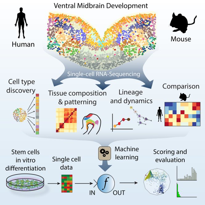
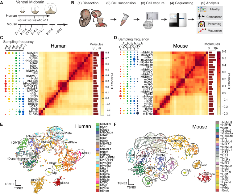
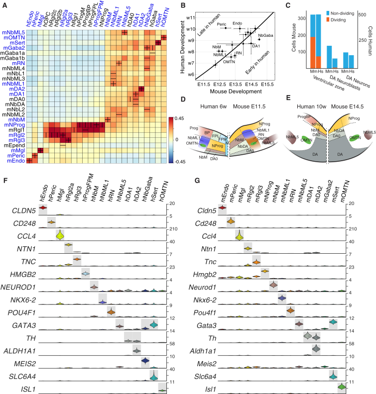
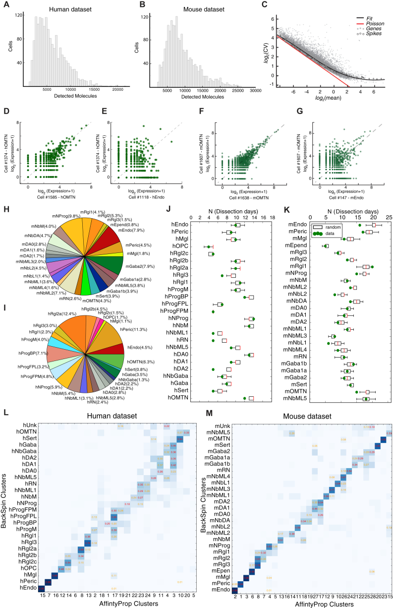
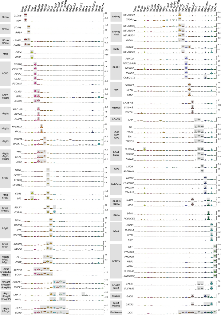
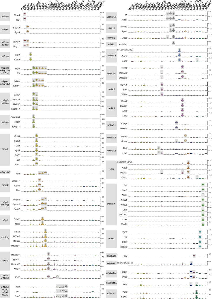
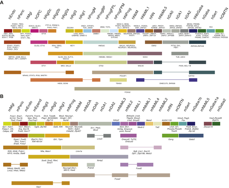
Understanding human embryonic ventral midbrain is of major interest for Parkinson's disease. However, the cell types, their gene expression dynamics, and their relationship to commonly used rodent models remain to be defined. We performed single-cell RNA sequencing to examine ventral midbrain development in human and mouse. We found 25 molecularly defined human cell types, including five subtypes of radial glia-like cells and four progenitors. In the mouse, two mature fetal dopaminergic neuron subtypes diversified into five adult classes during postnatal development. Cell types and gene expression were generally conserved across species, but with clear differences in cell proliferation, developmental timing, and dopaminergic neuron development. Additionally, we developed a method to quantitatively assess the fidelity of dopaminergic neurons derived from human pluripotent stem cells, at a single-cell level. Thus, our study provides insight into the molecular programs controlling human midbrain development and provides a foundation for the development of cell replacement therapies.
Overview
The claims and statistics of this paper haven’t been checked yet.
Run the check — free, no account needed. It reads the open-access text and checks the sources it cites.
Integrity signals
Registry
Cites
Statistics
Figures
Verification
Claims the paper makes
The claims and statistics of this paper haven’t been checked yet.
Run the check — free, no account needed. It reads the open-access text and checks the sources it cites.
Claims other papers make about this one
Each is checked against this paper’s own text: does it say what the citing paper says it does?
The source backs part of the claim, not all of it — or backs it for a different population, condition or comparison.
In the citing paper
“Based on the concordance of other genes co-expressed in Vip+ neurons [13–16], we developed the following mRNA signature for Vip neurons: Vip+ [13–16], Gipr+ [15], Calb1+ [13,14], Sox6- [13,14], Aldh1a1- [13,14].”
From Classification of midbrain dopamine neurons using single-cell gene expression profiling approaches (2020) · cites this paper as [14]
Experimental — accuracy not yet measuredIn the citing paper
“Molecular heterogeneity within the murine DAergic system has been investigated by at least six independent groups, all attempting to classify DA neurons based on gene expression [13–18].”
From Classification of midbrain dopamine neurons using single-cell gene expression profiling approaches (2020) · cites this paper as [14]
In the citing paper
“Although these studies differed significantly in their experimental parameters and analysis methods, they identified partially overlapping subtypes, some of which were located in a manner agnostic to traditional anatomical boundaries (SN, VTA, & RR).”
From Classification of midbrain dopamine neurons using single-cell gene expression profiling approaches (2020) · cites this paper as [14]
In the citing paper
“These cells also display Aldh1a7 [14,17] and Ndnf [13,14] mRNA expression.”
From Classification of midbrain dopamine neurons using single-cell gene expression profiling approaches (2020) · cites this paper as [14]
In the citing paper
“Aldh1a1 and Sox6 mRNAs are the defining markers for this DA neuron subtype, a population overtly represented in two studies: DA1A in [13] and DA-SNC in [14].”
From Classification of midbrain dopamine neurons using single-cell gene expression profiling approaches (2020) · cites this paper as [14]
In the citing paper
“Otx2+, Aldh1a1+ DA neurons in the ventromedial VTA”
From Classification of midbrain dopamine neurons using single-cell gene expression profiling approaches (2020) · cites this paper as [14]
In the citing paper
“In immunolabeling studies, there is a substantial SOX6+ population that is ALDH1A1- [13,14,26]; these cells are located more dorsal to the ALDH1A1+ neurons in the SNc, the parabrachial pigmented (PBP) region of the VTA and the RR.”
From Classification of midbrain dopamine neurons using single-cell gene expression profiling approaches (2020) · cites this paper as [14]
In the citing paper
“Vgat+ DA subtype is located mostly in the VTA”
From Classification of midbrain dopamine neurons using single-cell gene expression profiling approaches (2020) · cites this paper as [14]
In the citing paper
“These cells also co-express high levels of many of the mRNAs aforementioned in the previous population (Ndnf, Igf1 and Sncg), but in contrast, some of these neurons also express Calb1 [13,14], Lypd1 [14], Tacr3 [13,18], and Cyp26b1 [18] mRNAs.”
From Classification of midbrain dopamine neurons using single-cell gene expression profiling approaches (2020) · cites this paper as [14]
In the citing paper
“Sox6+, Aldh1a1- DA neurons are located in the SNc, parabrachial VTA, and RR”
From Classification of midbrain dopamine neurons using single-cell gene expression profiling approaches (2020) · cites this paper as [14]
In the citing paper
“This group of cells is enriched for Vgat [13–15,18], Calb1 [13,14], Crhbp [15,18], Gad2 [15,18] and Wnt7b [15,18] mRNAs.”
From Classification of midbrain dopamine neurons using single-cell gene expression profiling approaches (2020) · cites this paper as [14]
In the citing paper
“Vip+ DA neurons are the most caudally located population”
From Classification of midbrain dopamine neurons using single-cell gene expression profiling approaches (2020) · cites this paper as [14]
In the citing paper
“In addition to lacking Aldh1a1 mRNA, they also do not express Anxa1 [14,17,18] and Aldh1a7 [14,17,18].”
From Classification of midbrain dopamine neurons using single-cell gene expression profiling approaches (2020) · cites this paper as [14]
In the citing paper
“identified three types of embryonic DA neurons (E11.5-E18.5) in the mouse: 1) an immature type expressing Th and other key markers of DA neurons”
From Classification of midbrain dopamine neurons using single-cell gene expression profiling approaches (2020) · cites this paper as [14]
In the citing paper
“3) neurons with a similar signature but also expressing Aldh1a1 and Lmo3”
From Classification of midbrain dopamine neurons using single-cell gene expression profiling approaches (2020) · cites this paper as [14]
In the citing paper
“2) a more mature neuron type expressing both Th and Dat”
From Classification of midbrain dopamine neurons using single-cell gene expression profiling approaches (2020) · cites this paper as [14]
Self-check
Percentages, sums and reported differences re-computed from the paper’s own numbers.
The self-check needs the paper’s full text.
Citations
Cites
Scikit-learn: Machine Learning in Python
Pedregosa, F., Varoquaux, G., Gramfort, A., Michel, V., Thirion, B., Grisel, O., Blondel, M., Müller, A., Nothman, J., Louppe, G., et al. (2012) · arXiv · doi:10.48550/arxiv.1201.0490 · Cited by 71,016
Fiji: an open-source platform for biological-image analysis
Schindelin, J., Arganda-Carreras, I., Frise, E., Kaynig, V., Longair, M., Pietzsch, T., Preibisch, S., Rueden, C., Saalfeld, S., Schmid, B., et al. (2012) · Nature Methods · doi:10.1038/nmeth.2019 · Cited by 66,890
(2008) · Journal of Machine Learning Research · Cited by 33,051
Embryonic Stem Cell Lines Derived from Human Blastocysts
Thomson, J. A., Itskovitz‐Eldor, J., Shapiro, S. S., Waknitz, M. A., Swiergiel, J. J., Marshall, V. S., & Jones, J. M. (1998) · Science · doi:10.1126/science.282.5391.1145 · Cited by 15,947
Database resources of the National Center for Biotechnology Information: update
Wheeler, D. L. (2003) · Nucleic Acids Research · doi:10.1093/nar/gkh073 · Cited by 10,695
Trapnell, C., Cacchiarelli, D., Grimsby, J., Pokharel, P., Li, S., Morse, M., Lennon, N. J., Livak, K. J., Mikkelsen, T. S., & Rinn, J. L. (2014) · Nature Biotechnology · doi:10.1038/nbt.2859 · Cited by 7,314
Spatial reconstruction of single-cell gene expression data
Satija, R., Farrell, J. A., Gennert, D., Schier, A. F., & Regev, A. (2015) · Nature Biotechnology · doi:10.1038/nbt.3192 · Cited by 7,055
A robust and high-throughput Cre reporting and characterization system for the whole mouse brain
Madisen, L., Zwingman, T. A., Sunkin, S. M., Oh, S. W., Zariwala, H. A., Gu, H., Ng, L. L., Palmiter, R. D., Hawrylycz, M. J., Jones, A. R., et al. (2010) · Nature Neuroscience · doi:10.1038/nn.2467 · Cited by 6,907
scikit-image: image processing in Python
(2014) · doi:10.7717/peerj.453 · Cited by 6,794
Genome-wide atlas of gene expression in the adult mouse brain
Lein, E. S., Hawrylycz, M. J., Ao, N., Ayres, M., Bensinger, A., Bernard, A., Boe, A. F., Boguski, M. S., Brockway, K. S., Byrnes, E. J., et al. (2007) · Nature · doi:10.1038/nature05453 · Cited by 5,572
Cited by
Sorted by citations
A Unique Microglia Type Associated with Restricting Development of Alzheimer’s Disease
Keren-Shaul, H., Spinrad, A., Weiner, A., Matcovitch-Natan, O., Dvir-Szternfeld, R., Ulland, T. K., David, E., Baruch, K., Lara-Astaiso, D., Toth, B., et al. (2017) · Cell · doi:10.1016/j.cell.2017.05.018 · Cited by 5,493
Single-cell RNA sequencing technologies and bioinformatics pipelines
Hwang, B., Lee, J. H., & Bang, D. (2018) · Experimental & Molecular Medicine · doi:10.1038/s12276-018-0071-8 · Cited by 1,866
Brann, D. H., Tsukahara, T., Weinreb, C., Lipovsek, M., Van den Berge, K., Gong, B., Chance, R., Macaulay, I. C., Chou, H.-J., Fletcher, R. B., et al. (2020) · Science Advances · doi:10.1126/sciadv.abc5801 · Cited by 1,198
Cross-tissue immune cell analysis reveals tissue-specific features in humans
Conde, C. D., Xu, C., Jarvis, L. B., Rainbow, D. B., Wells, S. B., Gomes, T., Howlett, S. K., Suchánek, O., Polański, K., King, H. W., et al. (2022) · Science · doi:10.1126/science.abl5197 · Cited by 1,246
Construction of a human cell landscape at single-cell level
Han, X., Zhou, Z., Fei, L., Sun, H., Wang, R., Chen, Y., Chen, H., Wang, J., Tang, H., Ge, W., et al. (2020) · Nature · doi:10.1038/s41586-020-2157-4 · Cited by 1,087
Metadata
- Authors
- La Manno, G., Gyllborg, D., Codeluppi, S., Nishimura, K., Salto, C., Zeisel, A., Borm, L. E., Stott, S. R. W., Toledo, E. M., Villaescusa, J. C., Lönnerberg, P., Ryge, J., Barker, R. A., Arenas, E., & Linnarsson, S.
- Affiliations
- Karolinska Institutet (SE)
- Science for Life Laboratory (SE)
- University of Cambridge (GB)
- Karolinska University Hospital (SE)
- École Polytechnique Fédérale de Lausanne (CH)
- Corresponding author
- Arenas, Ernest
- Linnarsson, Sten
- ORCID
- Venue
- Cell
- Publisher
- Elsevier BV
- Published
- 2016-10
- Volume
- 167
- Issue
- 2
- Pages
- 566-580.e19
- ISSN
- 0092-8674 (ISSN-L)
- PMC
- PMC5055122
- OpenAlex
- W2528543174
- Cited by
- 835
- References
- 71
- Citations by year
- 2026: 60 · 2025: 81 · 2024: 84 · 2023: 87 · 2022: 100 · 2021: 103 · 2020: 111 · 2019: 87 · 2018: 72 · 2017: 44 · 2016: 3
- FWCI
- 35.87
- Type
- journal article
- Language
- en
- Open access
- hybrid
- doi.org
- Licence
- Locations
- Cell (publishedVersion, cc-by)
- PubMed Central (publishedVersion, cc-by)
- PubMed (publishedVersion)
- Infoscience (Ecole Polytechnique Fédérale de Lausanne) (submittedVersion)
- Infoscience (Ecole Polytechnique Fédérale de Lausanne) (submittedVersion, cc-by-nc-nd)
- Lund University Publications (Lund University) (submittedVersion)
- Apollo (University of Cambridge) (submittedVersion, cc-by)
- Apollo (cc-by)
- Funders
- Medical Research Council (MC_PC_12009)
- Topics
- Life Sciences › Biochemistry, Genetics and Molecular Biology › Molecular Biology › Single-cell and spatial transcriptomics
- Life Sciences › Neuroscience › Developmental Neuroscience › Neurogenesis and neuroplasticity mechanisms
- Life Sciences › Biochemistry, Genetics and Molecular Biology › Molecular Biology › Pluripotent Stem Cells Research
- Keywords
- Parkinson's disease, dopaminergic neurons, radial glia-like cells, neural progenitor cells, cell type diversity, species conservation, postnatal development, human pluripotent stem cell-derived neurons, cell replacement therapy, single-cell transcriptomics
- MeSH terms
- Cellular Reprogramming Techniques, Machine Learning, Animals, Cell Line, Humans, Mesencephalon, Neuroglia, Sequence Analysis, RNA, Pluripotent Stem Cells, Mice, Neurogenesis, Neural Stem Cells, Single-Cell Analysis, Dopaminergic Neurons
About these checks
Registry, retraction, Cites and statistics results are computed, not judged, with one exception: a printed reference that no index lists is read once by a model before it counts as a citation error. Claims are extracted by a model and kept only when their quote appears verbatim in the text; each cited source is then checked, with a model’s help, for whether it says what the claim relies on. Those verdicts can be wrong — read the claim’s quote and the cited source before relying on one.
Statistics and arithmetic are recomputed, not judged.
Confidence bands are shown only for fields calibrated on our annotated test set. None is calibrated yet, so no band is shown.
What counts as a claim ›How we measure confidence ›How we check this ›
Are you an author, or do you see a problem with this page? Request a review
