

The DOI matches its Crossref record; 69 of the 71 references it cites were found in a registry or an index.
Molecular Diversity of Midbrain Development in Mouse, Human, and Stem Cells
Open accessAbstract
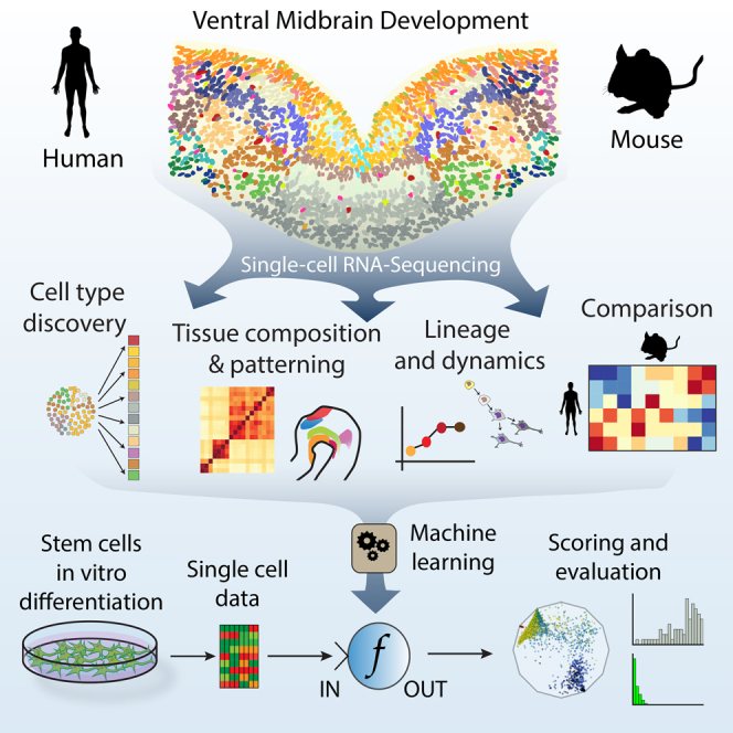
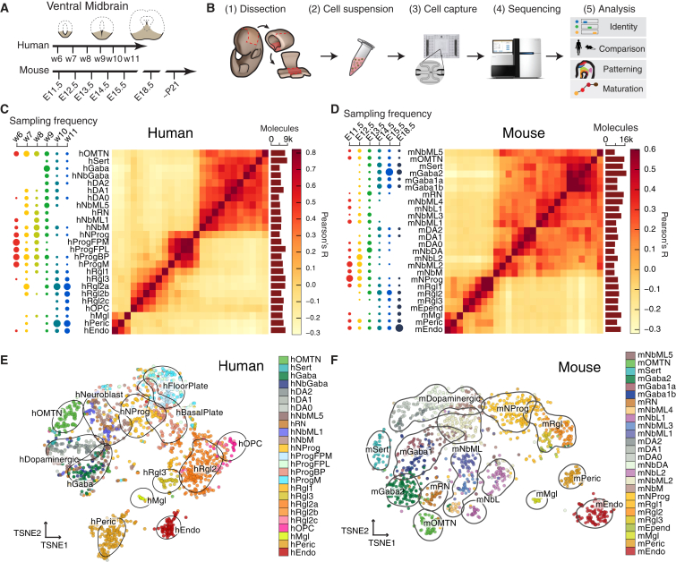
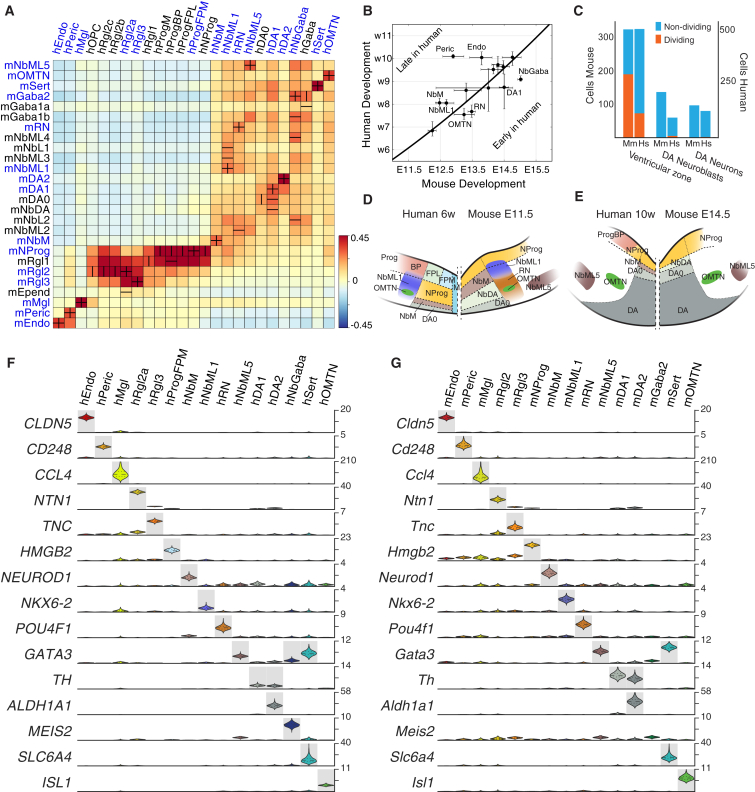
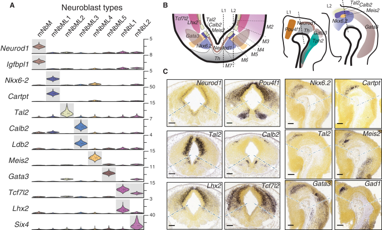
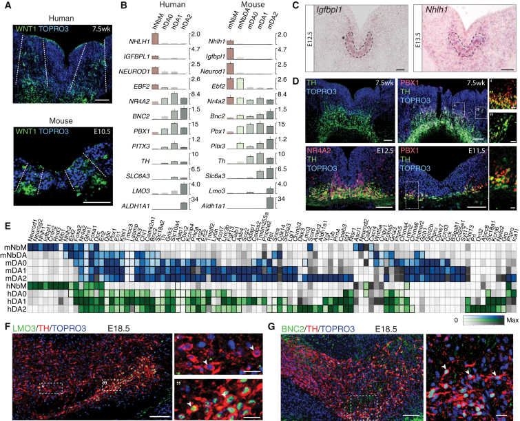
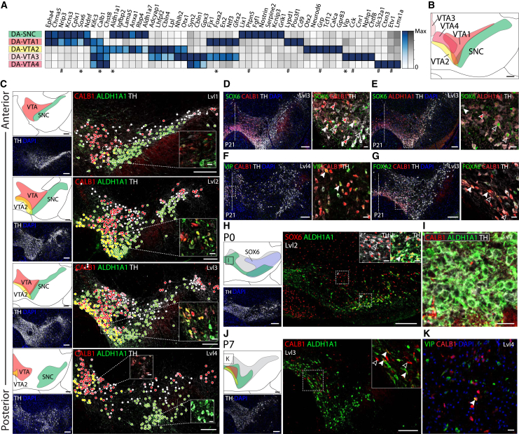
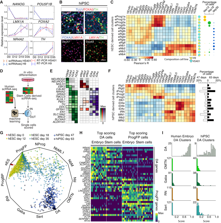
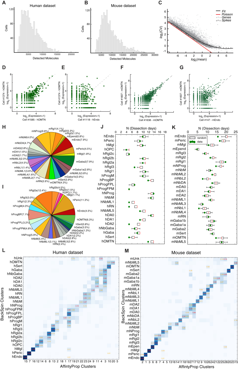
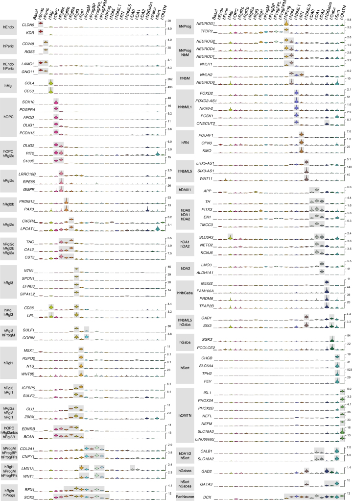
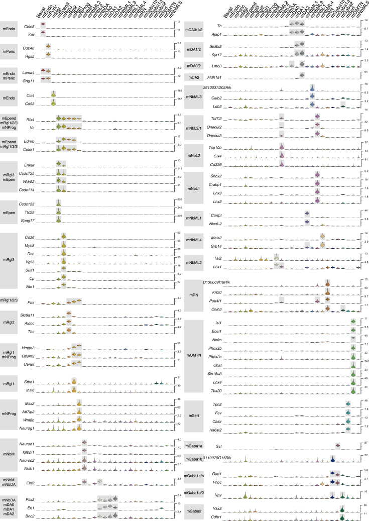
Understanding human embryonic ventral midbrain is of major interest for Parkinson's disease. However, the cell types, their gene expression dynamics, and their relationship to commonly used rodent models remain to be defined. We performed single-cell RNA sequencing to examine ventral midbrain development in human and mouse. We found 25 molecularly defined human cell types, including five subtypes of radial glia-like cells and four progenitors. In the mouse, two mature fetal dopaminergic neuron subtypes diversified into five adult classes during postnatal development. Cell types and gene expression were generally conserved across species, but with clear differences in cell proliferation, developmental timing, and dopaminergic neuron development. Additionally, we developed a method to quantitatively assess the fidelity of dopaminergic neurons derived from human pluripotent stem cells, at a single-cell level. Thus, our study provides insight into the molecular programs controlling human midbrain development and provides a foundation for the development of cell replacement therapies.
Overview
The claims and statistics of this paper haven’t been checked yet.
Run the check — free, no account needed. It reads the open-access text and checks the sources it cites.
Integrity signals
Registry
Cites
Statistics
Figures
Verification
Claims the paper makes
The claims and statistics of this paper haven’t been checked yet.
Run the check — free, no account needed. It reads the open-access text and checks the sources it cites.
Claims other papers make about this one
Each is checked against this paper’s own text: does it say what the citing paper says it does?
The source backs part of the claim, not all of it — or backs it for a different population, condition or comparison.
In the citing paper
“Although these studies differed significantly in their experimental parameters and analysis methods, they identified partially overlapping subtypes, some of which were located in a manner agnostic to traditional anatomical boundaries (SN, VTA, & RR).”
From Classification of midbrain dopamine neurons using single-cell gene expression profiling approaches (2020) · cites this paper as [14]
Experimental — accuracy not yet measuredThe source backs part of the claim, not all of it — or backs it for a different population, condition or comparison.
In the citing paper
“Based on the concordance of other genes co-expressed in Vip+ neurons [13–16], we developed the following mRNA signature for Vip neurons: Vip+ [13–16], Gipr+ [15], Calb1+ [13,14], Sox6- [13,14], Aldh1a1- [13,14].”
From Classification of midbrain dopamine neurons using single-cell gene expression profiling approaches (2020) · cites this paper as [14]
Experimental — accuracy not yet measuredIn the citing paper
“Molecular heterogeneity within the murine DAergic system has been investigated by at least six independent groups, all attempting to classify DA neurons based on gene expression [13–18].”
From Classification of midbrain dopamine neurons using single-cell gene expression profiling approaches (2020) · cites this paper as [14]
In the citing paper
“These cells also display Aldh1a7 [14,17] and Ndnf [13,14] mRNA expression.”
From Classification of midbrain dopamine neurons using single-cell gene expression profiling approaches (2020) · cites this paper as [14]
In the citing paper
“Aldh1a1 and Sox6 mRNAs are the defining markers for this DA neuron subtype, a population overtly represented in two studies: DA1A in [13] and DA-SNC in [14].”
From Classification of midbrain dopamine neurons using single-cell gene expression profiling approaches (2020) · cites this paper as [14]
In the citing paper
“Otx2+, Aldh1a1+ DA neurons in the ventromedial VTA”
From Classification of midbrain dopamine neurons using single-cell gene expression profiling approaches (2020) · cites this paper as [14]
In the citing paper
“In immunolabeling studies, there is a substantial SOX6+ population that is ALDH1A1- [13,14,26]; these cells are located more dorsal to the ALDH1A1+ neurons in the SNc, the parabrachial pigmented (PBP) region of the VTA and the RR.”
From Classification of midbrain dopamine neurons using single-cell gene expression profiling approaches (2020) · cites this paper as [14]
In the citing paper
“Vgat+ DA subtype is located mostly in the VTA”
From Classification of midbrain dopamine neurons using single-cell gene expression profiling approaches (2020) · cites this paper as [14]
In the citing paper
“These cells also co-express high levels of many of the mRNAs aforementioned in the previous population (Ndnf, Igf1 and Sncg), but in contrast, some of these neurons also express Calb1 [13,14], Lypd1 [14], Tacr3 [13,18], and Cyp26b1 [18] mRNAs.”
From Classification of midbrain dopamine neurons using single-cell gene expression profiling approaches (2020) · cites this paper as [14]
In the citing paper
“Sox6+, Aldh1a1- DA neurons are located in the SNc, parabrachial VTA, and RR”
From Classification of midbrain dopamine neurons using single-cell gene expression profiling approaches (2020) · cites this paper as [14]
In the citing paper
“This group of cells is enriched for Vgat [13–15,18], Calb1 [13,14], Crhbp [15,18], Gad2 [15,18] and Wnt7b [15,18] mRNAs.”
From Classification of midbrain dopamine neurons using single-cell gene expression profiling approaches (2020) · cites this paper as [14]
In the citing paper
“Vip+ DA neurons are the most caudally located population”
From Classification of midbrain dopamine neurons using single-cell gene expression profiling approaches (2020) · cites this paper as [14]
In the citing paper
“In addition to lacking Aldh1a1 mRNA, they also do not express Anxa1 [14,17,18] and Aldh1a7 [14,17,18].”
From Classification of midbrain dopamine neurons using single-cell gene expression profiling approaches (2020) · cites this paper as [14]
In the citing paper
“identified three types of embryonic DA neurons (E11.5-E18.5) in the mouse: 1) an immature type expressing Th and other key markers of DA neurons”
From Classification of midbrain dopamine neurons using single-cell gene expression profiling approaches (2020) · cites this paper as [14]
In the citing paper
“3) neurons with a similar signature but also expressing Aldh1a1 and Lmo3”
From Classification of midbrain dopamine neurons using single-cell gene expression profiling approaches (2020) · cites this paper as [14]
In the citing paper
“2) a more mature neuron type expressing both Th and Dat”
From Classification of midbrain dopamine neurons using single-cell gene expression profiling approaches (2020) · cites this paper as [14]
Self-check
Percentages, sums and reported differences re-computed from the paper’s own numbers.
The self-check needs the paper’s full text.
Citations
Cites
Scikit-learn: Machine Learning in Python
Pedregosa, F., Varoquaux, G., Gramfort, A., Michel, V., Thirion, B., Grisel, O., Blondel, M., Müller, A., Nothman, J., Louppe, G., et al. (2012) · arXiv · doi:10.48550/arxiv.1201.0490 · Cited by 71,016
Fiji: an open-source platform for biological-image analysis
Schindelin, J., Arganda-Carreras, I., Frise, E., Kaynig, V., Longair, M., Pietzsch, T., Preibisch, S., Rueden, C., Saalfeld, S., Schmid, B., et al. (2012) · Nature Methods · doi:10.1038/nmeth.2019 · Cited by 66,890
(2008) · Journal of Machine Learning Research · Cited by 33,051
Embryonic Stem Cell Lines Derived from Human Blastocysts
Thomson, J. A., Itskovitz‐Eldor, J., Shapiro, S. S., Waknitz, M. A., Swiergiel, J. J., Marshall, V. S., & Jones, J. M. (1998) · Science · doi:10.1126/science.282.5391.1145 · Cited by 15,947
Database resources of the National Center for Biotechnology Information: update
Wheeler, D. L. (2003) · Nucleic Acids Research · doi:10.1093/nar/gkh073 · Cited by 10,695
Trapnell, C., Cacchiarelli, D., Grimsby, J., Pokharel, P., Li, S., Morse, M., Lennon, N. J., Livak, K. J., Mikkelsen, T. S., & Rinn, J. L. (2014) · Nature Biotechnology · doi:10.1038/nbt.2859 · Cited by 7,314
Spatial reconstruction of single-cell gene expression data
Satija, R., Farrell, J. A., Gennert, D., Schier, A. F., & Regev, A. (2015) · Nature Biotechnology · doi:10.1038/nbt.3192 · Cited by 7,055
A robust and high-throughput Cre reporting and characterization system for the whole mouse brain
Madisen, L., Zwingman, T. A., Sunkin, S. M., Oh, S. W., Zariwala, H. A., Gu, H., Ng, L. L., Palmiter, R. D., Hawrylycz, M. J., Jones, A. R., et al. (2010) · Nature Neuroscience · doi:10.1038/nn.2467 · Cited by 6,907
scikit-image: image processing in Python
(2014) · doi:10.7717/peerj.453 · Cited by 6,794
Genome-wide atlas of gene expression in the adult mouse brain
Lein, E. S., Hawrylycz, M. J., Ao, N., Ayres, M., Bensinger, A., Bernard, A., Boe, A. F., Boguski, M. S., Brockway, K. S., Byrnes, E. J., et al. (2007) · Nature · doi:10.1038/nature05453 · Cited by 5,572
Cited by
Sorted by citations
A Unique Microglia Type Associated with Restricting Development of Alzheimer’s Disease
Keren-Shaul, H., Spinrad, A., Weiner, A., Matcovitch-Natan, O., Dvir-Szternfeld, R., Ulland, T. K., David, E., Baruch, K., Lara-Astaiso, D., Toth, B., et al. (2017) · Cell · doi:10.1016/j.cell.2017.05.018 · Cited by 5,493
Single-cell RNA sequencing technologies and bioinformatics pipelines
Hwang, B., Lee, J. H., & Bang, D. (2018) · Experimental & Molecular Medicine · doi:10.1038/s12276-018-0071-8 · Cited by 1,866
Brann, D. H., Tsukahara, T., Weinreb, C., Lipovsek, M., Van den Berge, K., Gong, B., Chance, R., Macaulay, I. C., Chou, H.-J., Fletcher, R. B., et al. (2020) · Science Advances · doi:10.1126/sciadv.abc5801 · Cited by 1,198
Cross-tissue immune cell analysis reveals tissue-specific features in humans
Conde, C. D., Xu, C., Jarvis, L. B., Rainbow, D. B., Wells, S. B., Gomes, T., Howlett, S. K., Suchánek, O., Polański, K., King, H. W., et al. (2022) · Science · doi:10.1126/science.abl5197 · Cited by 1,246
Construction of a human cell landscape at single-cell level
Han, X., Zhou, Z., Fei, L., Sun, H., Wang, R., Chen, Y., Chen, H., Wang, J., Tang, H., Ge, W., et al. (2020) · Nature · doi:10.1038/s41586-020-2157-4 · Cited by 1,087
Integrative single-cell analysis of transcriptional and epigenetic states in the human adult brain
Lake, B. B., Chen, S., Sos, B. C., Fan, J., Kaeser, G. E., Yung, Y. C., Duong, T. E., Gao, D., Chun, J., Kharchenko, P. V., et al. (2018) · Nature Biotechnology · doi:10.1038/nbt.4038 · Cited by 983
Dissecting Immune Circuits by Linking CRISPR-Pooled Screens with Single-Cell RNA-Seq
Jaitin, D. A., Weiner, A., Yofe, I., Lara-Astiaso, D., Keren-Shaul, H., David, E., Salame, T. M., Tanay, A., van Oudenaarden, A., & Amit, I. (2016) · Cell · doi:10.1016/j.cell.2016.11.039 · Cited by 777
Brain cell type–specific enhancer–promoter interactome maps and disease - risk association
Nott, A., Holtman, I. R., Coufal, N. G., Schlachetzki, J. C. M., Yu, M., Hu, R., Han, C. Z., Pena, M., Xiao, J., Wu, Y., et al. (2019) · Science · doi:10.1126/science.aay0793 · Cited by 699
Comparative cellular analysis of motor cortex in human, marmoset and mouse
Bakken, T. E., Jorstad, N. L., Hu, Q., Lake, B. B., Tian, W., Kalmbach, B. E., Crow, M., Hodge, R. D., Krienen, F. M., Sorensen, S. A., et al. (2021) · Nature · doi:10.1038/s41586-021-03465-8 · Cited by 812
Demontis, D., Walters, G. B., Athanasiadis, G., Walters, R., Therrien, K., Nielsen, T. T., Farajzadeh, L., Voloudakis, G., Bendl, J., Zeng, B., et al. (2022) · Nature Genetics · doi:10.1016/j.euroneuro.2022.07.018 · Cited by 863
SAVER: gene expression recovery for single-cell RNA sequencing
Huang, M. C., Wang, J., Torre, E. A., Dueck, H., Shaffer, S. M., Bonasio, R., Murray, J. I., Raj, A., Li, M., & Zhang, N. R. (2018) · Nature Methods · doi:10.1038/s41592-018-0033-z · Cited by 739
scmap: projection of single-cell RNA-seq data across data sets
(2018) · doi:10.1038/nmeth.4644 · Cited by 735
Developmental and oncogenic programs in H3K27M gliomas dissected by single-cell RNA-seq
Filbin, M. G., Tirosh, I., Hovestadt, V., Shaw, M. L., Escalante, L. E., Mathewson, N. D., Neftel, C., Frank, N., Pelton, K., Hebert, C. M., et al. (2018) · Science · doi:10.1126/science.aao4750 · Cited by 694
Genetic identification of brain cell types underlying schizophrenia
Major Depressive Disorder Working Group of the Psychiatric Genomics Consortium, Skene, N. G., Bryois, J., Bakken, T. E., Breen, G., Crowley, J. J., Gaspar, H. A., Giusti-Rodriguez, P., Hodge, R. D., Miller, J. A., et al. (2018) · Nature Genetics · doi:10.1038/s41588-018-0129-5 · Cited by 651
Self-citationSimultaneous single-cell profiling of lineages and cell types in the vertebrate brain
Raj, B., Wagner, D. E., McKenna, A., Pandey, S., Klein, A. M., Shendure, J., Gagnon, J. A., & Schier, A. F. (2018) · Nature Biotechnology · doi:10.1038/nbt.4103 · Cited by 610
Kamath, T., Abdulraouf, A., Burris, S. J., Langlieb, J., Gazestani, V., Nadaf, N. M., Balderrama, K., Vanderburg, C., & Macosko, E. Z. (2022) · Nature Neuroscience · doi:10.1038/s41593-022-01061-1 · Cited by 552
Molecular architecture of the developing mouse brain
La Manno, G., Siletti, K., Furlan, A., Gyllborg, D., Vinsland, E., Mossi Albiach, A., Mattsson Langseth, C., Khven, I., Lederer, A. R., Dratva, L. M., et al. (2021) · Nature · doi:10.1038/s41586-021-03775-x · Cited by 563
Self-citationClark, B. S., Stein-O’Brien, G., Shiau, F., Cannon, G. H., Davis-Marcisak, E. F., Sherman, T. D., Santiago, C. P., Hoang, T. V., Rajaii, F., James-Esposito, R. E., et al. (2019) · Neuron · doi:10.1016/j.neuron.2019.04.010 · Cited by 527
Häring, M., Zeisel, A., Hochgerner, H., Rinwa, P., Jakobsson, J. E. T., Lönnerberg, P., La Manno, G., Sharma, N., Borgius, L., Kiehn, O., et al. (2018) · Nature Neuroscience · doi:10.1038/s41593-018-0141-1 · Cited by 470
Self-citationTosches, M. A., Yamawaki, T. M., Naumann, R. K., Jacobi, A. A., Tushev, G., & Laurent, G. (2018) · Science · doi:10.1126/science.aar4237 · Cited by 506
Single-Cell Transcriptome Analysis Maps the Developmental Track of the Human Heart
Cui, Y., Zheng, Y., Liu, X., Yan, L., Fan, X., Yong, J., Hu, Y., Dong, J., Li, Q., Wu, X., et al. (2019) · Cell Reports · doi:10.1016/j.celrep.2019.01.079 · Cited by 471
Molecular Classification and Comparative Taxonomics of Foveal and Peripheral Cells in Primate Retina
Peng, Y., Shekhar, K., Yan, W., Herrmann, D., Sappington, A., Bryman, G. S., van Zyl, T. A., Do, M. T. H., Regev, A., & Sanes, J. R. (2019) · Cell · doi:10.1016/j.cell.2019.01.004 · Cited by 470
Cross-Species Single-Cell Analysis Reveals Divergence of the Primate Microglia Program
Geirsdottir, L., David, E., Keren-Shaul, H., Weiner, A., Bohlen, S. C., Neuber, J., Balic, A., Giladi, A., Sheban, F., Dutertre, C.-A., et al. (2019) · Cell · doi:10.1016/j.cell.2019.11.010 · Cited by 461
Parker, K. R., Migliorini, D., Perkey, E., Yost, K. E., Bhaduri, A., Bagga, P., Haris, M., Wilson, N. E., Liu, F., Gabunia, K., et al. (2020) · Cell · doi:10.1016/j.cell.2020.08.022 · Cited by 464
Whole-organism clone tracing using single-cell sequencing
Alemany, A., Florescu, M., Baron, C. S., Peterson-Maduro, J., & van Oudenaarden, A. (2018) · Nature · doi:10.1038/nature25969 · Cited by 447
Crow, M., Paul, A., Ballouz, S., Huang, Z. J., & Gillis, J. (2018) · Nature Communications · doi:10.1038/s41467-018-03282-0 · Cited by 365
The promise of spatial transcriptomics for neuroscience in the era of molecular cell typing
Lein, E., Borm, L. E., & Linnarsson, S. (2017) · Science · doi:10.1126/science.aan6827 · Cited by 383
Self-citationPoulin, J.-F., Caronia, G., Hofer, C., Cui, Q., Helm, B., Ramakrishnan, C., Chan, C. S., Dombeck, D. A., Deisseroth, K., & Awatramani, R. (2018) · Nature Neuroscience · doi:10.1038/s41593-018-0203-4 · Cited by 423
Volpato, V., & Webber, C. (2020) · Disease Models & Mechanisms · doi:10.1242/dmm.042317 · Cited by 379
Preissl, S., Fang, R., Huang, H., Zhao, Y., Raviram, R., Gorkin, D. U., Zhang, Y., Sos, B. C., Afzal, V., Dickel, D. E., et al. (2018) · Nature Neuroscience · doi:10.1038/s41593-018-0079-3 · Cited by 332
Physical Activity and Brain Health
Liegro, C. M. D., Schiera, G., Proia, P., & Liegro, I. D. (2019) · Genes · doi:10.3390/genes10090720 · Cited by 451
Cervo, P. R. D. V., Romanov, R. A., Spigolon, G., Masini, D., Martín‐Montañez, E., Toledo, E. M., Manno, G. L., Feyder, M. T., Pifl, C., Ng, Y. H., et al. (2017) · Nature Biotechnology · doi:10.1038/nbt.3835 · Cited by 357
Self-citationIntegration of spatial and single-cell transcriptomic data elucidates mouse organogenesis
Lohoff, T., Ghazanfar, S., Missarova, A., Koulena, N., Pierson, N. G., Griffiths, J. A., Bardot, E., Eng, C.-H. L., Tyser, R. C. V., Argelaguet, R., et al. (2021) · Nature Biotechnology · doi:10.1038/s41587-021-01006-2 · Cited by 333
Single-cell transcriptomics reveals expansion of cytotoxic CD4 T cells in supercentenarians
Hashimoto, K., Kouno, T., Ikawa, T., Hayatsu, N., Miyajima, Y., Yabukami, H., Terooatea, T., Sasaki, T., Suzuki, T., Valentine, M., et al. (2019) · Proceedings of the National Academy of Sciences · doi:10.1073/pnas.1907883116 · Cited by 343
Genetic mapping of cell type specificity for complex traits
Watanabe, K., Mirkov, M. U., de Leeuw, C., van den Heuvel, M. P., & Posthuma, D. (2019) · Nature Communications · doi:10.1038/s41467-019-11181-1 · Cited by 329
Single-Cell RNA Sequencing Reveals a Dynamic Stromal Niche That Supports Tumor Growth
Davidson, S., Efremova, M., Riedel, A., Mahata, B., Pramanik, J., Huuhtanen, J., Kar, G., Vento‐Tormo, R., Hagai, T., Chen, X. X., et al. (2020) · Cell Reports · doi:10.1016/j.celrep.2020.107628 · Cited by 330
Single-cell and spatial transcriptomics: deciphering brain complexity in health and disease
Piwecka, M., Rajewsky, N., & Rybak-Wolf, A. (2023) · Nature Reviews Neurology · doi:10.1038/s41582-023-00809-y · Cited by 320
Single-cell transcriptomic analyses provide insights into the developmental origins of neuroblastoma
Jansky, S., Sharma, A. K., Körber, V., Quintero, A., Toprak, U. H., Wecht, E. M., Gartlgruber, M., Greco, A., Chomsky, E., Grünewald, T. G. P., et al. (2021) · Nature Genetics · doi:10.1038/s41588-021-00806-1 · Cited by 298
Molecular landscapes of human hippocampal immature neurons across lifespan
Zhou, Y. F., Su, Y., Li, S., Kennedy, B. C., Zhang, D. Y., Bond, A. M., Sun, Y. Y., Jacob, F., Lu, L., Hu, P., et al. (2022) · Nature · doi:10.1038/s41586-022-04912-w · Cited by 314
Population-scale single-cell RNA-seq profiling across dopaminergic neuron differentiation
Jerber, J., Seaton, D. D., Cuomo, A. S. E., Kumasaka, N., Haldane, J., Steer, J., Patel, M. J., Pearce, D., Andersson, M. H. L., Bonder, M. J., et al. (2021) · Nature Genetics · doi:10.1038/s41588-021-00801-6 · Cited by 293
Tiklová, K., Björklund, Å. K., Lahti, L., Fiorenzano, A., Nolbrant, S., Gillberg, L., Volakakis, N., Yokota, C., Hilscher, M. M., Hauling, T., et al. (2019) · Nature Communications · doi:10.1038/s41467-019-08453-1 · Cited by 276
Patch-Seq Links Single-Cell Transcriptomes to Human Islet Dysfunction in Diabetes
Camuñas-Soler, J., Dai, X.-Q., Hang, Y., Bautista, A., Lyon, J. G., Suzuki, K., Kim, S. K., Quake, S. R., & MacDonald, P. E. (2020) · Cell Metabolism · doi:10.1016/j.cmet.2020.04.005 · Cited by 286
Transcriptional Convergence of Oligodendrocyte Lineage Progenitors during Development
Marques, S., van Bruggen, D., Vanichkina, D. P., Floriddia, E. M., Munguba, H., Väremo, L., Giacomello, S., Falcão, A. M., Meijer, M., Björklund, Å. K., et al. (2018) · Developmental Cell · doi:10.1016/j.devcel.2018.07.005 · Cited by 290
Self-citationMulti-ancestry genome-wide association meta-analysis of Parkinson’s disease
Kim, J. J., Vitale, D., Otani, D. V., Lian, M. M., Heilbron, K., Aslibekyan, S. W., Auton, A., Babalola, E., Bell, R. K., Bielenberg, J., et al. (2023) · Nature Genetics · doi:10.1038/s41588-023-01584-8 · Cited by 302
Comprehensive cell atlas of the first-trimester developing human brain
Braun, E., Danan-Gotthold, M., Borm, L. E., Vinsland, E., Lee, K. W., Lönnerberg, P., Hu, L., Li, X., He, X., Andrusivová, Ž., et al. (2023) · Science · doi:10.1126/science.adf1226 · Cited by 300
Self-citationBrann, D. H., Tsukahara, T., Weinreb, C., Lipovsek, M., den Berge, K. V., Gong, B., Chance, R., Macaulay, I. C., Chou, H.-J., Fletcher, R., et al. (2020) · bioRxiv (Cold Spring Harbor Laboratory) · doi:10.1101/2020.03.25.009084 · Cited by 306
Zero-preserving imputation of single-cell RNA-seq data
Linderman, G. C., Zhao, J., Roulis, M., Bielecki, P., Flavell, R. A., Nadler, B., & Kluger, Y. (2022) · Nature Communications · doi:10.1038/s41467-021-27729-z · Cited by 383
Ecker, J. R., Geschwind, D. H., Kriegstein, A. R., Ngai, J., Osten, P., Polioudakis, D., Regev, A., Šestan, N., Wickersham, I. R., & Zeng, H. (2017) · Neuron · doi:10.1016/j.neuron.2017.10.007 · Cited by 258
Artegiani, B., Lyubimova, A., Muraro, M., van Es, J. H., van Oudenaarden, A., & Clevers, H. (2017) · Cell Reports · doi:10.1016/j.celrep.2017.11.050 · Cited by 259
Neural Stem Cell Transplantation for Neurodegenerative Diseases
De Gioia, R., Biella, F., Citterio, G., Rizzo, F., Abati, E., Nizzardo, M., Bresolin, N., Comi, G. P., & Corti, S. (2020) · International Journal of Molecular Sciences · doi:10.3390/ijms21093103 · Cited by 247
Human brain organogenesis: Toward a cellular understanding of development and disease
Kelley, K. W., & Pașca, S. P. (2021) · Cell · doi:10.1016/j.cell.2021.10.003 · Cited by 244
Impact of the Human Cell Atlas on medicine
Rood, J. E., Maartens, A., Hupalowska, A., Teichmann, S. A., & Regev, A. (2022) · Nature Medicine · doi:10.1038/s41591-022-02104-7 · Cited by 222
Classification of midbrain dopamine neurons using single-cell gene expression profiling approaches
Poulin, J.-F., Gaertner, Z., Moreno-Ramos, O. A., & Awatramani, R. (2020) · Trends in Neurosciences · doi:10.1016/j.tins.2020.01.004 · Cited by 238
Watanabe, K., Jansen, P. R., Savage, J. E., Nandakumar, P., Wang, X., Agee, M., Aslibekyan, S. W., Auton, A., Bell, R. K., Bryc, K., et al. (2022) · Nature Genetics · doi:10.1038/s41588-022-01124-w · Cited by 240
Loera-Valencia, R., Goikolea, J., Parrado-Fernandez, C., Merino-Serrais, P., & Maioli, S. (2019) · The Journal of Steroid Biochemistry and Molecular Biology · doi:10.1016/j.jsbmb.2019.03.003 · Cited by 214
Rifes, P., Isaksson, M., Rathore, G. S., Aldrin-Kirk, P., Møller, O. K., Barzaghi, G., Lee, J., Egerod, K. L., Rausch, D. M., Parmar, M., et al. (2020) · Nature Biotechnology · doi:10.1038/s41587-020-0525-0 · Cited by 226
Human iPSC banking: barriers and opportunities
Huang, C., Liu, C.-L., Ting, C.-Y., Chiu, Y.-T., Cheng, Y., Nicholson, M. W., & Hsieh, P. C. H. (2019) · Journal of Biomedical Science · doi:10.1186/s12929-019-0578-x · Cited by 222
Liu, I., Jiang, L., Samuelsson, E. R., Salas, S. M., Beck, A., Hack, O. A., Jeong, D., Shaw, M. L., Englinger, B., LaBelle, J. J., et al. (2022) · Nature Genetics · doi:10.1038/s41588-022-01236-3 · Cited by 225
Cell-type specialization is encoded by specific chromatin topologies
Winick‐Ng, W., Kukalev, A., Harabula, I., Zea-Redondo, L., Szabó, D., Meijer, M., Serebreni, L., Zhang, Y., Bianco, S., Chiariello, A. M., et al. (2021) · Nature · doi:10.1038/s41586-021-04081-2 · Cited by 214
Nievergelt, C. M., Maihofer, A. X., Atkinson, E. G., Chen, C., Choi, K. W., Coleman, J. R. I., Daskalakis, N. P., Duncan, L. E., Polimanti, R. A., Aaronson, C. J., et al. (2024) · Nature Genetics · doi:10.1038/s41588-024-01707-9 · Cited by 234
Kelley, K. W., Nakao-Inoue, H., Molofsky, A. V., & Oldham, M. C. (2018) · Nature Neuroscience · doi:10.1038/s41593-018-0216-z · Cited by 198
Interplay between MicroRNAs and Oxidative Stress in Neurodegenerative Diseases
Konovalova, J., Gerasymchuk, D., Parkkinen, I., Chmielarz, P., & Domanskyi, A. (2019) · International Journal of Molecular Sciences · doi:10.3390/ijms20236055 · Cited by 204
Genetic mapping of etiologic brain cell types for obesity
Timshel, P. N., Thompson, J. J., & Pers, T. H. (2020) · eLife · doi:10.7554/elife.55851 · Cited by 202
Introduction to Single‐Cell RNA Sequencing
Olsen, T. K., & Baryawno, N. (2018) · Current Protocols in Molecular Biology · doi:10.1002/cpmb.57 · Cited by 201
A Single-Cell Transcriptional Atlas of the Developing Murine Cerebellum
Carter, R. A., Bihannic, L., Rosencrance, C. D., Hadley, J. L., Tong, Y., Phoenix, T. N., Natarajan, S., Easton, J. B., Northcott, P. A., & Gawad, C. (2018) · Current Biology · doi:10.1016/j.cub.2018.07.062 · Cited by 190
Data denoising with transfer learning in single-cell transcriptomics
Wang, J., Agarwal, D., Huang, M. C., Hu, G., Zhou, Z., Ye, C., & Zhang, N. R. (2019) · Nature Methods · doi:10.1038/s41592-019-0537-1 · Cited by 190
Kupari, J., Usoskin, D., Parisien, M., Lou, D., Hu, Y., Fatt, M. P., Lönnerberg, P., Spångberg, M., Eriksson, B., Barkas, N., et al. (2021) · Nature Communications · doi:10.1038/s41467-021-21725-z · Cited by 195
Self-citationA roadmap for the Human Developmental Cell Atlas
Haniffa, M. A., Taylor, D. M., Linnarsson, S., Aronow, B. J., Bader, G. D., Barker, R. A., Cámara, P. G., Camp, J. G., Chédotal, A., Copp, A. J., et al. (2021) · Nature · doi:10.1038/s41586-021-03620-1 · Cited by 193
Self-citationWidespread Transcriptional Scanning in the Testis Modulates Gene Evolution Rates
Xia, B., Yan, Y., Baron, M., Wagner, F., Barkley, D., Chiodin, M., Kim, S. Y., Keefe, D. L., Alukal, J. P., Boeke, J. D., et al. (2020) · Cell · doi:10.1016/j.cell.2019.12.015 · Cited by 190
Sabaté‐Soler, S., Nickels, S., Saraiva, C., Berger, E., Dubonyte, U., Barmpa, K., Lan, Y. J., Kouno, T., Jarazo, J., Robertson, G., et al. (2022) · Glia · doi:10.1002/glia.24167 · Cited by 197
Ion Channel Degeneracy, Variability, and Covariation in Neuron and Circuit Resilience
Goaillard, J., & Marder, E. E. (2021) · Annual Review of Neuroscience · doi:10.1146/annurev-neuro-092920-121538 · Cited by 188
The changing mouse embryo transcriptome at whole tissue and single-cell resolution
He, P., Williams, B. A., Trout, D. E., Marinov, G. K., Amrhein, H., Berghella, L., Goh, S.-T., Plajzer-Frick, I., Afzal, V., Pennacchio, L. A., et al. (2020) · Nature · doi:10.1038/s41586-020-2536-x · Cited by 174
Genetic predispositions of Parkinson’s disease revealed in patient-derived brain cells
Tran, J., Anastacio, H. T. D., & Bardy, C. (2020) · npj Parkinson s Disease · doi:10.1038/s41531-020-0110-8 · Cited by 190
Cadwell, C. R., Scala, F., Li, S., Livrizzi, G., Shen, S., Sandberg, R., Jiang, X., & Tolias, A. S. (2017) · Nature Protocols · doi:10.1038/nprot.2017.120 · Cited by 147
Unique functional responses differentially map onto genetic subtypes of dopamine neurons
Azcorra, M., Gaertner, Z. A., Davidson, C., He, Q., Kim, H., Nagappan, S., Hayes, C. K., Ramakrishnan, C., Fenno, L. E., Kim, Y. S., et al. (2023) · Nature Neuroscience · doi:10.1038/s41593-023-01401-9 · Cited by 172
Stein-O’Brien, G., Clark, B. S., Sherman, T. D., Zibetti, C., Hu, Q., Sealfon, R. S. G., Liu, S., Qian, J. X., Colantuoni, C., Blackshaw, S., et al. (2019) · Cell Systems · doi:10.1016/j.cels.2019.04.004 · Cited by 172
Dopamine, Updated: Reward Prediction Error and Beyond
Lerner, T. N., Holloway, A. L., & Seiler, J. L. (2020) · Current Opinion in Neurobiology · doi:10.1016/j.conb.2020.10.012 · Cited by 195
Heitzeneder, S., Bosse, K. R., Zhu, Z., Zhelev, D. V., Majzner, R. G., Radosevich, M., Dhingra, S., Sotillo, E., Buongervino, S. N., Pascual‐Pasto, G., et al. (2021) · Cancer Cell · doi:10.1016/j.ccell.2021.12.005 · Cited by 174
CRISPR-based functional genomics for neurological disease
Kampmann, M. (2020) · Nature Reviews Neurology · doi:10.1038/s41582-020-0373-z · Cited by 163
Single-cell-resolution transcriptome map of human, chimpanzee, bonobo, and macaque brains
Khrameeva, E., Kurochkin, I., Han, D., Guijarro, P., Kanton, S., Santel, M., Qian, Z., Rong, S., Mazin, P., Sabirov, M., et al. (2020) · Genome Research · doi:10.1101/gr.256958.119 · Cited by 162
Transcriptomic comparison of human and mouse brain microvessels
Song, H. W., Foreman, K. L., Gastfriend, B. D., Kuo, J. S., Palecek, S. P., & Shusta, E. V. (2020) · Scientific Reports · doi:10.1038/s41598-020-69096-7 · Cited by 166
Fiorenzano, A., Sozzi, E., Birtele, M., Kajtez, J., Giacomoni, J., Nilsson, F., Bruzelius, A., Sharma, Y., Zhang, Y., Mattsson, B., et al. (2021) · Nature Communications · doi:10.1038/s41467-021-27464-5 · Cited by 160
Universal Alternative Splicing of Noncoding Exons
Deveson, I. W., Brunck, M. E. G., Blackburn, J., Tseng, E., Hon, T., Clark, T. A., Clark, M. B., Crawford, J., Dinger, M. E., Nielsen, L. K., et al. (2018) · Cell Systems · doi:10.1016/j.cels.2017.12.005 · Cited by 155
Gojo, J., Englinger, B., Jiang, L., Hübner, J.-M., Shaw, M. L., Hack, O. A., Madlener, S., Kirchhofer, D., Liu, I., Pyrdol, J. W., et al. (2020) · Cancer Cell · doi:10.1016/j.ccell.2020.06.004 · Cited by 150
Yim, A., Smith, C., & Brown, A. M. (2022) · Immunological Reviews · doi:10.1111/imr.13081 · Cited by 161
Cell-type-specific 3D epigenomes in the developing human cortex
Song, M., Pebworth, M.-P., Yang, X., Abnousi, A., Fan, C., Wen, J., Rosen, J. D., Choudhary, M. N. K., Cui, X., Jones, I. R., et al. (2020) · Nature · doi:10.1038/s41586-020-2825-4 · Cited by 145
Single-Cell RNA Sequencing: Unraveling the Brain One Cell at a Time
Ofengeim, D., Γιαγτζόγλου, Ν., Huh, D., Zou, C., & Yuan, J. (2017) · Trends in Molecular Medicine · doi:10.1016/j.molmed.2017.04.006 · Cited by 150
Rotenone exerts developmental neurotoxicity in a human brain spheroid model
Pamies, D., Block, K., Lau, P., Gribaldo, L., Pardo, C. A., Barreras, P., Smirnova, L., Wiersma, D., Zhao, L., Harris, G., et al. (2018) · Toxicology and Applied Pharmacology · doi:10.1016/j.taap.2018.02.003 · Cited by 155
Evaluating measures of association for single-cell transcriptomics
Skinnider, M. A., Squair, J. W., & Foster, L. J. (2019) · Nature Methods · doi:10.1038/s41592-019-0372-4 · Cited by 145
Fernandes, H. J. R., Patikas, N., Foskolou, S., Field, S. F., Park, J.-E., Byrne, M. L., Bassett, A. R., & Metzakopian, E. (2020) · Cell Reports · doi:10.1016/j.celrep.2020.108263 · Cited by 141
SIDR: simultaneous isolation and parallel sequencing of genomic DNA and total RNA from single cells
Han, K. Y., Kim, K.-T., Joung, J.-G., Son, D.-S., Kim, Y. J., Jo, A., Jeon, H.-J., Moon, H.-S., Yoo, C. E., Chung, W., et al. (2018) · Genome Research · doi:10.1101/gr.223263.117 · Cited by 143
Best Practices for Translational Disease Modeling Using Human iPSC-Derived Neurons
Engle, S. J., Blaha, L. C., & Kleiman, R. J. (2018) · Neuron · doi:10.1016/j.neuron.2018.10.033 · Cited by 148
Emerging links between cerebrovascular and neurodegenerative diseases—a special role for pericytes
Lendahl, U., Nilsson, P., & Betsholtz, C. (2019) · EMBO Reports · doi:10.15252/embr.201948070 · Cited by 148
Ensembles of endothelial and mural cells promote angiogenesis in prenatal human brain
Crouch, E. E., Bhaduri, A., Andrews, M. G., Cebrián‐Silla, A., Diafos, L. N., Birrueta, J. O., Wedderburn-Pugh, K., Valenzuela, E. J., Bennett, N. K., Eze, U. C., et al. (2022) · Cell · doi:10.1016/j.cell.2022.09.004 · Cited by 139
What does time mean in development?
Ebisuya, M., & Briscoe, J. (2018) · Development · doi:10.1242/dev.164368 · Cited by 136
Hook, P. W., McClymont, S. A., Cannon, G. H., Law, W. D., Morton, A. J., Goff, L. A., & McCallion, A. S. (2018) · The American Journal of Human Genetics · doi:10.1016/j.ajhg.2018.02.001 · Cited by 136
Andrey, G., Schöpflin, R., Jerković, I., Heinrich, V., Ibrahim, D. M., Paliou, C., Hochradel, M., Timmermann, B., Haas, S. A., Vingron, M., et al. (2016) · Genome Research · doi:10.1101/gr.213066.116 · Cited by 126
Direct neuronal reprogramming: Fast forward from new concepts toward therapeutic approaches
Bocchi, R., Masserdotti, G., & Götz, M. (2021) · Neuron · doi:10.1016/j.neuron.2021.11.023 · Cited by 135
Vitamin D and schizophrenia: 20 years on
Cui, X., McGrath, J. J., Burne, T. H. J., & Eyles, D. W. (2021) · Molecular Psychiatry · doi:10.1038/s41380-021-01025-0 · Cited by 135
Metadata
- Authors
- La Manno, G., Gyllborg, D., Codeluppi, S., Nishimura, K., Salto, C., Zeisel, A., Borm, L. E., Stott, S. R. W., Toledo, E. M., Villaescusa, J. C., Lönnerberg, P., Ryge, J., Barker, R. A., Arenas, E., & Linnarsson, S.
- Affiliations
- Karolinska Institutet (SE)
- Science for Life Laboratory (SE)
- University of Cambridge (GB)
- Karolinska University Hospital (SE)
- École Polytechnique Fédérale de Lausanne (CH)
- Corresponding author
- Arenas, Ernest
- Linnarsson, Sten
- ORCID
- Venue
- Cell
- Publisher
- Elsevier BV
- Published
- 2016-10
- Volume
- 167
- Issue
- 2
- Pages
- 566-580.e19
- ISSN
- 0092-8674 (ISSN-L)
- PMC
- PMC5055122
- OpenAlex
- W2528543174
- Cited by
- 835
- References
- 71
- Citations by year
- 2026: 60 · 2025: 81 · 2024: 84 · 2023: 87 · 2022: 100 · 2021: 103 · 2020: 111 · 2019: 87 · 2018: 72 · 2017: 44 · 2016: 3
- FWCI
- 35.87
- Type
- journal article
- Language
- en
- Open access
- hybrid
- doi.org
- Licence
- Locations
- Cell (publishedVersion, cc-by)
- PubMed Central (publishedVersion, cc-by)
- PubMed (publishedVersion)
- Infoscience (Ecole Polytechnique Fédérale de Lausanne) (submittedVersion)
- Infoscience (Ecole Polytechnique Fédérale de Lausanne) (submittedVersion, cc-by-nc-nd)
- Lund University Publications (Lund University) (submittedVersion)
- Apollo (University of Cambridge) (submittedVersion, cc-by)
- Apollo (cc-by)
- Funders
- Medical Research Council (MC_PC_12009)
- Topics
- Life Sciences › Biochemistry, Genetics and Molecular Biology › Molecular Biology › Single-cell and spatial transcriptomics
- Life Sciences › Neuroscience › Developmental Neuroscience › Neurogenesis and neuroplasticity mechanisms
- Life Sciences › Biochemistry, Genetics and Molecular Biology › Molecular Biology › Pluripotent Stem Cells Research
- Keywords
- Parkinson's disease, dopaminergic neurons, radial glia-like cells, neural progenitor cells, cell type diversity, species conservation, postnatal development, human pluripotent stem cell-derived neurons, cell replacement therapy, single-cell transcriptomics
- MeSH terms
- Cellular Reprogramming Techniques, Machine Learning, Animals, Cell Line, Humans, Mesencephalon, Neuroglia, Sequence Analysis, RNA, Pluripotent Stem Cells, Mice, Neurogenesis, Neural Stem Cells, Single-Cell Analysis, Dopaminergic Neurons
About these checks
Registry, retraction, Cites and statistics results are computed, not judged, with one exception: a printed reference that no index lists is read once by a model before it counts as a citation error. Claims are extracted by a model and kept only when their quote appears verbatim in the text; each cited source is then checked, with a model’s help, for whether it says what the claim relies on. Those verdicts can be wrong — read the claim’s quote and the cited source before relying on one.
Statistics and arithmetic are recomputed, not judged.
Confidence bands are shown only for fields calibrated on our annotated test set. None is calibrated yet, so no band is shown.
What counts as a claim ›How we measure confidence ›How we check this ›
Are you an author, or do you see a problem with this page? Request a review
