

The DOI matches its Crossref record; 67 of the 73 references it cites were found in a registry or an index.
SARS-CoV-2 Omicron-B.1.1.529 leads to widespread escape from neutralizing antibody responses
Open accessAbstract
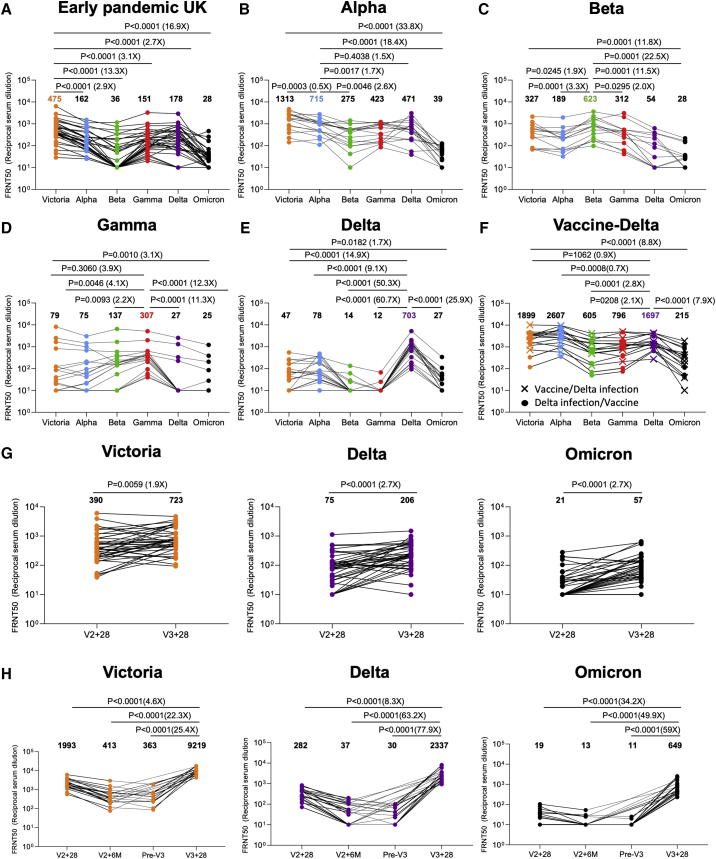
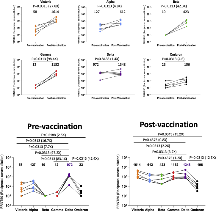
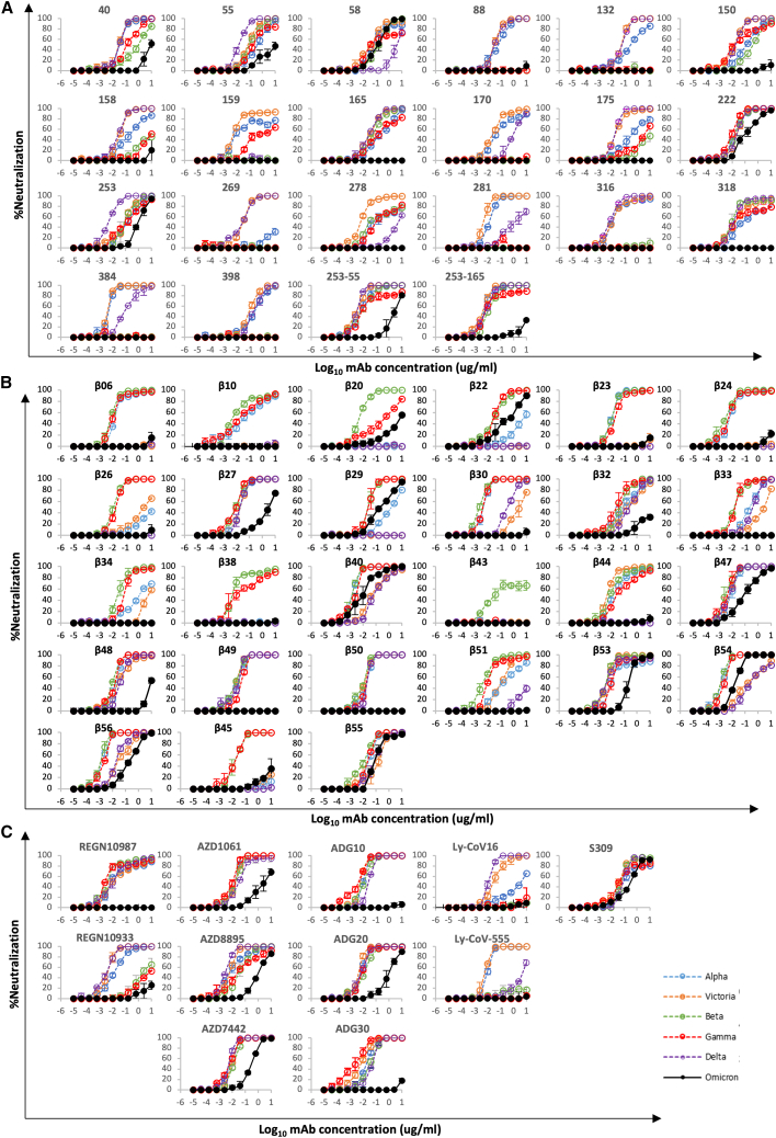
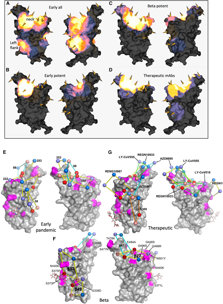
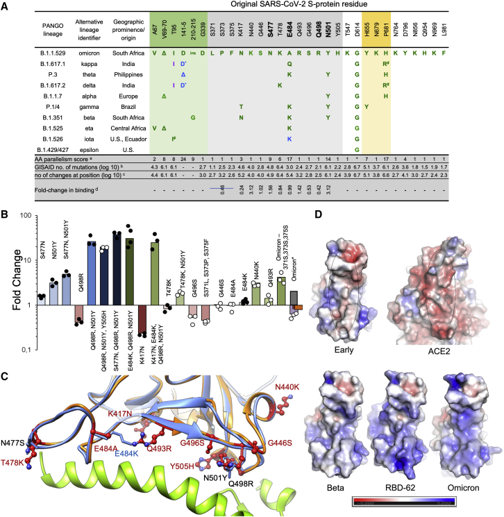
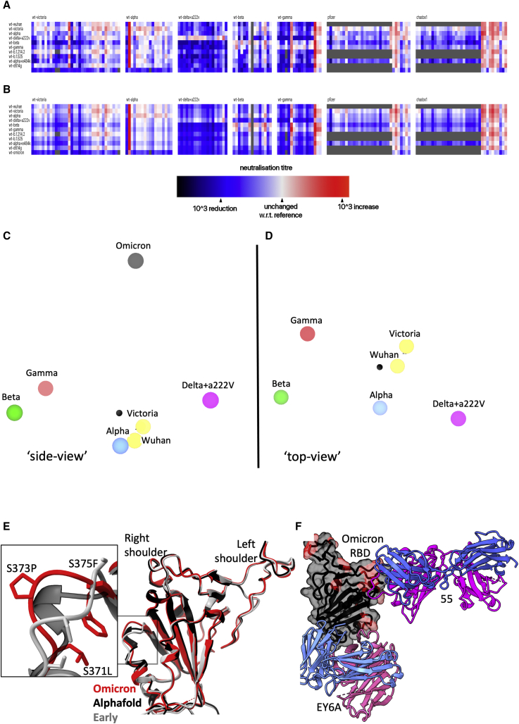
November 2021, the sequence of a new SARS-CoV-2 viral isolate Omicron-B.1.1.529 was announced, containing far more mutations in Spike (S) than previously reported variants. Neutralization titers of Omicron by sera from vaccinees and convalescent subjects infected with early pandemic Alpha, Beta, Gamma, or Delta are substantially reduced, or the sera failed to neutralize. Titers against Omicron are boosted by third vaccine doses and are high in both vaccinated individuals and those infected by Delta. Mutations in Omicron knock out or substantially reduce neutralization by most of the large panel of potent monoclonal antibodies and antibodies under commercial development. Omicron S has structural changes from earlier viruses and uses mutations that confer tight binding to ACE2 to unleash evolution driven by immune escape. This leads to a large number of mutations in the ACE2 binding site and rebalances receptor affinity to that of earlier pandemic viruses.
Overview
The claims and statistics of this paper haven’t been checked yet.
Run the check — free, no account needed. It reads the open-access text and checks the sources it cites.
Integrity signals
Registry
Cites
Statistics
Figures
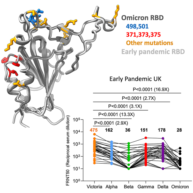
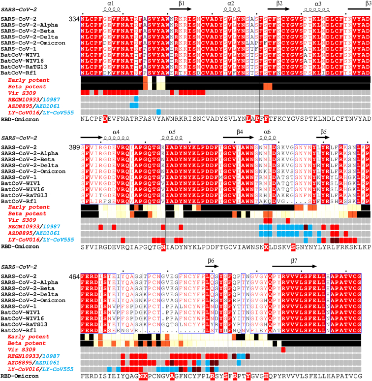
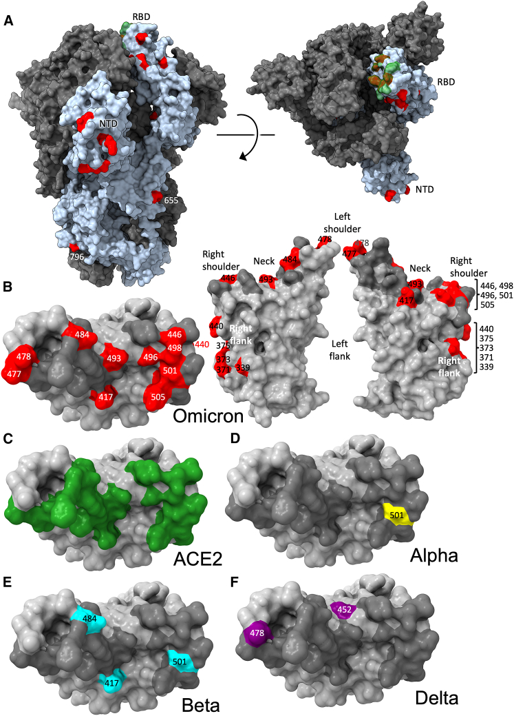
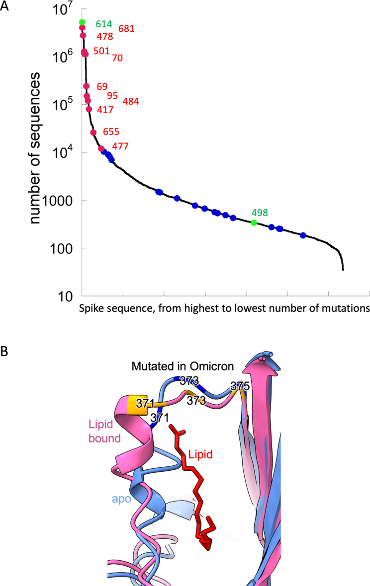
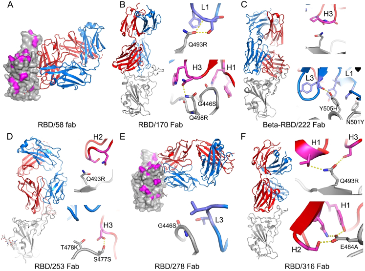
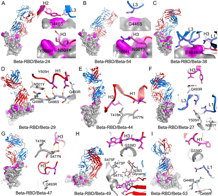
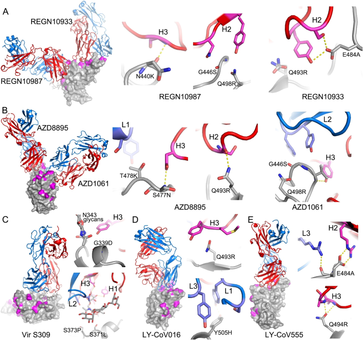
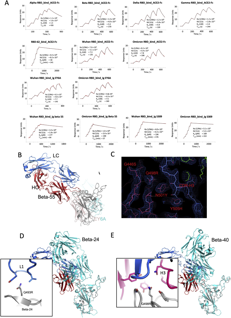
Verification
Claims the paper makes
The claims and statistics of this paper haven’t been checked yet.
Run the check — free, no account needed. It reads the open-access text and checks the sources it cites.
Claims other papers make about this one
Each is checked against this paper’s own text: does it say what the citing paper says it does?
The source backs part of the claim, not all of it — or backs it for a different population, condition or comparison.
In the citing paper
“Recent studies have revealed that repeated immunization extends neutralization to non-homologous variants, possibly through affinity maturation of memory B cell populations12,25.”
From Efficacy of an unmodified bivalent mRNA vaccine against SARS-CoV-2 variants in female small animal models (2023) · cites this paper as [12]
Experimental — accuracy not yet measured
Self-check
Percentages, sums and reported differences re-computed from the paper’s own numbers.
The self-check needs the paper’s full text.
Citations
Cites
Highly accurate protein structure prediction with AlphaFold
Jumper, J., Evans, R., Pritzel, A., Green, T., Figurnov, M., Ronneberger, O., Tunyasuvunakool, K., Bates, R., Žídek, A., Potapenko, A., et al. (2021) · Nature · doi:10.1038/s41586-021-03819-2 · Cited by 43,764
Features and development of Coot
Emsley, P., Lohkamp, B., Scott, W. G., & Cowtan, K. (2010) · Acta Crystallographica Section D Biological Crystallography · doi:10.1107/s0907444910007493 · Cited by 29,915
Phaser crystallographic software
McCoy, A. J., Grosse-Kunstleve, R. W., Adams, P. D., Winn, M. D., Storoni, L. C., & Read, R. J. (2007) · Journal of Applied Crystallography · doi:10.1107/s0021889807021206 · Cited by 20,612
Inference of Macromolecular Assemblies from Crystalline State
Krissinel, E., & Henrick, K. (2007) · Journal of Molecular Biology · doi:10.1016/j.jmb.2007.05.022 · Cited by 10,007
UCSF ChimeraX : Structure visualization for researchers, educators, and developers
Pettersen, E. F., Goddard, T. D., Huang, C. C., Meng, E. C., Couch, G. S., Croll, T. I., Morris, J. H., & Ferrin, T. E. (2020) · Protein Science · doi:10.1002/pro.3943 · Cited by 9,521
Cryo-EM structure of the 2019-nCoV spike in the prefusion conformation
Wrapp, D., Wang, N., Corbett, K. S., Goldsmith, J. A., Hsieh, C.-L., Abiona, O., Graham, B. S., & McLellan, J. S. (2020) · Science · doi:10.1126/science.abb2507 · Cited by 8,897
Structure, function and antigenicity of the SARS-CoV-2 spike glycoprotein
Walls, A. C., Park, Y.-J., Tortorici, M. A., Wall, A., McGuire, A. T., & Veesler, D. (2020) · doi:10.1101/2020.02.19.956581 · Cited by 8,586
Liebschner, D., Afonine, P. V., Baker, M. L., Bunkóczi, G., Chen, V. B., Croll, T. I., Hintze, B., Hung, L.-W., Jain, S., McCoy, A. J., et al. (2019) · Acta Crystallographica Section D Structural Biology · doi:10.1107/s2059798319011471 · Cited by 7,953
Self-citationDeciphering key features in protein structures with the new ENDscript server
Robert, X., & Gouet, P. (2014) · Nucleic Acids Research · doi:10.1093/nar/gku316 · Cited by 7,053
Structure of the SARS-CoV-2 spike receptor-binding domain bound to the ACE2 receptor
Lan, J., Ge, J., Yu, J., Shan, S., Zhou, H., Fan, S., Zhang, Q., Shi, X., Wang, Q., Zhang, L., et al. (2020) · Nature · doi:10.1038/s41586-020-2180-5 · Cited by 5,983
Research works printed but not linked: 2
- Cele, 2021, SARS-CoV-2 evolved during advanced HIV disease immunosuppression has Beta-like escape of vaccine and Delta infection elicited immunity, medRxiv, medRxiv
- Schubert, 2021, Human serum from SARS-CoV-2 vaccinated and COVID-19 patients shows reduced binding to the RBD of SARS-CoV-2 Omicron variant in comparison to the original Wuhan strain and the Beta and Delta variants, medRxiv
Cited by
Sorted by citations
Items 901–931 of 931
pH-induced structural changes in SARS-CoV-2 spike variantsPreprint
Oliveira, A. S. F., Tulli, L., Kearns, F. L., Casalino, L., Rosenfeld, M. A., Berger, I., Schaffitzel, C., Davidson, A. D., Haddrell, A. E., Reid, J. P., et al. (2025) · bioRxiv (Cold Spring Harbor Laboratory) · doi:10.1101/2025.11.17.688702 · Cited by 1
Additional file 1 of Broadly neutralizing human antibodies against Omicron subvariants of SARS-CoV-2
Chiang, H.-L., Liang, K.-H., Lu, R.-M., Kuo, T.-W., Lin, Y., & Wu, H.-C. (2023) · Figshare · doi:10.6084/m9.figshare.23812506
Lee, I.-J., Sun, C.-P., Wu, P.-Y., Lan, Y.-H., Wang, I.-H., Liu, W.-C., Yuan, J. P., Chang, Y.-W., Tseng, S.-C., Tsung, S.-I., et al. (2022) · Figshare · doi:10.6084/m9.figshare.20267321
Qin, Q., Jiang, X., Huo, L., Qian, J., Yu, H., Zhu, H., Du, W., Cao, Y., Zhang, X., & Huang, Q. (2024) · Figshare · doi:10.6084/m9.figshare.25201944
Additional file 2 of Broadly neutralizing human antibodies against Omicron subvariants of SARS-CoV-2
Chiang, H.-L., Liang, K.-H., Lu, R.-M., Kuo, T.-W., Lin, Y., & Wu, H.-C. (2023) · Figshare · doi:10.6084/m9.figshare.23812509
Kumari, M., Lu, R.-M., Li, M.-C., Huang, J., Hsu, F.-F., Ko, S.-H., Ke, F.-Y., Su, S.-C., Liang, K.-H., Yuan, J. P., et al. (2022) · Figshare · doi:10.6084/m9.figshare.21085242
Schubert, M., Bertoglio, F., Steinke, S., Heine, P. A., Ynga-Durand, M. A., Maaß, H., Sammartino, J. C., Cassaniti, I., Zuo, F., Du, L., et al. (2022) · Figshare · doi:10.6084/m9.figshare.19297886
Shi, H., Sun, J., Zeng, Y., Wang, X., Liu, S., Zhang, L., & Shao, E. (2024) · Figshare · doi:10.6084/m9.figshare.26640601
Jedlitzke, B., Wissett, O., Nguyen, T. H., Ghedin, E., & Sormanni, P. (2025) · bioRxiv (Cold Spring Harbor Laboratory) · doi:10.64898/2025.12.23.696170
Yalcin, I., Hu, L., West, B. R., Narayan, K. M., Holmes, A., & Wingertzahn, M. A. (2026) · Infectious Diseases and Therapy · doi:10.1007/s40121-026-01315-6
Self-citationMеlisovich, A. (2026) · American Journal of Medical Science and Technology · doi:10.11648/j.ajmst.20260202.13
Onari, T., & Ashiuchi, M. (2026) · Journal of Coatings Technology and Research · doi:10.1007/s11998-025-01239-9
Harit, A., Mor, M., Yefet, R., Izhaki-Tavor, L. S., Gal-Tanamy, M., Freund, N. T., & Dessau, M. (2026) · Proceedings of the National Academy of Sciences · doi:10.1073/pnas.2523864123 · Cited by 1
Chen, X., Phan, T., Lee, W. L., Clark, K., Rhode, S. F., Brozak, S. J., Pell, B., Palden, T., Leifels, M., Gitter, A., et al. (2026) · International Journal of Hygiene and Environmental Health · doi:10.1016/j.ijheh.2026.114790
Ruiz-Rodríguez, P., Sanz-Carbonell, A., Pérez‐Cataluña, A., Cano-Jiménez, P., Ruiz‐Roldán, L., Alandes, R. A., Valiente-Mullor, C., Gimeno, M. C., Comas, I., Sánchez, G., et al. (2026) · medRxiv · doi:10.64898/2026.03.31.26346553
Stringent selection drives convergence toward omicron-like SARS-CoV-2 receptor-binding motifs
Shoshany, A., RuoJin, T., Padilla‐Blanco, M., Hruška, A., Konar, A., Baxová, K. L., Zoler, E., Mokrejš, M., Schreiber, G., & Zahradník, J. (2026) · Nature Communications · doi:10.1038/s41467-026-72312-z
Self-citationEngineering nasally delivered IgM antibodies for respiratory viral infections
Ramesh, A. K., & An, Z. (2026) · hLife · doi:10.1016/j.hlife.2026.03.004
Schlaepfer, J., Neck, S. D., Penrice-Randal, R., Sharma, P., Kirby, A., Tatham, L. M., Toledo, E. G., Herriott, J., Kijak, E., Sharp, J., et al. (2026) · bioRxiv (Cold Spring Harbor Laboratory) · doi:10.64898/2026.05.27.728081
Self-citationSvitich, O. A., Васильевич, З. В., Leneva, I. A., Mikhailova, N. A., Ivanina, A. V., Файзулоев, Е. Б., Gracheva, A. V., Korchevaya, E. R., Владимировна, С. А., Kostinov, M. P., et al. (2026) · Russian Journal of Infection and Immunity · doi:10.15789/2220-7619-teo-18067
Kontou, E., Detsika, M. G., Salamaliki, C., Bletsa, A., Ampelakiotou, K., Zoulas, D., Athanasiou, N., Totsikas, C., Tzavara, C., Dimopoulou, I., et al. (2026) · Vaccine X · doi:10.1016/j.jvacx.2026.100862
SARS-CoV-2 NSP8-Derived Peptide Effectively Suppresses the Activity of Helicase NSP13
Pashova, S., Petkov, P., Hristova, R., Krachmarova, E., Nacheva, G. A., Gospodinov, A. G., Lilkova, E., Ilieva, N., Rangelov, M. A., Todorova, N. H., et al. (2026) · Computational and Structural Biotechnology Journal · doi:10.34133/csbj.0174
Not All Heroes Wear Capes—Some Just Glow: Exploring Carbon Dots as Nanoagents Against COVID-19
Karagianni, A., Zourou, A., Tuekprakhon, A., Boyar, N., Stamataki, Z., & Kordatos, K. (2026) · Processes · doi:10.3390/pr14142372
Self-citationBabashamsi, M. M., Bagherzadeh, K., Babashamsi, M., Hosseininejad-Chafi, M., Kazemi‐Lomedasht, F., Kazemi, M. S., Parsian, H., & Behdani, M. (2026) · International Journal of Biological Macromolecules · doi:10.1016/j.ijbiomac.2026.153690
Omuseni, E., Nyataya, J., Sifuna, P., Awinda, G., Lemtudo, A. P., Masakhwe, C., Mutai, B. K., Maina, G. M., Kariuki, S., & Waitumbi, J. N. (2026) · PLOS Global Public Health · doi:10.1371/journal.pgph.0006706
ACE2-fused nanobody targeting a cryptic RBD epitope broadly neutralizes SARS-like viruses
Zeng, W., Zhang, G., Ma, H., Xiong, L., Xu, M., Wang, S., Huang, X., Chen, S., Chi, X., Chiu, S., et al. (2026) · Journal of Virology · doi:10.1128/jvi.00608-26
Shim, K., Choi, J., Hwang, E.-H., Kim, G., Bae, G., Jeon, E., Baek, S. H., Kim, H., Kim, S., Kim, D.-S., et al. (2026) · Journal of General Virology · doi:10.1099/jgv.0.002314
Jia, Q., Liu, C., Zhao, C., & Yao, Z. (2026) · Acta Pharmaceutica Sinica B · doi:10.1016/j.apsb.2026.09.025
Self-citationWang, M., Wang, W., Chen, Z., Lu, L., Liu, Z., & Jiang, S. (2026) · Microbiology and Molecular Biology Reviews · doi:10.1128/mmbr.00100-25
Mayr, W., Knell, A. I., Boehm, A. K., Jäger, M., Ennemoser, L., Griesmacher, A., Bellmann‐Weiler, R., Koch, R., Tilg, H., Posch, W., et al. (2026) · Vaccine · doi:10.1016/j.vaccine.2026.129224
Metadata
- Authors
- Dejnirattisai, W., Huo, J., Zhou, D., Zahradník, J., Supasa, P., Liu, C., Duyvesteyn, H. M. E., Ginn, H. M., Mentzer, A. J., Tuekprakhon, A., Nutalai, R., Wang, B., Dijokaite, A., Khan, S., Avinoam, O., Bahar, M., Skelly, D., Adele, S., Johnson, S. A., Amini, A., Ritter, T. G., Mason, C., Dold, C., Pan, D., Assadi, S., Bellass, A., Omo-Dare, N., Koeckerling, D., Flaxman, A., Jenkin, D., Aley, P. K., Voysey, M., Costa Clemens, S. A., Naveca, F. G., Nascimento, V., Nascimento, F., Fernandes da Costa, C., Resende, P. C., Pauvolid-Correa, A., Siqueira, M. M., Baillie, V., Serafin, N., Kwatra, G., Da Silva, K., Madhi, S. A., Nunes, M. C., Malik, T., Openshaw, P. J. M., Baillie, J. K., Semple, M. G., Townsend, A. R., Huang, K.-Y. A., Tan, T. K., Carroll, M. W., Klenerman, P., Barnes, E., Dunachie, S. J., Constantinides, B., Webster, H., Crook, D., Pollard, A. J., Lambe, T., Conlon, C., Deeks, A. S., Frater, J., Frending, L., Gardiner, S., Jämsén, A., Jeffery, K., Malone, T., Phillips, E., Rothwell, L., Stafford, L., Baillie, J. K., Semple, M. G., Openshaw, P. J., Carson, G., Alex, B., Andrikopoulos, P., Bach, B., Barclay, W. S., Bogaert, D., Chand, M., Chechi, K., Cooke, G. S., da Silva Filipe, A., de Silva, T., Docherty, A. B., dos Santos Correia, G., Dumas, M.-E., Dunning, J., Fletcher, T., Green, C. A., Greenhalf, W., Griffin, J. L., Gupta, R. K., Harrison, E. M., Hiscox, J. A., Wai Ho, A. Y., Horby, P. W., Ijaz, S., Khoo, S., Klenerman, P., Law, A., Lewis, M. R., Liggi, S., Lim, W. S., Maslen, L., Mentzer, A. J., Merson, L., Meynert, A. M., Moore, S. C., Noursadeghi, M., Olanipekun, M., Osagie, A., Palmarini, M., Palmieri, C., Paxton, W. A., Pollakis, G., Price, N., Rambaut, A., Robertson, D. L., Russell, C. D., Sancho-Shimizu, V., Sands, C. J., Scott, J. T., Sigfrid, L., Solomon, T., Sriskandan, S., Stuart, D., Summers, C., Swann, O. V., Takats, Z., Takis, P., Tedder, R. S., Thompson, A. R., Thomson, E. C., Thwaites, R. S., Turtle, L. C., Zambon, M., Hardwick, H., Donohue, C., Griffiths, F., Oosthuyzen, W., Donegan, C., Spencer, R. G., Norman, L., Pius, R., Drake, T. M., Fairfield, C. J., Knight, S. R., Mclean, K. A., Murphy, D., Shaw, C. A., Dalton, J., Girvan, M., Saviciute, E., Roberts, S., Harrison, J., Marsh, L., Connor, M., Halpin, S., Jackson, C., Gamble, C., Plotkin, D., Lee, J., Leeming, G., Law, A., Wham, M., Clohisey, S., Hendry, R., Scott-Brown, J., Shaw, V., McDonald, S. E., Keating, S., Ahmed, K. A., Armstrong, J. A., Ashworth, M., Asiimwe, I. G., Bakshi, S., Barlow, S. L., Booth, L., Brennan, B., Bullock, K., Catterall, B. W., Clark, J. J., Clarke, E. A., Cole, S., Cooper, L., Cox, H., Davis, C., Dincarslan, O., Dunn, C., Dyer, P., Elliott, A., Evans, A., Finch, L., Fisher, L. W., Foster, T., Garcia-Dorival, I., Gunning, P., Hartley, C., Jensen, R. L., Jones, C. B., Jones, T. R., Khandaker, S., King, K., Kiy, R. T., Koukorava, C., Lake, A., Lant, S., Latawiec, D., Lavelle-Langham, L., Lefteri, D., Lett, L., Livoti, L. A., Mancini, M., McDonald, S., McEvoy, L., McLauchlan, J., Metelmann, S., Miah, N. S., Middleton, J., Mitchell, J., Moore, S. C., Murphy, E. G., Penrice-Randal, R., Pilgrim, J., Prince, T., Reynolds, W., Ridley, P. M., Sales, D., Shaw, V. E., Shears, R. K., Small, B., Subramaniam, K. S., Szemiel, A., Taggart, A., Tanianis-Hughes, J., Thomas, J., Trochu, E., van Tonder, L., Wilcock, E., Zhang, J. E., Flaherty, L., Maziere, N., Cass, E., Carracedo, A. D., Carlucci, N., Holmes, A., Massey, H., Murphy, L., McCafferty, S., Clark, R., Fawkes, A., Morrice, K., Maclean, A., Wrobel, N., Donnelly, L., Coutts, A., Hafezi, K., MacGillivray, L., Gilchrist, T., Adeniji, K., Agranoff, D., Agwuh, K., Ail, D., Aldera, E. L., Alegria, A., Allen, S., Angus, B., Ashish, A., Atkinson, D., Bari, S., Barlow, G., Barnass, S., Barrett, N., Bassford, C., Basude, S., Baxter, D., Beadsworth, M., Bernatoniene, J., Berridge, J., Berry, C., Best, N., Bothma, P., Chadwick, D., Brittain-Long, R., Bulteel, N., Burden, T., Burtenshaw, A., Caruth, V., Chadwick, D., Chambler, D., Chee, N., Child, J., Chukkambotla, S., Clark, T., Collini, P., Cosgrove, C., Cupitt, J., Cutino-Moguel, M.-T., Dark, P., Dawson, C., Dervisevic, S., Donnison, P., Douthwaite, S., Drummond, A., DuRand, I., Dushianthan, A., Dyer, T., Evans, C., Eziefula, C., Fegan, C., Finn, A., Fullerton, D., Garg, S., Garg, S., Garg, A., Gkrania-Klotsas, E., Godden, J., Goldsmith, A., Graham, C., Hardy, E., Hartshorn, S., Harvey, D., Havalda, P., Hawcutt, D. B., Hobrok, M., Hodgson, L., Hormis, A., Jacobs, M., Jain, S., Jennings, P., Kaliappan, A., Kasipandian, V., Kegg, S., Kelsey, M., Kendall, J., Kerrison, C., Kerslake, I., Koch, O., Koduri, G., Koshy, G., Laha, S., Laird, S., Larkin, S., Leiner, T., Lillie, P., Limb, J., Linnett, V., Little, J., Lyttle, M., MacMahon, M., MacNaughton, E., Mankregod, R., Masson, H., Matovu, E., McCullough, K., McEwen, R., Meda, M., Mills, G., Minton, J., Mirfenderesky, M., Mohandas, K., Mok, Q., Moon, J., Moore, E., Morgan, P., Morris, C., Mortimore, K., Moses, S., Mpenge, M., Mulla, R., Murphy, M., Nagel, M., Nagarajan, T., Nelson, M., Norris, L., O’Shea, M. K., Otahal, I., Ostermann, M., Pais, M., Palmieri, C., Panchatsharam, S., Papakonstantinou, D., Paraiso, H., Patel, B., Pattison, N., Pepperell, J., Peters, M., Phull, M., Pintus, S., Pooni, J. S., Planche, T., Post, F., Price, D., Prout, R., Rae, N., Reschreiter, H., Reynolds, T., Richardson, N., Roberts, M., Roberts, D., Rose, A., Rousseau, G., Ruge, B., Ryan, B., Saluja, T., Schmid, M. L., Shah, A., Shanmuga, P., Sharma, A., Shawcross, A., Sizer, J., Shankar-Hari, M., Smith, R., Snelson, C., Spittle, N., Staines, N., Stambach, T., Stewart, R., Subudhi, P., Szakmany, T., Tatham, K., Thomas, J., Thompson, C., Thompson, R., Tridente, A., Tupper-Carey, D., Twagira, M., Vallotton, N., Vancheeswaran, R., Vincent-Smith, L., Visuvanathan, S., Vuylsteke, A., Waddy, S., Wake, R., Walden, A., Welters, I., Whitehouse, T., Whittaker, P., Whittington, A., Papineni, P., Wijesinghe, M., Williams, M., Wilson, L., Cole, S., Winchester, S., Wiselka, M., Wolverson, A., Wootton, D. G., Workman, A., Yates, B., Young, P., Paterson, N. G., Williams, M. A., Hall, D. R., Fry, E. E., Mongkolsapaya, J., Ren, J., Schreiber, G., Stuart, D. I., & Screaton, G. R.
- Affiliations
- Centre for Human Genetics (GB)
- Weizmann Institute of Science (IL)
- Diamond Light Source (GB)
- University of Oxford (GB)
- Medawar Building for Pathogen Research (GB)
- Oxford University Hospitals NHS Trust (GB)
- University of Leicester (GB)
- University Hospitals of Leicester NHS Trust (GB)
- Fiocruz Amazônia (BR)
- Fundação Oswaldo Cruz (BR)
- South African Medical Research Council (ZA)
- Public Health England (GB)
- John Radcliffe Hospital (GB)
- Chang Gung University (TW)
- Siriraj Hospital (TH)
- Roslin Institute (GB)
- University of Liverpool (GB)
- Imperial College London (GB)
- Corresponding author
- Mongkolsapaya, Juthathip
- Ren, Jingshan
- Schreiber, Gideon
- Screaton, Gavin R.
- ORCID
- Dejnirattisai, Wanwisa
- Huo, Jiandong
- Zhou, Daming
- Zahradník, Jiří
- Supasa, Piyada
- Liu, Chang
- Duyvesteyn, Helen M.E.
- Ginn, Helen M.
- Tuekprakhon, Aekkachai
- Nutalai, Rungtiwa
- Wang, Beibei
- Khan, Suman
- Avinoam, Ori
- Bahar, Mohammad
- Skelly, Donal
- Adele, Sandra
- Amini, Ali
- Ritter, Thomas G.
- Mason, Chris
- Dold, Christina
- Pan, Daniel
- Bellass, Adam
- Koeckerling, David
- Flaxman, Amy
- Jenkin, Daniel
- Aley, Parvinder K.
- Voysey, Merryn
- Naveca, Felipe Gomes
- Nascimento, Valdinete
- Nascimento, Fernanda
- Resende, Paola Cristina
- Siqueira, Marilda M.
- Baillie, Vicky
- Kwatra, Gaurav
- Madhi, Shabir A.
- Nunes, Marta C.
- Malik, Tariq
- Townsend, Alain R.
- Huang, Kuan-Ying A.
- Tan, Tiong Kit
- Carroll, Miles W.
- Barnes, Eleanor
- Dunachie, Susanna J.
- Constantinides, Bede
- Webster, Hermione
- Crook, Derrick
- Pollard, Andrew J.
- Lambe, Teresa
- Deeks, Alexandra S.
- Frater, John
- Gardiner, Siobhan
- Jeffery, Katie
- Phillips, Eloise
- Stafford, Lizzie
- Alex, Beatrice
- Andrikopoulos, Petros
- Bach, Benjamin
- Bogaert, Debby
- Chand, Meera
- Chechi, Kanta
- Docherty, Annemarie B.
- Dumas, Marc-Emmanuel
- Dunning, Jake
- Fletcher, Tom
- Greenhalf, William
- Griffin, Julian L.
- Gupta, Rishi K.
- Harrison, Ewen M.
- Hiscox, Julian A.
- Horby, Peter W.
- Ijaz, Samreen
- Khoo, Saye
- Lewis, Matthew R.
- Lim, Wei Shen
- Maslen, Lynn
- Merson, Laura
- Noursadeghi, Mahdad
- Olanipekun, Michael
- Palmarini, Massimo
- Paxton, William A.
- Pollakis, Georgios
- Rambaut, Andrew
- Robertson, David L.
- Russell, Clark D.
- Sands, Caroline J.
- Sigfrid, Louise
- Solomon, Tom
- Sriskandan, Shiranee
- Summers, Charlotte
- Swann, Olivia V.
- Takis, Panteleimon
- Tedder, Richard S.
- Thomson, Emma C.
- Thwaites, Ryan S.
- Turtle, Lance CW.
- Zambon, Maria
- Donohue, Chloe
- Donegan, Cara
- Spencer, Rebecca G.
- Drake, Thomas M.
- Fairfield, Cameron J.
- Knight, Stephen R.
- Mclean, Kenneth A.
- Shaw, Catherine A.
- Girvan, Michelle
- Halpin, Sophie
- Jackson, Clare
- Gamble, Carrol
- Plotkin, Daniel
- Lee, James
- Leeming, Gary
- Clohisey, Sara
- Hendry, Ross
- Scott-Brown, James
- Ahmed, Katie A.
- Armstrong, Jane A.
- Ashworth, Milton
- Asiimwe, Innocent G.
- Bakshi, Siddharth
- Bullock, Katie
- Catterall, Benjamin WA.
- Clark, Jordan J.
- Cox, Helen
- Davis, Christopher
- Finch, Lorna
- Foster, Terry
- Hartley, Catherine
- Jensen, Rebecca L.
- Jones, Christopher B.
- Khandaker, Shadia
- King, Katharine
- Kiy, Robyn T.
- Koukorava, Chrysa
- Lant, Suzannah
- Lefteri, Daniella
- Lett, Lauren
- McEvoy, Laurence
- McLauchlan, John
- Miah, Nahida S.
- Murphy, Ellen G.
- Penrice-Randal, Rebekah
- Pilgrim, Jack
- Prince, Tessa
- Ridley, P. Matthew
- Shears, Rebecca K.
- Small, Benjamin
- Subramaniam, Krishanthi S.
- Taggart, Aislynn
- Tanianis-Hughes, Jolanta
- Zhang, J. Eunice
- Carracedo, Alejandra Doce
- Holmes, Anthony
- Murphy, Lee
- McCafferty, Sarah
- Clark, Richard
- Fawkes, Angie
- Wrobel, Nicola
- Donnelly, Lorna
- Coutts, Audrey
- MacGillivray, Louise
- Adeniji, Kayode
- Agranoff, Daniel
- Aldera, Erin L.
- Angus, Brian
- Barlow, Gavin
- Barrett, Nicholas
- Baxter, David
- Bernatoniene, Jolanta
- Brittain-Long, Robin
- Collini, Paul
- Dark, Paul
- Dervisevic, Samir
- Douthwaite, Sam
- Evans, Cariad
- Eziefula, Chi
- Finn, Adam
- Graham, Clive
- Hartshorn, Stuart
- Hawcutt, Daniel B.
- Hodgson, Luke
- Hormis, Anil
- Jacobs, Michael
- Kasipandian, Vidya
- Koch, Oliver
- Koduri, Gouri
- Laird, Steven
- Lillie, Patrick
- Lyttle, Mark
- MacMahon, Michael
- Masson, Huw
- Meda, Manjula
- Mills, Gary
- Minton, Jane
- Mirfenderesky, Mariyam
- Mok, Quen
- Morris, Craig
- Moses, Samuel
- Murphy, Michael
- O’Shea, Matthew K.
- Ostermann, Marlies
- Papakonstantinou, Danai
- Pepperell, Justin
- Peters, Mark
- Phull, Mandeep
- Planche, Tim
- Post, Frank
- Rae, Nikolas
- Reynolds, Tim
- Sharma, Anil
- Shawcross, Anna
- Smith, Richard
- Tatham, Kate
- Tridente, Ascanio
- Vancheeswaran, Rama
- Waddy, Sam
- Wake, Rachel
- Welters, Ingeborg
- Whitehouse, Tony
- Papineni, Padmasayee
- Winchester, Stephen
- Wiselka, Martin
- Yates, Bryan
- Paterson, Neil G.
- Williams, Mark A.
- Hall, David R.
- Fry, Elizabeth E.
- Mongkolsapaya, Juthathip
- Ren, Jingshan
- Schreiber, Gideon
- Stuart, David I.
- Screaton, Gavin R.
- Venue
- Cell
- Publisher
- Elsevier BV
- Published
- 2022-02
- Volume
- 185
- Issue
- 3
- Pages
- 467-484.e15
- ISSN
- 0092-8674 (ISSN-L)
- PMC
- PMC8723827
- OpenAlex
- W4206153788
- Cited by
- 930
- References
- 73
- Citations by year
- 2026: 30 · 2025: 73 · 2024: 121 · 2023: 246 · 2022: 455
- FWCI
- 109.39
- Type
- journal article
- Language
- en
- Open access
- hybrid
- doi.org
- Licence
- Locations
- Cell (publishedVersion, cc-by)
- PubMed Central (publishedVersion, cc-by)
- PubMed (publishedVersion)
- Edinburgh Research Explorer (University of Edinburgh) (publishedVersion, cc-by)
- LA Referencia (Red Federada de Repositorios Institucionales de Publicaciones Científicas) (publishedVersion, other-oa)
- White Rose Research Online (University of Leeds, The University of Sheffield, University of York) (acceptedVersion)
- HAL (submittedVersion)
- Arca - Repositório Institucional da Fiocruz (submittedVersion, other-oa)
- Figshare (submittedVersion, cc-by)
- Research Explorer (The University of Manchester) (submittedVersion, cc-by)
- University of Birmingham Research Portal (University of Birmingham) (submittedVersion, other-oa)
- Spiral (Imperial College London) (submittedVersion, other-oa)
- Apollo (University of Cambridge) (submittedVersion, cc-by)
- Apollo (cc-by)
- Funders
- Israel Science Foundation (3814/19)
- National Institute for Health Research (63/125,984, 277,546, PCT/GB2012/000467)
- MRC (MC_PC_19059)
- Fundação Oswaldo Cruz (VPPCB-007- FIO-18-2-30)
- NIHR Oxford Biomedical Research Centre
- Department of Health and Social Care
- Wellcome Trust (203141/Z/16/Z, 101122/Z/13/Z, 090532/Z/09/Z)
- Bill & Melinda Gates Foundation (INV-016202)
- Conselho Nacional de Desenvolvimento Científico e Tecnológico (403276/2020-9)
- National Institutes of Health (U19 I082360)
- Oak Foundation
- Coalition for Epidemic Preparedness Innovations
- Chinese Academy of Medical Sciences
- Public Health England
- UK Research and Innovation
- CAMS (2018-I2M-2-002)
- Fundação de Amparo à Pesquisa do Estado do Amazonas
- Topics
- Health Sciences › Medicine › Infectious Diseases › SARS-CoV-2 and COVID-19 Research
- Life Sciences › Biochemistry, Genetics and Molecular Biology › Molecular Biology › vaccines and immunoinformatics approaches
- Life Sciences › Agricultural and Biological Sciences › Animal Science and Zoology › Animal Virus Infections Studies
- Keywords
- Omicron variant, spike protein mutations, neutralizing antibodies, immune evasion, vaccine escape, monoclonal antibodies, ACE2 binding site, receptor binding affinity, SARS-CoV-2 variants, COVID-19 pandemic
About these checks
Registry, retraction, Cites and statistics results are computed, not judged, with one exception: a printed reference that no index lists is read once by a model before it counts as a citation error. Claims are extracted by a model and kept only when their quote appears verbatim in the text; each cited source is then checked, with a model’s help, for whether it says what the claim relies on. Those verdicts can be wrong — read the claim’s quote and the cited source before relying on one.
Statistics and arithmetic are recomputed, not judged.
Confidence bands are shown only for fields calibrated on our annotated test set. None is calibrated yet, so no band is shown.
What counts as a claim ›How we measure confidence ›How we check this ›
Are you an author, or do you see a problem with this page? Request a review
