

The DOI matches its Crossref record; 75 of the 75 references it cites were found in a registry or an index.
Carbon source availability drives nutrient utilization in CD8+ T cells
Open accessAbstract
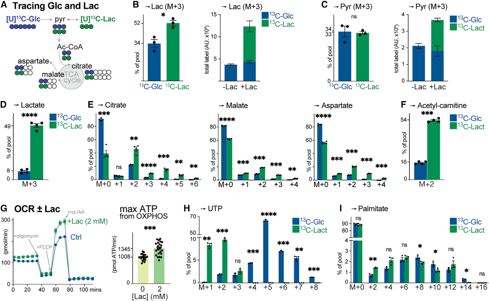
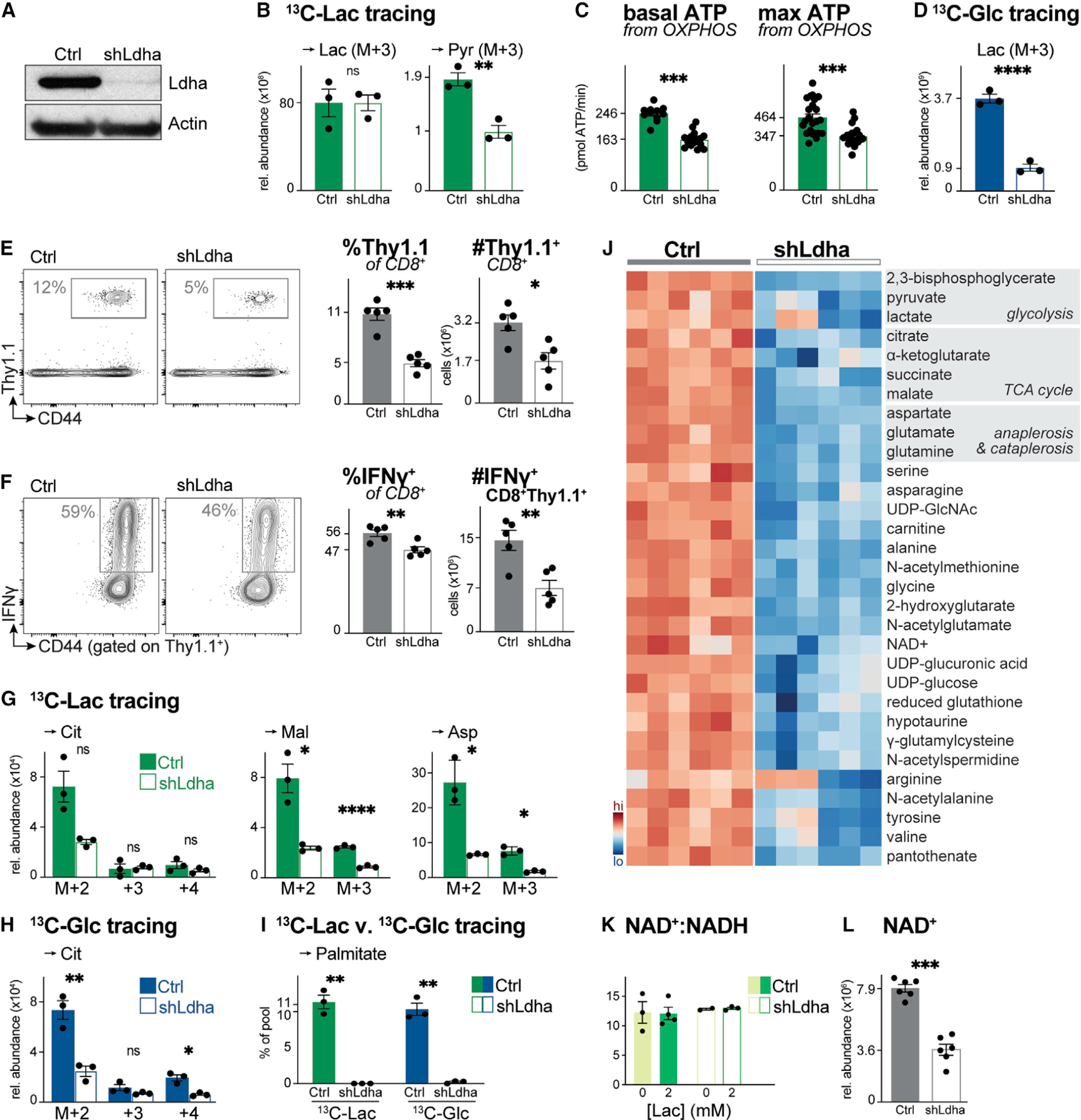
How environmental nutrient availability impacts T cell metabolism and function remains poorly understood. Here, we report that the presence of physiologic carbon sources (PCSs) in cell culture medium broadly impacts glucose utilization by CD8 + T cells, independent of transcriptional changes in metabolic reprogramming. The presence of PCSs reduced glucose contribution to the TCA cycle and increased effector function of CD8 + T cells, with lactate directly fueling the TCA cycle. In fact, CD8 + T cells responding to Listeria infection preferentially consumed lactate over glucose as a TCA cycle substrate in vitro , with lactate enhancing T cell bioenergetic and biosynthetic capacity. Inhibiting lactate-dependent metabolism in CD8 + T cells by silencing lactate dehydrogenase A (Ldha) impaired both T cell metabolic homeostasis and proliferative expansion in vivo . Together, our data indicate that carbon source availability shapes T cell glucose metabolism and identifies lactate as a bioenergetic and biosynthetic fuel for CD8 + effector T cells.
Overview
The claims and statistics of this paper haven’t been checked yet.
Run the check — free, no account needed. It reads the open-access text and checks the sources it cites.
Integrity signals
Registry
Cites
Statistics
Figures
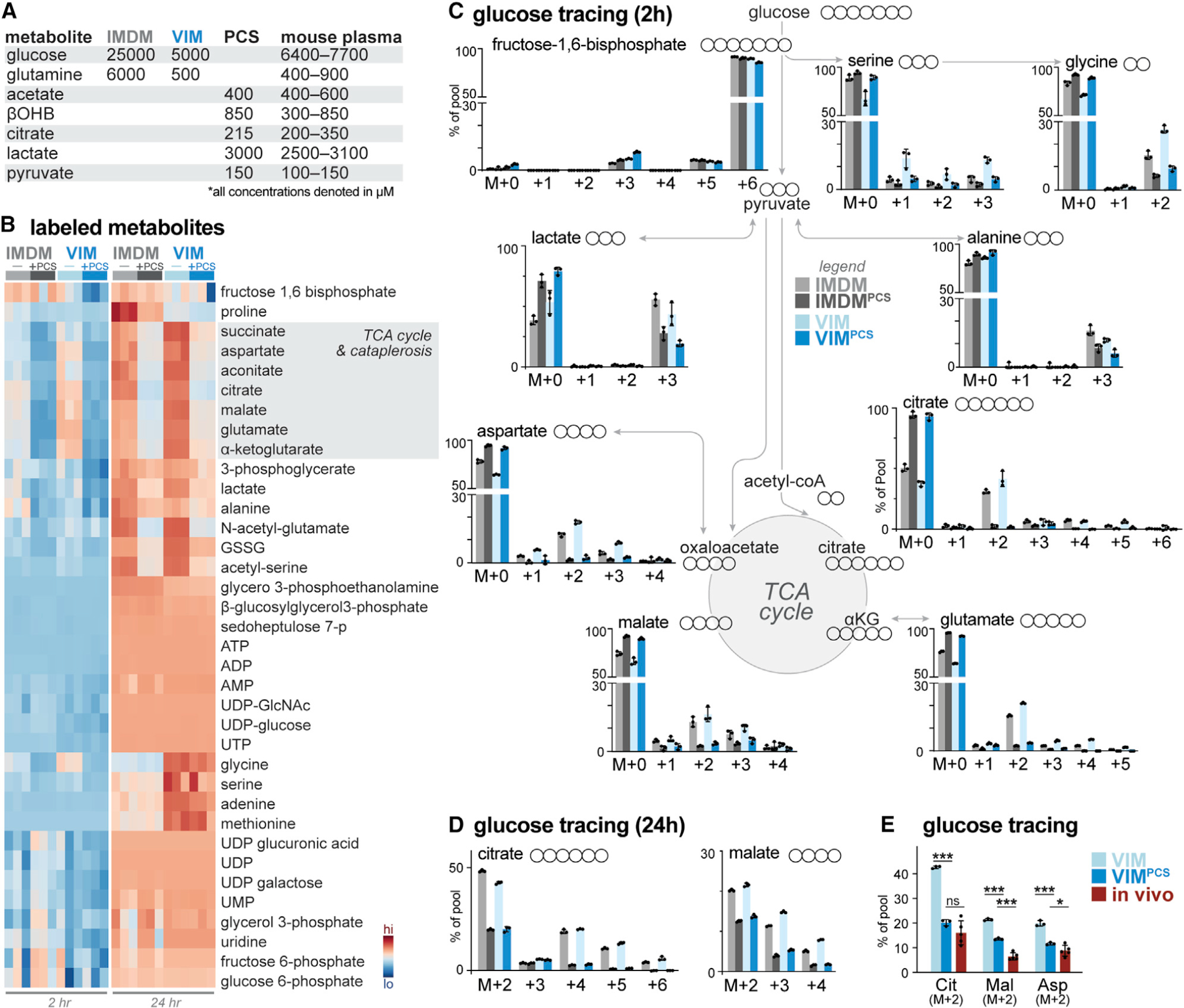
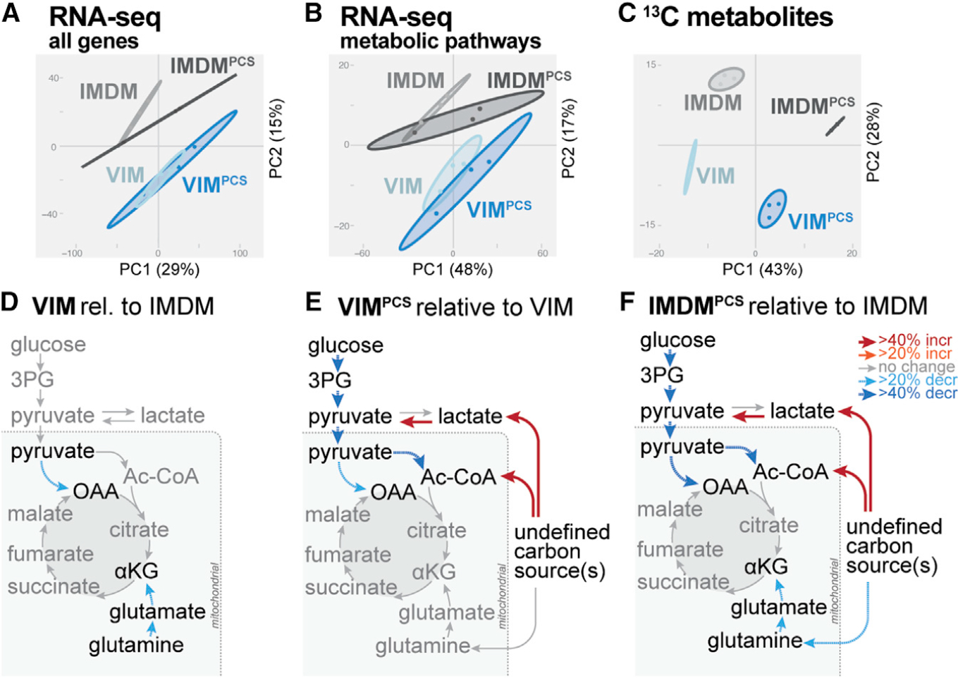
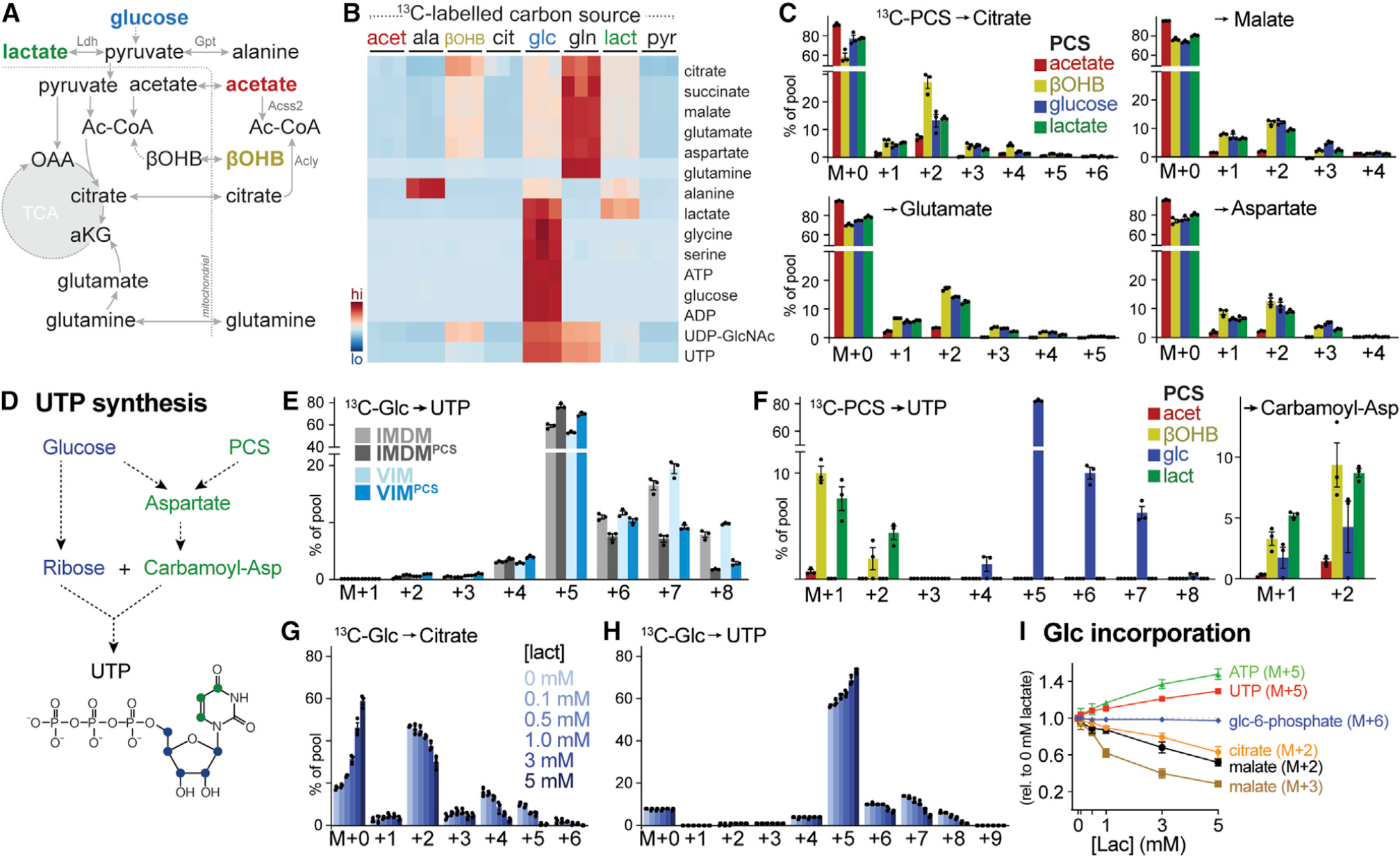
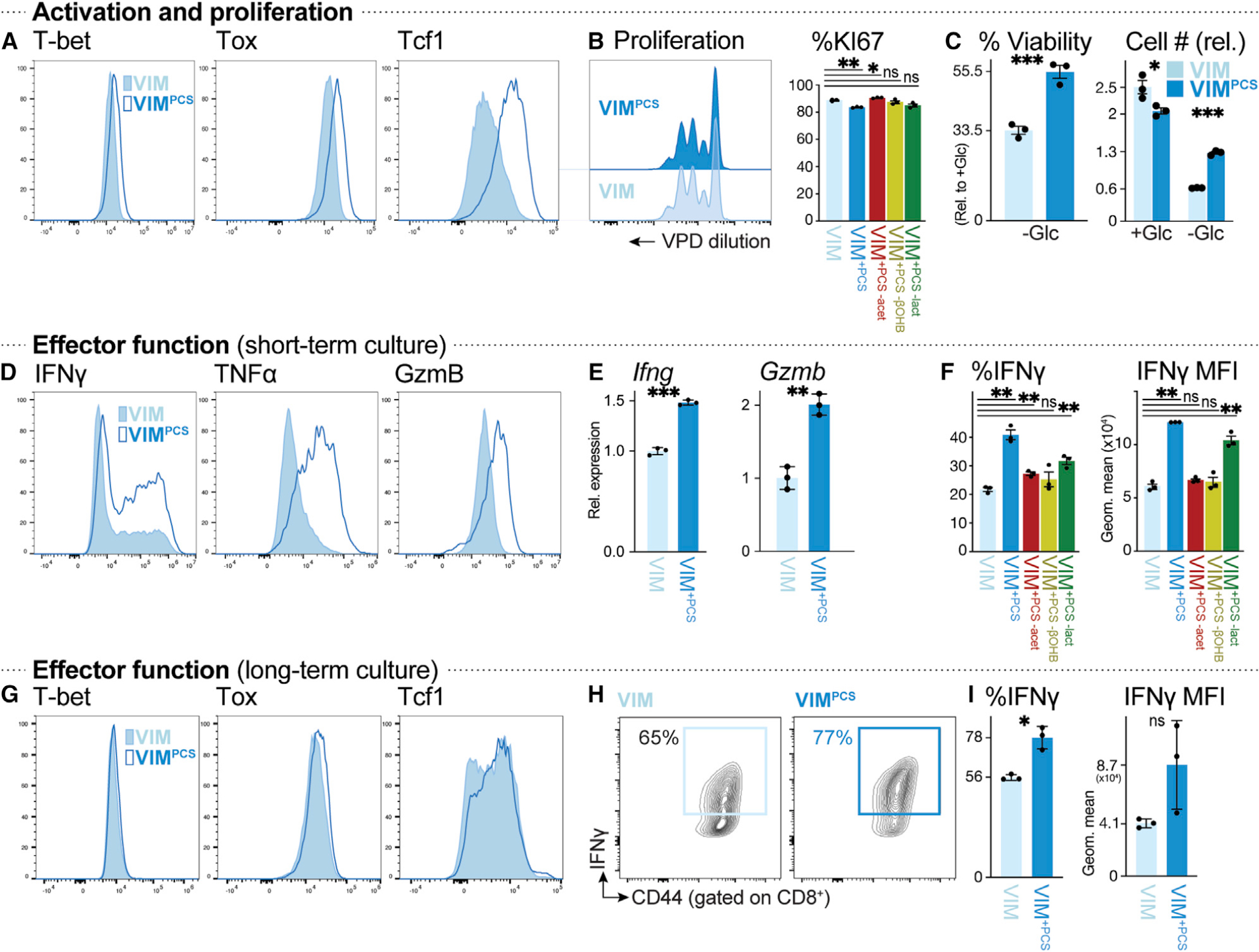
Verification
Claims the paper makes
The claims and statistics of this paper haven’t been checked yet.
Run the check — free, no account needed. It reads the open-access text and checks the sources it cites.
Claims other papers make about this one
Each is checked against this paper’s own text: does it say what the citing paper says it does?
The source backs part of the claim, not all of it — or backs it for a different population, condition or comparison.
In the citing paper
“Also, CD8+ T cells preferentially utilize non-glucose physiologic carbon sources as metabolic fuel, and their activation and effector function are regulated by carbon availability.9”
From SGLT2 inhibitor promotes ketogenesis to improve MASH by suppressing CD8+ T cell activation (2024) · cites this paper as [9]
Experimental — accuracy not yet measuredThe source backs part of the claim, not all of it — or backs it for a different population, condition or comparison.
In the citing paper
“Also, CD8+ T cells preferentially utilize non-glucose physiologic carbon sources as metabolic fuel, and their activation and effector function are regulated by carbon availability.9”
From SGLT2 inhibitor promotes ketogenesis to improve MASH by suppressing CD8+ T cell activation (2024) · cites this paper as [9]
Experimental — accuracy not yet measured
Self-check
Percentages, sums and reported differences re-computed from the paper’s own numbers.
The self-check needs the paper’s full text.
Citations
Cites
A RAPID METHOD OF TOTAL LIPID EXTRACTION AND PURIFICATION
Bligh, E. G., & Dyer, W. J. (1959) · Canadian Journal of Biochemistry and Physiology · doi:10.1139/o59-099 · Cited by 54,751 · cited 1×
“…saline and centrifuged before being snap frozen on dry-ice and stored at −80°C. Metabolites were extracted by modified Bligh-Dyer extraction (Bligh and Dyer, 1959) by the addition of ice-cold methanol (A456, Fisher Scientific) directly to frozen cells, to which one volume of chloroform…”
Understanding the Warburg Effect: The Metabolic Requirements of Cell Proliferation
Vander Heiden, M. G., Cantley, L. C., & Thompson, C. B. (2009) · Science · doi:10.1126/science.1160809 · Cited by 16,197
A guide to immunometabolism for immunologists
O'Neill, L. A. J., Kishton, R. J., & Rathmell, J. (2016) · Nature Reviews Immunology · doi:10.1038/nri.2016.70 · Cited by 3,315
Posttranscriptional Control of T Cell Effector Function by Aerobic Glycolysis
Chang, C.-H., Curtis, J. D., Maggi, L. B., Faubert, B., Villarino, A. V., O’Sullivan, D., Huang, S. C.-C., van der Windt, G. J. W., Blagih, J., Qiu, J., et al. (2013) · Cell · doi:10.1016/j.cell.2013.05.016 · Cited by 2,204
Self-citationThe transcription factor Myc controls metabolic reprogramming upon T lymphocyte activation
Wang, R., Dillon, C. P., Shi, L. Z., Milasta, S., Carter, R., Finkelstein, D., McCormick, L. L., Fitzgerald, P., Chi, H., Munger, J., et al. (2011) · Immunity · doi:10.1016/j.immuni.2011.09.021 · Cited by 2,140 · cited 2×
“Upon activation, CD8 + T cells undergo extensive metabolic reprogramming characterized by transcriptional changes in metabolic gene expression (Wang et al., 2011).”
LDHA-Associated Lactic Acid Production Blunts Tumor Immunosurveillance by T and NK Cells
Brand, A., Singer, K., Koehl, G. E., Kolitzus, M., Schoenhammer, G., Thiel, A., Matos, C., Bruss, C., Klobuch, S., Peter, K., et al. (2016) · Cell Metabolism · doi:10.1016/j.cmet.2016.08.011 · Cited by 2,039
Inhibitory effect of tumor cell-derived lactic acid on human T cells
Fischer, K., Hoffmann, P., Voelkl, S., Meidenbauer, N., Ammer, J., Edinger, M., Gottfried, E., Schwarz, S., Rothe, G., Hoves, S., et al. (2007) · Blood · doi:10.1182/blood-2006-07-035972 · Cited by 1,946
Glucose feeds the TCA cycle via circulating lactate
Hui, S., Ghergurovich, J. M., Morscher, R. J., Jang, C., Teng, X., Lu, W., Esparza, L. A., Reya, T., Zhan, L., Yanxiang Guo, J., et al. (2017) · Nature · doi:10.1038/nature24057 · Cited by 1,721
Transcriptional control of effector and memory CD8+ T cell differentiation
Kaech, S. M., & Cui, W. (2012) · Nature Reviews Immunology · doi:10.1038/nri3307 · Cited by 1,628
The CD28 Signaling Pathway Regulates Glucose Metabolism
Frauwirth, K. A., Riley, J. L., Harris, M. H., Parry, R. V., Rathmell, J. C., Plas, D. R., Elstrom, R. L., June, C. H., & Thompson, C. B. (2002) · Immunity · doi:10.1016/s1074-7613(02)00323-0 · Cited by 1,475 · cited 1×
“…taken up by activated T cells and is a major substrate fueling central carbon metabolism (i.e., glycolysis; the pentose phosphate pathway [PPP]; serine, glycine, and one-carbon [SGOC] metabolism; the TCA cycle; and nucleotide metabolism) in T cells (Frauwirth et al., 2002; Wang et al., 2011).”
Cited by
Sorted by citations
CD8+ T cells in the cancer-immunity cycle
Giles, J. R., Globig, A.-M., Kaech, S. M., & Wherry, E. J. (2023) · Immunity · doi:10.1016/j.immuni.2023.09.005 · Cited by 507
Lactic acid and lactate: revisiting the physiological roles in the tumor microenvironment
Apostolova, P., & Pearce, E. L. (2022) · Trends in Immunology · doi:10.1016/j.it.2022.10.005 · Cited by 367
Lactate: A key regulator of the immune response
Llibre, A., Kucuk, S., Gope, A., Certo, M., & Mauro, C. (2025) · Immunity · doi:10.1016/j.immuni.2025.02.008 · Cited by 316
Zhao, Y., Liu, Z., Liu, G., Zhang, Y., Liu, S., Gan, D., Chang, W., Peng, X., Sung, E. S., Gilbert, K., et al. (2023) · Cell Metabolism · doi:10.1016/j.cmet.2023.09.004 · Cited by 231
Self-citationThe emerging role of lactate in tumor microenvironment and its clinical relevance
Chen, S., Xu, Y., Zhuo, W., & Zhang, L. (2024) · Cancer Letters · doi:10.1016/j.canlet.2024.216837 · Cited by 205
Dysfunction of exhausted T cells is enforced by MCT11-mediated lactate metabolism
Peralta, R. M., Xie, B., Lontos, K., Nieves-Rosado, H. M., Spahr, K., Joshi, S. K., Ford, B. R., Quann, K., Frisch, A. T., Dean, V. G., et al. (2024) · Nature Immunology · doi:10.1038/s41590-024-01999-3 · Cited by 222
Emerging roles of lactate in acute and chronic inflammation
Fang, Y., Li, Z., Yang, L., Li, W., Wang, Y., Kong, Z., Miao, J., Chen, Y., Bian, Y., & Zeng, L. (2024) · Cell Communication and Signaling · doi:10.1186/s12964-024-01624-8 · Cited by 186
Gu, X.-Y., Yang, J., Lai, R., Zhou, Z., Tang, D., Hu, L., & Zhao, L. (2025) · Frontiers in Immunology · doi:10.3389/fimmu.2025.1563303 · Cited by 174
The effect of physical exercise on anticancer immunity
Fiuza‐Luces, C., Valenzuela, P. L., Gálvez, B. G., Ramírez, M. R., López‐Soto, A., Simpson, R. J., & Lucía, A. (2023) · Nature reviews. Immunology · doi:10.1038/s41577-023-00943-0 · Cited by 164
Ketolysis drives CD8+ T cell effector function through effects on histone acetylation
Luda, K. M., Longo, J., Kitchen-Goosen, S. M., Duimstra, L. R., Ma, E. H., Watson, M. J., Oswald, B. M., Fu, Z., Madaj, Z., Kupai, A., et al. (2023) · Immunity · doi:10.1016/j.immuni.2023.07.002 · Cited by 140
Self-citationLithium carbonate revitalizes tumor-reactive CD8+ T cells by shunting lactic acid into mitochondria
Ma, J., Tang, L., Tan, Y., Xiao, J., Wei, K., Zhang, X., Ma, Y., Tong, S., Chen, J., Zhou, N., et al. (2024) · Nature Immunology · doi:10.1038/s41590-023-01738-0 · Cited by 127
Mitochondrial ATP generation is more proteome efficient than glycolysis
Shen, Y., Dinh, H. V., Cruz, E. R., Chen, Z., Bartman, C., Xiao, T., Call, C. M., Ryseck, R.-P., Pratas, J., Weilandt, D., et al. (2024) · Nature Chemical Biology · doi:10.1038/s41589-024-01571-y · Cited by 118
Metabolic Messengers: ketone bodies
Nelson, A. B., Queathem, E. D., Puchalska, P., & Crawford, P. A. (2023) · Nature Metabolism · doi:10.1038/s42255-023-00935-3 · Cited by 118
Liu, Y., Wang, F., Peng, D., Zhang, D., Liu, L., Wei, J., Yuan, J., Zhao, L., Jiang, H., Zhang, T., et al. (2024) · Science Translational Medicine · doi:10.1126/scitranslmed.adk7399 · Cited by 127
Nutrient-driven histone code determines exhausted CD8 + T cell fates
Ma, S., Dahabieh, M. S., Mann, T. H., Zhao, S., McDonald, B., Song, W., Chung, H., Farsakoglu, Y., Garcia-Rivera, L., Hoffmann, F. A., et al. (2024) · Science · doi:10.1126/science.adj3020 · Cited by 118
Self-citationSubudhi, I., Konieczny, P., Prystupa, A., Castillo, R., Sze-Tu, E., Xing, Y., Rosenblum, D., Reznikov, I., Sidhu, I., Loomis, C. A., et al. (2024) · Immunity · doi:10.1016/j.immuni.2024.04.022 · Cited by 110
Nutrients: Signal 4 in T cell immunity
Raynor, J. L., & Chi, H. (2024) · The Journal of Experimental Medicine · doi:10.1084/jem.20221839 · Cited by 99
Cai, H., Chen, X., Liu, Y., Chen, Y.-B., Zhong, G., Chen, X., Rong, S., Zeng, H., Zhang, L., Li, Z., et al. (2025) · Nature Communications · doi:10.1038/s41467-025-58563-2 · Cited by 105
Lactate and Lactylation: Dual Regulators of T-Cell-Mediated Tumor Immunity and Immunotherapy
Hao, Z.-N., Tan, X.-P., Zhang, Q., Li, J., Xia, R., & Ma, Z. (2024) · Biomolecules · doi:10.3390/biom14121646 · Cited by 96
Lipid metabolism reprogramming of CD8+ T cell and therapeutic implications in cancer
Wang, R., Liu, Z., Fan, Z., & Zhan, H. (2023) · Cancer Letters · doi:10.1016/j.canlet.2023.216267 · Cited by 80
Targeting lactate metabolism for cancer immunotherapy - a matter of precision
Heuser-Loy, C., Renner, K., Kreutz, M. P., & Gattinoni, L. (2022) · Seminars in Cancer Biology · doi:10.1016/j.semcancer.2022.12.001 · Cited by 77
Pillai, R., LeBoeuf, S. E., Hao, Y., New, C., Blum, J. L. E., Rashidfarrokhi, A., Huang, S. M., Bahamon, C., Wu, W., Karadal-Ferrena, B., et al. (2024) · Science Advances · doi:10.1126/sciadv.adm9859 · Cited by 81
SGLT2 inhibitor promotes ketogenesis to improve MASH by suppressing CD8+ T cell activation
Liu, W., You, D., Lin, J., Zou, H., Zhang, L., Luo, S., Yuan, Y., Wang, Z., Qi, J., Wang, W., et al. (2024) · Cell Metabolism · doi:10.1016/j.cmet.2024.08.005 · Cited by 61
NaCl enhances CD8+ T cell effector functions in cancer immunotherapy
Scirgolea, C., Sottile, R., Luca, M. D., Susana, A., Carnevale, S., Puccio, S., Ferrari, V., Lise, V., Contarini, G., Scarpa, A., et al. (2024) · Nature Immunology · doi:10.1038/s41590-024-01923-9 · Cited by 60
The tumor microenvironment is an ecosystem sustained by metabolic interactions
Kay, E. J., & Zanivan, S. R. (2025) · Cell Reports · doi:10.1016/j.celrep.2025.115432 · Cited by 63
13 C metabolite tracing reveals glutamine and acetate as critical in vivo fuels for CD8 T cells
Ma, E. H., Dahabieh, M. S., DeCamp, L. M., Kaymak, I., Kitchen-Goosen, S. M., Oswald, B. M., Longo, J., Roy, D. G., Verway, M. J., Johnson, R. M., et al. (2024) · Science Advances · doi:10.1126/sciadv.adj1431 · Cited by 53
Self-citationMitochondria sense bacterial lactate and drive release of neutrophil extracellular traps
Wise, A. D., TenBarge, E. G., Mendonça, J. D. C., Mennen, E. C., McDaniel, S. R., Reber, C. P., Holder, B. E., Bunch, M. L., Belevska, E., Marshall, M. G., et al. (2025) · Cell Host & Microbe · doi:10.1016/j.chom.2025.02.003 · Cited by 54
Chen, X., Liu, L., Kang, S., Gnanaprakasam, J. R., & Wang, R. (2023) · Science Advances · doi:10.1126/sciadv.add9554 · Cited by 54
Immunometabolism of CD8+ T cell differentiation in cancer
Shi, H., Chen, S., & Chi, H. (2024) · Trends in cancer · doi:10.1016/j.trecan.2024.03.010 · Cited by 58
Boufaied, N., Chetta, P., Hallal, T., Cacciatore, S., Lalli, D., Luthold, C., Homsy, K., Imada, E. L., Syamala, S., Photopoulos, C., et al. (2024) · Cancer Research · doi:10.1158/0008-5472.can-23-0519 · Cited by 59
ACLY and ACSS2 link nutrient-dependent chromatin accessibility to CD8 T cell effector responses
Kaymak, I., Watson, M. J., Oswald, B. M., Ma, S., Johnson, B. K., DeCamp, L. M., Mabvakure, B. M., Luda, K. M., Ma, E. H., Lau, K. H., et al. (2024) · The Journal of Experimental Medicine · doi:10.1084/jem.20231820 · Cited by 53
Self-citationThe immunometabolic ecosystem in cancer
Bantug, G. R., & Hess, C. (2023) · Nature Immunology · doi:10.1038/s41590-023-01675-y · Cited by 53
Targeting metabolic dysfunction of CD8 T cells and natural killer cells in cancer
Viel, S., Vivier, É., Walzer, T., & Marçais, A. (2024) · Nature Reviews Drug Discovery · doi:10.1038/s41573-024-01098-w · Cited by 53
Frisch, A. T., Wang, Y., Xie, B., Yang, A., Ford, B. R., Joshi, S. K., Kedziora, K. M., Peralta, R. M., Wilfahrt, D. N., Mullett, S. J., et al. (2025) · Cell Metabolism · doi:10.1016/j.cmet.2024.12.007 · Cited by 50
LDHA as a regulator of T cell fate and its mechanisms in disease
Dai, M., Wang, L., Yang, J., Chen, J., Dou, X., Chen, R., Ge, Y., & Lin, Y. (2023) · Biomedicine & Pharmacotherapy · doi:10.1016/j.biopha.2022.114164 · Cited by 50
Xu, N., Zhu, Y., Han, Y., Liu, Q., Tong, L., Li, Y., Chen, Z., Shao, S., He, W., Li, M., et al. (2025) · Nature Metabolism · doi:10.1038/s42255-025-01347-1 · Cited by 47
García‐Giménez, J. L., Cánovas‐Cervera, I., Nácher-Sendra, E., Dolz-Andrés, E., Sánchez-Bernabéu, Á., Agúndez, A. B., Hernández‐Gil, J., Mena, S., & Pallardó, F. V. (2025) · Free Radical Biology and Medicine · doi:10.1016/j.freeradbiomed.2025.04.004 · Cited by 36
Screening in serum-derived medium reveals differential response to compounds targeting metabolism
Abbott, K. L., Ali, A., Casalena, D. E., Do, B. T., Ferreira, R., Cheah, J. H., Soule, C. K., Deik, A., Kunchok, T., Schmidt, D. R., et al. (2023) · Cell chemical biology · doi:10.1016/j.chembiol.2023.08.007 · Cited by 35
Mitochondrial dysfunctions in T cells: focus on inflammatory bowel disease
Lee, H., Jeon, J., & Kim, E. S. (2023) · Frontiers in Immunology · doi:10.3389/fimmu.2023.1219422 · Cited by 33
Oswald, B. M., DeCamp, L. M., Longo, J., Dahabieh, M. S., Bunda, N., Johnson, B. K., Watson, M. J., Ma, S., Preston, S. E. J., Sheldon, R. D., et al. (2025) · Nature Metabolism · doi:10.1038/s42255-025-01415-6 · Cited by 35
Self-citationFarsani, S. S. M., & Verma, V. (2023) · Frontiers in Oncology · doi:10.3389/fonc.2023.1175532 · Cited by 33
Targeting glycolysis: exploring a new frontier in glioblastoma therapy
Yang, L., Li, S., Yu, L., Leng, J., & Li, N. (2025) · Frontiers in Immunology · doi:10.3389/fimmu.2024.1522392 · Cited by 28
The role of tumor metabolism in modulating T-Cell activity and in optimizing immunotherapy
Ganjoo, S., Gupta, P., Corbali, H. I., Nanez, S., Riad, T. S., Duong, L. K., Barsoumian, H. B., Masrorpour, F., Jiang, H., Welsh, J. W., et al. (2023) · Frontiers in Immunology · doi:10.3389/fimmu.2023.1172931 · Cited by 32
Oncometabolites in cancer: from cancer cells to the tumor microenvironment
Chen, L., & Huang, M. (2024) · Holistic Integrative Oncology · doi:10.1007/s44178-024-00096-7 · Cited by 28
Zeng, Y., Huang, Y., Tan, Q., Peng, L., Wang, J., Tong, F., & Dong, X. (2024) · Molecular Medicine Reports · doi:10.3892/mmr.2024.13413 · Cited by 31
Metabolic reprogramming of immune cells in MASH
Ma, C., Wang, S., Dong, B., & Tian, Y. (2025) · Hepatology · doi:10.1097/hep.0000000000001371 · Cited by 32
Glucose-dependent glycosphingolipid biosynthesis fuels CD8+ T cell function and tumor control
Longo, J., DeCamp, L. M., Oswald, B. M., Teis, R., Reyes-Oliveras, A., Dahabieh, M. S., Ellis, A. E., Vincent, M. P., Damico, H., Gallik, K. L., et al. (2025) · Cell Metabolism · doi:10.1016/j.cmet.2025.07.006 · Cited by 30
Self-citationTumor-intrinsic metabolic reprogramming and how it drives resistance to anti-PD-1/PD-L1 treatment
Laubach, K., Turan, T., Mathew, R. S., Wilsbacher, J. L., Engelhardt, J., & Samayoa, J. A. (2023) · Cancer Drug Resistance · doi:10.20517/cdr.2023.60 · Cited by 27
Metabolic plasticity of T cell fate decision
Pan, X., Wang, J., Zhang, L., Li, G., & Huang, B. (2023) · Chinese Medical Journal · doi:10.1097/cm9.0000000000002989 · Cited by 26
A diverse proteome is present and enzymatically active in metabolite extracts
House, R. J., Soper-Hopper, M. T., Vincent, M. P., Ellis, A. E., Capan, C. D., Madaj, Z., Wolfrum, E., Isaguirre, C., Castello, C. D., Johnson, A., et al. (2024) · Nature Communications · doi:10.1038/s41467-024-50128-z · Cited by 32
Self-citationBritt, E. C., Qing, X., Votava, J. A., Lika, J., Wagner, A. S., Shen, S., Arp, N. L., Khan, H., Schieke, S. M., Fletcher, C. D. �., et al. (2024) · Proceedings of the National Academy of Sciences · doi:10.1073/pnas.2321212121 · Cited by 25
Pivotal role of exogenous pyruvate in human natural killer cell metabolism
Coquillat, N. K., Picq, L., Hamond, A., Megy, P., Benezech, S., Drouillard, A., Lager-Lachaud, N., Cahoreau, E., Moreau, M., Fallone, L., et al. (2025) · Nature Metabolism · doi:10.1038/s42255-024-01188-4 · Cited by 24
The prostacyclin receptor PTGIR is a NRF2-dependent regulator of CD8+ T cell exhaustion
Dahabieh, M. S., DeCamp, L. M., Oswald, B. M., Kitchen-Goosen, S. M., Fu, Z. F., Vos, M., Compton, S. E., Longo, J., Foy, N. M., Williams, K. S., et al. (2025) · Nature Immunology · doi:10.1038/s41590-025-02185-9 · Cited by 24
Self-citationThe ATP-exporting channel Pannexin 1 promotes CD8+ T cell effector and memory responses
Vardam-Kaur, T., Banuelos, A., Gabaldon-Parish, M., Macedo, B. D. G., Salgado, C. L., Wanhainen, K. M., Zhou, M. H., van Dijk, S., Santiago-Carvalho, I., Beniwal, A., et al. (2024) · iScience · doi:10.1016/j.isci.2024.110290 · Cited by 25
Lactate in skin homeostasis: metabolism, skin barrier, and immunomodulation
Ruan, D., Hu, T., Yang, X., Mo, X., & Ju, Q. (2025) · Frontiers in Immunology · doi:10.3389/fimmu.2025.1510559 · Cited by 23
The dual role of citrate in cancer
Icard, P., Simula, L., Zahn, G., Alifano, M., & Mycielska, M. E. (2023) · Biochimica et Biophysica Acta (BBA) - Reviews on Cancer · doi:10.1016/j.bbcan.2023.188987 · Cited by 25
Nutrient allocation fuels T cell-mediated immunity
Longo, J., Watson, M. J., Williams, K. S., Sheldon, R. D., & Jones, R. G. (2025) · Cell Metabolism · doi:10.1016/j.cmet.2025.09.008 · Cited by 19
Self-citationThe metabolic cross-talk between cancer and T cells
Miguel, S. C.-D., Lucianer, G., & Elia, I. (2023) · Trends in Biochemical Sciences · doi:10.1016/j.tibs.2023.03.004 · Cited by 22
d'Aria, S., Maquet, C., Li, S., Dhup, S., Lepez, A., Köhler, A., Hée, V. F. V., Dadhich, R. K., Frenière, M., Andris, F., et al. (2024) · Proceedings of the National Academy of Sciences · doi:10.1073/pnas.2306763121 · Cited by 20
Jonker, P. B., & Muir, A. (2024) · Disease Models & Mechanisms · doi:10.1242/dmm.050814 · Cited by 21
Research progress on the regulatory role of lactate and lactylation in tumor microenvironment
Gao, C., Li, J., & Shan, B. (2025) · Biochimica et Biophysica Acta (BBA) - Reviews on Cancer · doi:10.1016/j.bbcan.2025.189339 · Cited by 22
PGC-1β maintains mitochondrial metabolism and restrains inflammatory gene expression
Guak, H., Sheldon, R. D., Beddows, I., Ark, A. V., Weiland, M. J., Shen, H., Jones, R. G., St‐Pierre, J., Ma, E. H., & Krawczyk, C. M. (2022) · Scientific Reports · doi:10.1038/s41598-022-20215-6 · Cited by 17
Self-citationExtracellular domains of CARs reprogramme T cell metabolism without antigen stimulation
Lakhani, A., Chen, X., Chen, L. C., Hong, M., Khericha, M., Chen, Y., Chen, Y. Y., & Park, J. O. (2024) · Nature Metabolism · doi:10.1038/s42255-024-01034-7 · Cited by 21
Mitochondrial fatty acid synthesis and MECR regulate CD4+ T cell function and oxidative metabolism
Steiner, K. K., Young, A., Patterson, A. R., Sugiura, A., Watson, M. J., Preston, S. E. J., Zhelonkin, A. R., Jennings, E. Q., Chi, C., Heintzman, D. R., et al. (2025) · The Journal of Immunology · doi:10.1093/jimmun/vkaf034 · Cited by 17
Self-citationCPT2-mediated Fatty Acid Oxidation Is Dispensable for Humoral Immunity
Li, M., Zhou, X., Zhu, X., Li, Y., Hitosugi, T., Li, Y., & Zeng, H. (2024) · The Journal of Immunology · doi:10.4049/jimmunol.2400285 · Cited by 16
Schofield, J. H., Longo, J., Sheldon, R. D., Albano, E., Ellis, A. E., Hawk, M. A., Murphy, S. P., Duong, L., Rahmy, S., Lü, X., et al. (2024) · Cell Reports · doi:10.1016/j.celrep.2024.113984 · Cited by 15
Self-citationThe potential role of CMC1 as an immunometabolic checkpoint in T cell immunity
Chen, Y., Gao, J., Ma, M., Wang, K., Liu, F., Yang, F., Zou, X., Cheng, Z., & Wu, D. (2024) · OncoImmunology · doi:10.1080/2162402x.2024.2344905 · Cited by 13
Metabolic Dialogue Shapes Immune Response in the Tumor Microenvironment
Gao, F., Shah, R., Xin, G., & Wang, R. (2025) · European Journal of Immunology · doi:10.1002/eji.202451102 · Cited by 15
Role of Neutrophils in Homeostasis and Diseases
Chang, X., Liu, Y., Qiu, J., & Hua, K. (2025) · MedComm · doi:10.1002/mco2.70390 · Cited by 15
Bal, S. K., Giuliani, S., Block, J., Repiščák, P., Hafemeister, C., Shahin, T., Kasap, N., Ransmayr, B., Miao, Y., van de Wetering, C., et al. (2023) · Blood · doi:10.1182/blood.2022018303 · Cited by 17
Why and how citrate may sensitize malignant tumors to immunotherapy
Icard, P., Prieto, M., Coquerel, A., Fournel, L., Gligorov, J., Noel, J., Mouren, A., Dohan, A. J. J., Alifano, M., & Simula, L. (2024) · Drug Resistance Updates · doi:10.1016/j.drup.2024.101177 · Cited by 14
Reprogramming T-cell metabolism to enhance adoptive cell therapies
Kates, M., & Saibil, S. D. (2024) · International Immunology · doi:10.1093/intimm/dxae007 · Cited by 13
Lysine lactylation-based insight to understanding the characterization of cervical cancer
He, C., Zhang, J., Bai, X., Lu, C., & Zhang, K. (2024) · Biochimica et Biophysica Acta (BBA) - Molecular Basis of Disease · doi:10.1016/j.bbadis.2024.167356 · Cited by 13
Patterson, A. R., Needle, G. A., Sugiura, A., Jennings, E. Q., Chi, C., Steiner, K. K., Fisher, E. L., Robertson, G. L., Bodnya, C., Markle, J., et al. (2024) · Science Immunology · doi:10.1126/sciimmunol.adh0368 · Cited by 12
Self-citationRegulatory T cells in the context: deciphering the dynamic interplay with the tissue environment
Huang, X. J., & Rudensky, A. Y. (2024) · Current Opinion in Immunology · doi:10.1016/j.coi.2024.102453 · Cited by 11
Glucose metabolism in B cell malignancies: a focus on glycolysis branching pathways
Simon‐Molas, H., Prete, R. D., & Kabanova, A. (2023) · Molecular Oncology · doi:10.1002/1878-0261.13570 · Cited by 10
How lactate affects immune strategies in lymphoma
Zhou, Y., Lou, J., Yuqin, T., Ding, J., Wang, X., & Tang, B. (2024) · Frontiers in Molecular Biosciences · doi:10.3389/fmolb.2024.1480884 · Cited by 11
Gorría, T., Sierra-Boada, M., Rojas, M., Figueras, C., Marín, S., Madurga, S., Cascante, M., & Maurel, J. (2025) · Cancers · doi:10.3390/cancers17030498 · Cited by 10
The Landscape of Cancer Metabolism as a Therapeutic Target
Ohshima, K. (2025) · Pathology International · doi:10.1111/pin.70034 · Cited by 11
Scaglione, M., Knight, M., Trihemasava, K., Rome, K., Archambault, A., Oh, J., Tanaka, E., Hall, E., Le, T. N. V., Lines, C. L., et al. (2026) · Immunity · doi:10.1016/j.immuni.2026.04.004 · Cited by 10
Madaj, Z., Dahabieh, M. S., Kamalumpundi, V., Muhire, B. M., Pettinga, D., Siwicki, R. A., Ellis, A. E., Isaguirre, C. N., Galvis, M. L. E., DeCamp, L. M., et al. (2023) · RNA Biology · doi:10.1080/15476286.2023.2204586 · Cited by 9
Self-citationChen, X. L., Tharp, K. M., Ojalill, M., Ozmadenci, D., Boyer, A. D., Haanen, T. J., Lawson, C., Lee, J., Xia, M., Tahon, E., et al. (2026) · Cell Reports · doi:10.1016/j.celrep.2026.117009 · Cited by 8
Tiberti, S., Gennari, S., Romeo, M. A., Sheraj, I., Kassir, M. F., Fernández-García, J., Lauson, C. B. N., Noberini, R., Catozzi, C., Dallavilla, T., et al. (2026) · Science Immunology · doi:10.1126/sciimmunol.aeb1459 · Cited by 8
Claiborne, M. D. (2023) · Frontiers in Immunology · doi:10.3389/fimmu.2022.1061411 · Cited by 9
Supercharging cancer-fighting T cells with lithium carbonate
Xu, Y., Ma, K., Zhang, L., & Li, G. (2024) · Cell Metabolism · doi:10.1016/j.cmet.2024.02.006 · Cited by 8
Approaches to investigate tissue-resident innate lymphocytes metabolism at the single-cell level
Corkish, C., de Aguiar, C. F., & Finlay, D. K. (2024) · Nature Communications · doi:10.1038/s41467-024-54516-3 · Cited by 8
Targeting organelle function in T cells for cancer immunotherapy
Baldwin, J., Heuser-Loy, C., & Gattinoni, L. (2025) · Nature reviews. Immunology · doi:10.1038/s41577-025-01223-9 · Cited by 8
Benavente, S. (2023) · Cancer Drug Resistance · doi:10.20517/cdr.2022.141 · Cited by 7
Population Structure and Genetic Diversity of Yunling Cattle Determined by Whole-Genome Resequencing
Chen, J., Zhang, L., Gao, L., Wei, Z., Dang, D., & Yang, L. (2023) · Genes · doi:10.3390/genes14122141 · Cited by 7
Mitochondrial protein OPA1 is required for the expansion of effector CD8 T cells
Willett, B. A. S., Thompson, S. B., Chen, V., Darehshouri, A., Jackson, C. L., Brunetti, T. M., D’Alessandro, A., Klarquist, J. S., Nemkov, T. G., & Kedl, R. M. (2025) · Cell Reports · doi:10.1016/j.celrep.2025.115610 · Cited by 9
Immunometabolism at the crossroads of obesity and cancer—a Keystone Symposia report
Cable, J., Rathmell, J. C., Pearce, E. L., Ho, P., Haigis, M. C., Mamedov, M. R., Wu, M., Kaech, S. M., Lynch, L., Febbraio, M. A., et al. (2023) · Annals of the New York Academy of Sciences · doi:10.1111/nyas.14976 · Cited by 6
Self-citationSeeing the world through the eyes of cultured cells
Tan, J., Kunzmann, G. B., Virtue, S., Cantor, J. R., & Fazakerley, D. J. (2025) · Cell Metabolism · doi:10.1016/j.cmet.2025.01.027 · Cited by 6
Li, X., Du, G., Li, L., & Ke, P. (2023) · Frontiers in Oncology · doi:10.3389/fonc.2023.1253783 · Cited by 6
Metabolic Screening of T Lymphocytes During Activation via SEAHORSE Extracellular Flux (XF) Analysis
Gubert, G. F., Hochrein, S. M., Sinning, K., & Vaeth, M. (2025) · Methods in molecular biology · doi:10.1007/978-1-0716-4414-0_17 · Cited by 5
Min, F., Li, J., Wang, Z., Shao, H., Liu, W., Wang, J., Tong, P., Li, X., Wu, Y., & Chen, H. (2025) · Journal of Agricultural and Food Chemistry · doi:10.1021/acs.jafc.5c09540 · Cited by 5
Metabolic traits of T cells and the implications in autoimmune diseases
Zhou, Y., Zhou, Y., Zhu, Q., Guo, W., & Li, C. (2026) · Autoimmunity Reviews · doi:10.1016/j.autrev.2026.103989 · Cited by 6
Strategies for uncovering stable isotope tracing patterns between cell populations
MacPherson, S., Duncan, K. D., Goodlett, D. R., & Lum, J. J. (2023) · Current Opinion in Biotechnology · doi:10.1016/j.copbio.2023.102991 · Cited by 5
Blocking tumor-intrinsic MNK1 kinase restricts metabolic adaptation and diminishes liver metastasis
Preston, S. E. J., Dahabieh, M. S., González, R. E. F., Gonçalves, C., Richard, V. R., Leibovitch, M., Dakin, E., Papadopoulos, T., Naranjo, C. L., McCallum, P. A., et al. (2024) · Science Advances · doi:10.1126/sciadv.adi7673 · Cited by 6
Self-citationvan de Wall, S., Crooks, S. D., Varga, S. M., Badovinac, V. P., & Harty, J. T. (2023) · The Journal of Immunology · doi:10.4049/jimmunol.2200931 · Cited by 5
NRF2-dependent regulation of the prostacyclin receptor PTGIR drives CD8 T cell exhaustionPreprint
Dahabieh, M. S., DeCamp, L. M., Oswald, B. M., Kitchen-Goosen, S. M., Fu, Z. F., Vos, M., Compton, S. E., Longo, J., Williams, K. S., Ellis, A. E., et al. (2024) · bioRxiv (Cold Spring Harbor Laboratory) · doi:10.1101/2024.06.23.600279 · Cited by 5
Self-citation
Metadata
- Authors
- Kaymak, I., Luda, K. M., Duimstra, L. R., Ma, E. H., Longo, J., Dahabieh, M. S., Faubert, B., Oswald, B. M., Watson, M. J., Kitchen-Goosen, S. M., DeCamp, L. M., Compton, S. E., Fu, Z., DeBerardinis, R. J., Williams, K. S., Sheldon, R. D., & Jones, R. G.
- Affiliations
- Van Andel Institute (US)
- University of Copenhagen (DK)
- University of Chicago (US)
- Children's Medical Center (US)
- Corresponding author
- Jones, Russell G.
- ORCID
- Venue
- Cell Metabolism
- Publisher
- Elsevier BV
- Published
- 2022-09
- Volume
- 34
- Issue
- 9
- Pages
- 1298-1311.e6
- ISSN
- 1550-4131 (ISSN-L)
- PMC
- PMC10068808
- OpenAlex
- W4292111508
- Cited by
- 160
- References
- 75
- Citations by year
- 2026: 40 · 2025: 43 · 2024: 41 · 2023: 31 · 2022: 5
- FWCI
- 11.46
- Type
- journal article
- Language
- en
- Open access
- hybrid
- doi.org
- Licence
- Locations
- Cell Metabolism (publishedVersion, cc-by-nc-nd)
- PubMed (publishedVersion)
- Research at the University of Copenhagen (University of Copenhagen) (publishedVersion, cc-by-nc-nd)
- PubMed Central (acceptedVersion, cc-by-nc-nd)
- Funders
- Van Andel Research Institute
- Division of Intramural Research, National Institute of Allergy and Infectious Diseases
- National Cancer Institute Division of Cancer Prevention
- National Cancer Institute Division of Cancer Epidemiology and Genetics (R35CA22044901)
- The Paul G Allen Frontiers Group
- Howard Hughes Medical Institute
- Fonds de Recherche du Québec - Santé
- National Cancer Institute (T32CA251066-01A1)
- National Institute of Allergy and Infectious Diseases (R01AI165722)
- Topics
- Life Sciences › Immunology and Microbiology › Immunology › T-cell and B-cell Immunology
- Life Sciences › Biochemistry, Genetics and Molecular Biology › Cancer Research › Cancer, Hypoxia, and Metabolism
- Life Sciences › Immunology and Microbiology › Immunology › Immune cells in cancer
- Keywords
- CD8+ T cells, effector T cells, carbon source availability, nutrient utilization, T cell metabolism, glucose metabolism, T cell activation
- MeSH terms
- Nutrients, Carbon, Glucose, CD8-Positive T-Lymphocytes, Lactic Acid
About these checks
Registry, retraction, Cites and statistics results are computed, not judged, with one exception: a printed reference that no index lists is read once by a model before it counts as a citation error. Claims are extracted by a model and kept only when their quote appears verbatim in the text; each cited source is then checked, with a model’s help, for whether it says what the claim relies on. Those verdicts can be wrong — read the claim’s quote and the cited source before relying on one.
Statistics and arithmetic are recomputed, not judged.
Confidence bands are shown only for fields calibrated on our annotated test set. None is calibrated yet, so no band is shown.
What counts as a claim ›How we measure confidence ›How we check this ›
Are you an author, or do you see a problem with this page? Request a review
