

The DOI matches its Crossref record; 1 of the 1 references it cites were found in a registry or an index.
Advancements in fluorescence lifetime imaging microscopy Instrumentation: Towards high speed and 3D
Open accessAbstract
Fluorescence lifetime imaging microscopy (FLIM) is a powerful imaging tool offering molecular specific insights into samples through the measurement of fluorescence decay time, with promising applications in diverse research fields. However, to acquire two-dimensional lifetime images, conventional FLIM relies on extensive scanning in both the spatial and temporal domain, resulting in much slower acquisition rates compared to intensity-based approaches. This problem is further magnified in three-dimensional imaging, as it necessitates additional scanning along the depth axis. Recent advancements have aimed to enhance the speed and three-dimensional imaging capabilities of FLIM. This review explores the progress made in addressing these challenges and discusses potential directions for future developments in FLIM instrumentation.
Overview
The claims and statistics of this paper haven’t been checked yet.
Run the check — free, no account needed. It reads the open-access text and checks the sources it cites.
Integrity signals
Registry
Cites
Statistics
Figures
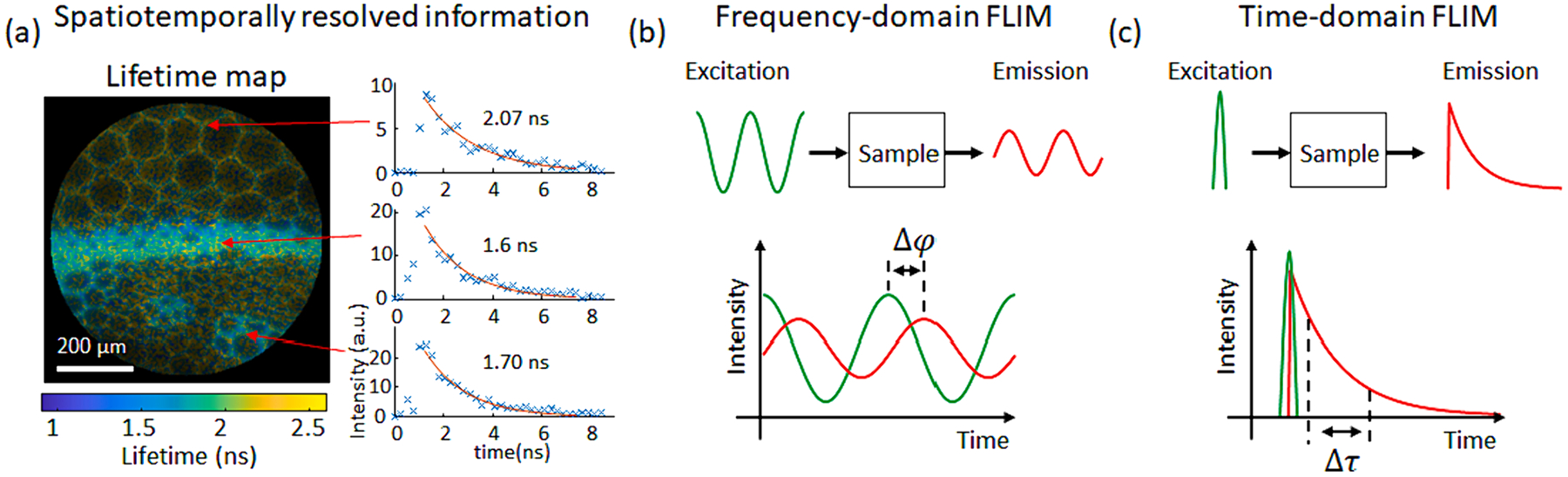
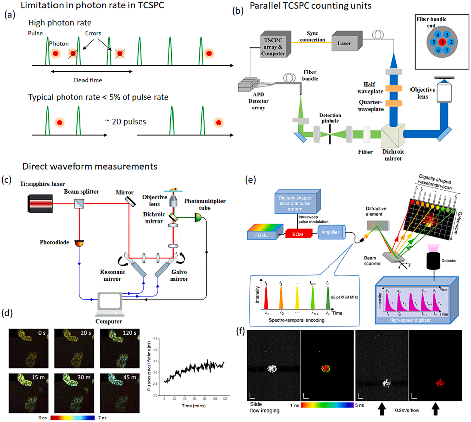
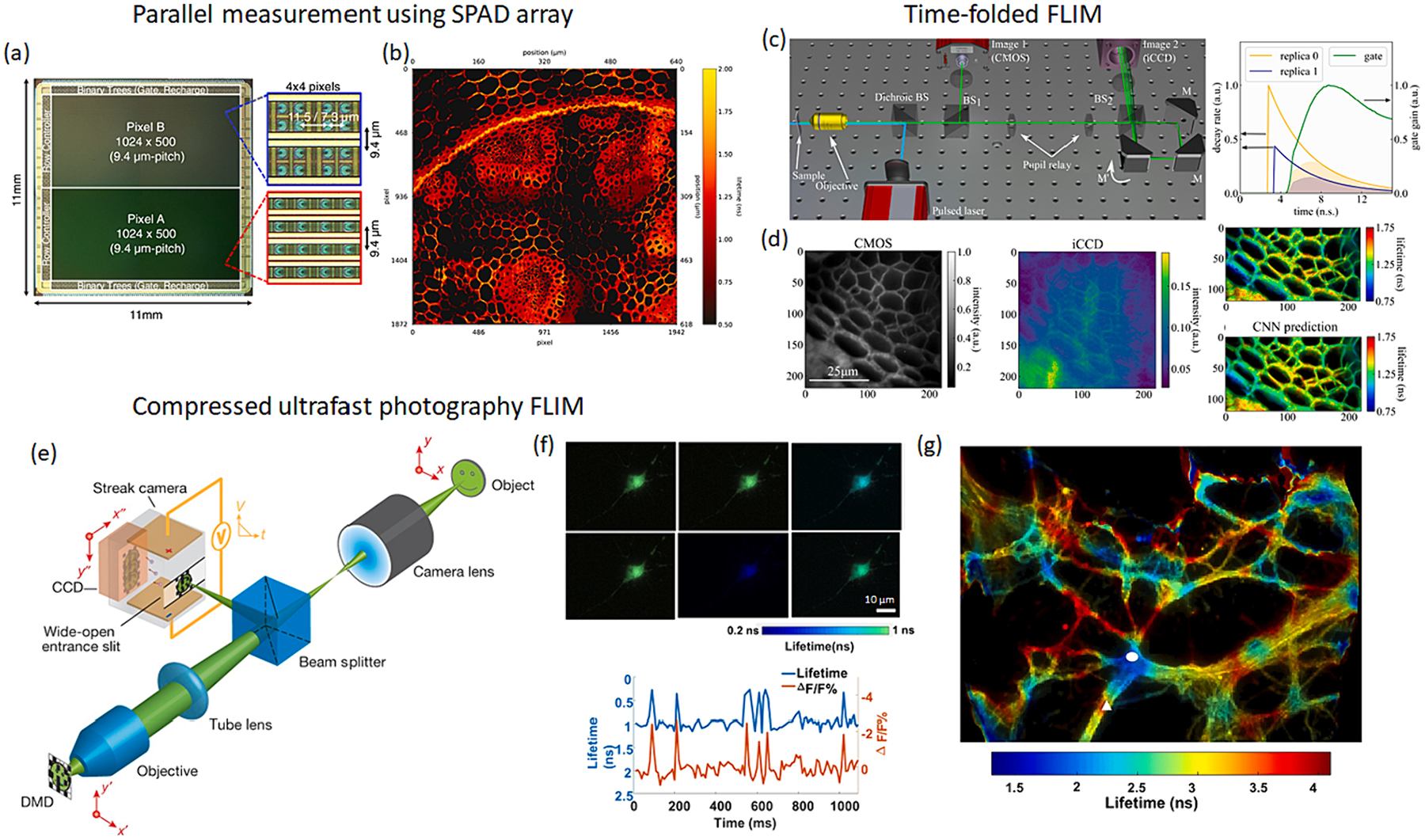
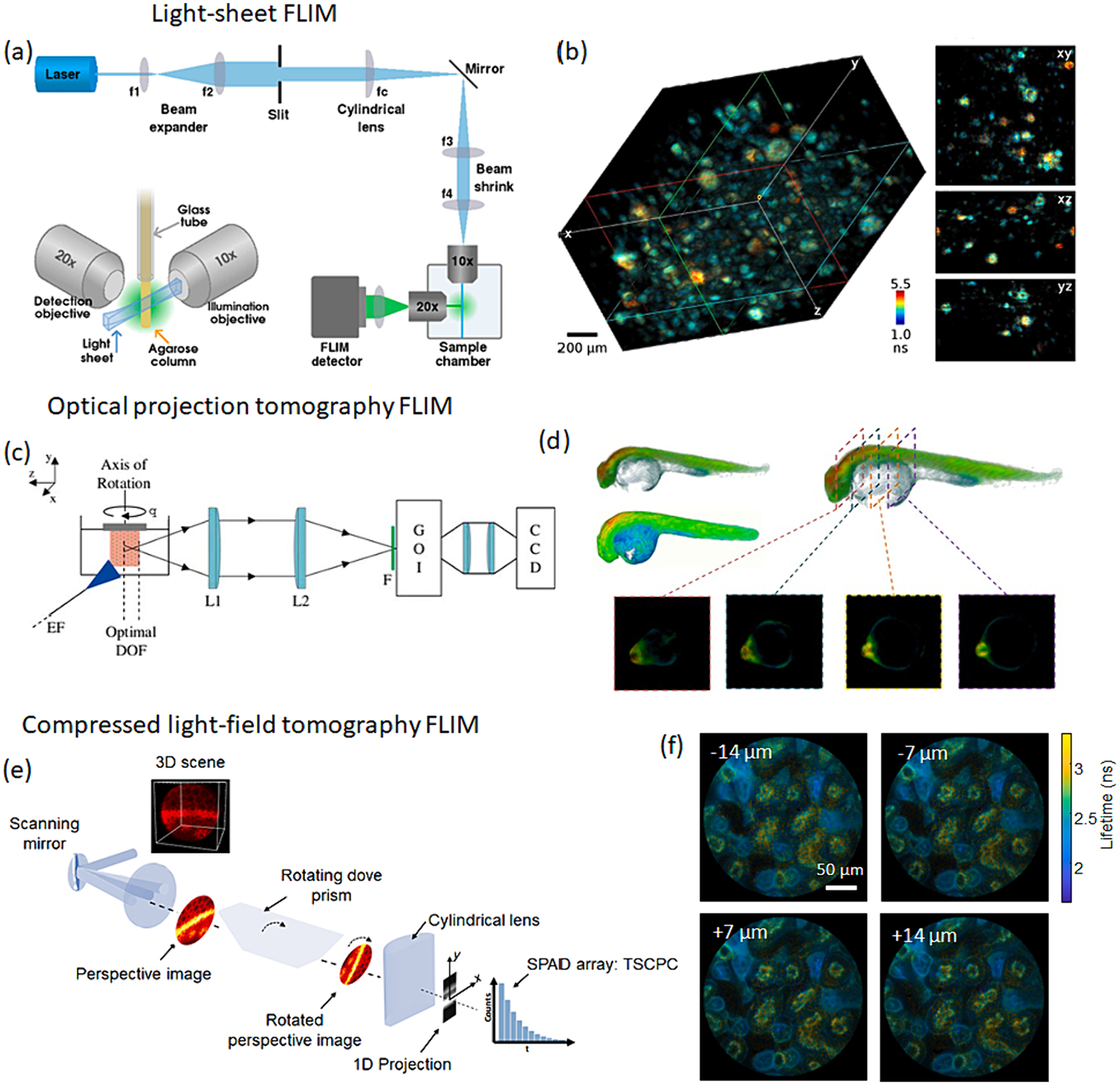
Verification
Claims the paper makes
The claims and statistics of this paper haven’t been checked yet.
Run the check — free, no account needed. It reads the open-access text and checks the sources it cites.
Claims other papers make about this one
Each is checked against this paper’s own text: does it say what the citing paper says it does?
The source backs part of the claim, not all of it — or backs it for a different population, condition or comparison.
In the citing paper
“fluorescence lifetime imaging microscopy (FLIM) has also emerged as a refined method to assess proliferation in 3D models with improved specificity and reduced signal interference [270,271].”
From Modeling Tumor Microenvironment Complexity In Vitro: Spheroids as Physiologically Relevant Tumor Models and Strategies for Their Analysis (2025) · cites this paper as [271]
Experimental — accuracy not yet measured
Self-check
Percentages, sums and reported differences re-computed from the paper’s own numbers.
The self-check needs the paper’s full text.
Citations
Cites
Highly Nonlinear Photodamage in Two-Photon Fluorescence Microscopy
Hopt, A., & Neher, E. (2001) · Biophysical Journal · doi:10.1016/s0006-3495(01)76173-5 · Cited by 442
Research works printed but not linked: 70
- Berezin, 2010, Fluorescence lifetime measurements and biological imaging, Chem. Rev., 110, 2641, 10.1021/cr900343z · DOI
- Datta, 2020, Fluorescence lifetime imaging microscopy: fundamentals and advances in instrumentation, analysis, and applications, J. Biomed. Opt., 25, 10.1117/1.JBO.25.7.071203 · DOI
- Alfonso-Garcia, 2021, Mesoscopic fluorescence lifetime imaging: fundamental principles, clinical applications and future directions, J. Biophotonics, 14, 10.1002/jbio.202000472 · DOI
- Dmitriev, 2021, Luminescence lifetime imaging of three-dimensional biological objects, J. Cell Sci., 134, 1, 10.1242/jcs.254763 · DOI
- Le Marois, 2017, Quantitative live cell FLIM imaging in three dimensions, Multi-Parametric Live Cell Microscopy of 3D Tissue Models, 31, 10.1007/978-3-319-67358-5_3 · DOI
- Becker, 2012, Fluorescence lifetime imaging – techniques and applications, J. Microsc., 247, 119, 10.1111/j.1365-2818.2012.03618.x · DOI · Paper page
- Borst, 2010, Fluorescence lifetime imaging microscopy in life sciences, Meas. Sci. Technol., 21, 10.1088/0957-0233/21/10/102002 · DOI · Paper page
- Ebrecht, 2014, Fluorescence lifetime imaging microscopy in the medical sciences, Protoplasma, 251, 293, 10.1007/s00709-013-0598-4 · DOI
- Marcu, 2012, Fluorescence lifetime techniques in medical applications, Ann. Biomed. Eng., 40, 304, 10.1007/s10439-011-0495-y · DOI
- Eichorst JP, Teng KW, Clegg RM: Fluorescence lifetime imaging techniques: Frequency-domain FLIM. In Fluorescence Lifetime Spectroscopy and Imaging: Principles and Applications in Biomedical Diagnostics. Edited by L Marcu PF, and DS Elson CRC Press; 2014:165-186.
- Becker W: Fluorescence lifetime imaging techniques: time-correlated single-photon counting. In Fluorescence Lifetime Spectroscopy and Imaging. Edited by: CRC Press; 2014:220-251. · DOI
- Gratton, 2003, Fluorescence lifetime imaging for the two-photon microscope: time-domain and frequency-domain methods, J. Biomed. Opt., 8, 381, 10.1117/1.1586704 · DOI
- Visscher, 1994, Fluorescence saturation in confocal microscopy, J. Microscopy-Oxford, 175, 162, 10.1111/j.1365-2818.1994.tb03479.x · DOI
- Liu, 2019, Fast fluorescence lifetime imaging techniques: a review on challenge and development, J. Innovative Optical Health Sci., 12, 1930003, 10.1142/S1793545819300039 · DOI
- Becker, 2019, Fast-acquisition TCSPC FLIM with sub-25-ps IRF width, Multiphoton Microscopy in the Biomedical Sci. XIX: SPIE, 10.1117/12.2510272 · DOI
- Liu, 2018, Parallelized fluorescence lifetime imaging microscopy (FLIM) based on photon reassignment, Opt. Commun., 421, 83, 10.1016/j.optcom.2018.03.060 · DOI
- Bower, 2018, High-speed imaging of transient metabolic dynamics using two-photon fluorescence lifetime imaging microscopy, Optica, 5, 1290, 10.1364/OPTICA.5.001290 · DOI
- Bower, 2019, Tracking metabolic dynamics of apoptosis with high-speed two-photon fluorescence lifetime imaging microscopy, Biomed. Opt. Express, 10, 6408, 10.1364/BOE.10.006408 · DOI
- Karpf, 2020, Spectro-temporal encoded multiphoton microscopy and fluorescence lifetime imaging at kilohertz frame-rates, Nat. Commun., 11, 2062, 10.1038/s41467-020-15618-w · DOI
- Bruschini, 2019, Single-photon avalanche diode imagers in biophotonics: review and outlook, Light Sci. Appl., 8, 87, 10.1038/s41377-019-0191-5 · DOI
- Zickus, 2020, Fluorescence lifetime imaging with a megapixel SPAD camera and neural network lifetime estimation, Sci. Rep., 10, 20986, 10.1038/s41598-020-77737-0 · DOI
- Morimoto, 2020, Megapixel time-gated SPAD image sensor for 2D and 3D imaging applications, Optica, 7, 346, 10.1364/OPTICA.386574 · DOI
- Kapitany, 2023, Single-shot time-folded fluorescence lifetime imaging, Proc. Natl. Acad. Sci., 120, 10.1073/pnas.2214617120 · DOI
- Ma, 2021, High-speed compressed-sensing fluorescence lifetime imaging microscopy of live cells, Proc. Natl. Acad. Sci., 118, 10.1073/pnas.2004176118 · DOI
- Gao, 2014, Single-shot compressed ultrafast photography at one hundred billion frames per second, Nature, 516, 74, 10.1038/nature14005 · DOI
- Liu, 2019, Single-shot compressed optical-streaking ultra-high-speed photography, Opt. Lett., 44, 1387, 10.1364/OL.44.001387 · DOI
- Liu, 2021, Fast wide-field upconversion luminescence lifetime thermometry enabled by single-shot compressed ultrahigh-speed imaging, Nat. Commun., 12, 6401, 10.1038/s41467-021-26701-1 · DOI
- Bowman, 2019, Electro-optic imaging enables efficient wide-field fluorescence lifetime microscopy, Nat. Commun., 10, 4561, 10.1038/s41467-019-12535-5 · DOI
- Bowman, 2021, Resonant electro-optic imaging for microscopy at Nanosecond resolution, ACS Nano, 15, 16043, 10.1021/acsnano.1c04470 · DOI
- Bowman, 2023, Wide-field fluorescence lifetime imaging of neuron spiking and subthreshold activity in vivo, Sci., 380, 1270, 10.1126/science.adf9725 · DOI
- Grant, 2005, Optically sectioned fluorescence lifetime imaging using a nipkow disk microscope and a tunable ultrafast continuum excitation source, Opt. Lett., 30, 3353, 10.1364/OL.30.003353 · DOI
- Grant, 2007, High speed optically sectioned fluorescence lifetime imaging permits study of live cell signaling events, Opt. Express, 15, 15656, 10.1364/OE.15.015656 · DOI
- Mitchell, 2017, Functional in vivo imaging using fluorescence lifetime light-sheet microscopy, Opt. Lett., 42, 1269, 10.1364/OL.42.001269 · DOI
- Funane, 2018, Selective plane illumination microscopy (SPIM) with time-domain fluorescence lifetime imaging microscopy (FLIM) for volumetric measurement of cleared mouse brain samples, Rev. Sci. Instrum., 89, 10.1063/1.5018846 · DOI
- Hirvonen, 2020, Lightsheet fluorescence lifetime imaging microscopy with wide-field time-correlated single photon counting, J. Biophotonics, 13, 10.1002/jbio.201960099 · DOI
- Samimi K, Desa DE, Lin W, Weiss K, Li J, Huisken J, Miskolci V, Huttenlocher A, Chacko JV, Velten A, et al.: Light sheet autofluorescence lifetime imaging with a single photon avalanche diode array. bioRxiv 2023. · DOI
- Greger, 2011, Three-dimensional fluorescence lifetime imaging with a single plane illumination microscope provides an improved signal to noise ratio, Opt. Express, 19, 20743, 10.1364/OE.19.020743 · DOI
- Kumar AT: Tomographic fluorescence lifetime imaging. In Fluorescence Lifetime Spectroscopy and Imaging. Edited by: CRC Press; 2014:461-476.
- McGinty, 2008, Fluorescence lifetime optical projection tomography, J Biophotonics, 1, 390, 10.1002/jbio.200810044 · DOI
- McGinty, 2011, In vivo fluorescence lifetime optical projection tomography, Biomed. Opt. Express, 2, 1340, 10.1364/BOE.2.001340 · DOI
- Andrews, 2016, Visualising apoptosis in live zebrafish using fluorescence lifetime imaging with optical projection tomography to map FRET biosensor activity in space and time, J. Biophotonics, 9, 414, 10.1002/jbio.201500258 · DOI
- Sun, 2023, Optical projection tomography reconstruction with few views using highly-generalizable deep learning at sinogram domain, Biomed. Opt. Express, 14, 6260, 10.1364/BOE.500152 · DOI
- Ma, 2023, Light-field tomographic fluorescence lifetime imaging microscopy, PREPRINT at Res. Square
- Feng, 2021, Ultrafast light field tomography for snapshot transient and non-line-of-sight imaging, Nat. Commun., 12, 2179, 10.1038/s41467-021-22461-0 · DOI
- Feng, 2022, Compact light field photography towards versatile three-dimensional vision, Nat. Commun., 13, 3333, 10.1038/s41467-022-31087-9 · DOI
- Hirvonen, 2020, Fast timing techniques in FLIM applications, Front. Phys., 8, 161, 10.3389/fphy.2020.00161 · DOI
- Park, 2021, Review of bio-optical imaging systems with a high space-bandwidth product, Adv. Photonics, 3, 044001, 10.1117/1.AP.3.4.044001 · DOI
- Wetzstein, 2020, Inference in artificial intelligence with deep optics and photonics, Nature, 588, 39, 10.1038/s41586-020-2973-6 · DOI
- Tian, 2021, Deep Learning in biomedical optics, Lasers Surg. Med., 53, 748, 10.1002/lsm.23414 · DOI
- Brady, 2020, Deep learning for camera data acquisition, control, and image estimation, Adv. Opt. Photon., 12, 787, 10.1364/AOP.398263 · DOI
- Sorrells, 2021, Single-photon peak event detection (SPEED): a computational method for fast photon counting in fluorescence lifetime imaging microscopy, Opt. Express, 29, 37759, 10.1364/OE.439675 · DOI
- Ochoa, 2020, High compression deep learning based single-pixel hyperspectral macroscopic fluorescence lifetime imaging in vivo, Biomed. Opt. Express, 11, 5401, 10.1364/BOE.396771 · DOI
- Pian, 2017, Compressive hyperspectral time-resolved wide-field fluorescence lifetime imaging, Nat. Photonics, 11, 411, 10.1038/nphoton.2017.82 · DOI
- Lee, 2018, Progress in single-photon avalanche diode image sensors in standard CMOS: from two-dimensional monolithic to three-dimensional-stacked technology, Jpn. J. Appl. Phys., 10.7567/JJAP.57.1002A3 · DOI
- Hutchings, 2019, A reconfigurable 3-D-stacked SPAD imager with in-pixel histogramming for flash LIDAR or high-speed time-of-flight imaging, IEEE J. Solid State Circuits, 54, 2947, 10.1109/JSSC.2019.2939083 · DOI
- Erdogan, 2022, A high dynamic range 128 × 120 3-D stacked CMOS SPAD image sensor SoC for fluorescence microendoscopy, IEEE J. Solid State Circuits, 57, 1649, 10.1109/JSSC.2022.3150721 · DOI
- Matheson, 2023, Handheld wide-field fluorescence lifetime imaging system based on a distally mounted SPAD array, Opt. Express, 31, 22766, 10.1364/OE.482273 · DOI
- Choi, 2012, 3D-resolved fluorescence and phosphorescence lifetime imaging using temporal focusing wide-field two-photon excitation, Opt. Express, 20, 26219, 10.1364/OE.20.026219 · DOI
- Jia, 2021, Spatiotemporal control of biology: synthetic photochemistry toolbox with far-red and near-infrared light, ACS Chem. Biol., 17, 3255, 10.1021/acschembio.1c00518 · DOI
- Wong, 2022, Extending optical chemical tools and technologies to mice by shifting to the shortwave infrared region, Curr. Opin. Chem. Biol., 68, 10.1016/j.cbpa.2022.102131 · DOI · Paper page
- Cao, 2020, Recent progress in NIR-II contrast agent for biological imaging, Front. Bioeng. Biotechnol., 7, 487, 10.3389/fbioe.2019.00487 · DOI
- Carr, 2018, Shortwave infrared fluorescence imaging with the clinically approved near-infrared dye indocyanine green, Proc. Natl. Acad. Sci., 115, 4465, 10.1073/pnas.1718917115 · DOI · Paper page
- Chavez, 2023, Characterization of fluorescence lifetime of organic fluorophores for molecular imaging in the shortwave infrared window, J. Biomed. Opt., 28, 10.1117/1.JBO.28.9.094806 · DOI
- Thorburn, 2022, Ge-on-si single-photon avalanche diode detectors for short-wave infrared wavelengths, J. Phy: Photonics, 4
- Boas, 2001, Imaging the body with diffuse optical tomography, IEEE Signal Process Mag., 18, 57, 10.1109/79.962278 · DOI
- Kumar, 2008, A time domain fluorescence tomography system for small animal imaging, IEEE Trans. Med. Imaging, 27, 1152, 10.1109/TMI.2008.918341 · DOI
- Nothdurft, 2009, In vivo fluorescence lifetime tomography, J. Biomed. Opt., 10.1117/1.3086607 · DOI
- Chen, 2000, Optical computed tomography in a turbid medium using early arriving photons, J. Biomed. Opt., 5, 144, 10.1117/1.429981 · DOI
- Liu, 1993, Ultrafast laser-pulse transmission and imaging through biological tissues, Appl. Opt., 32, 554, 10.1364/AO.32.000554 · DOI
- Niedre, 2008, Early photon tomography allows fluorescence detection of lung carcinomas and disease progression in mice in vivo, Proc. Natl. Acad. Sci., 105, 19126, 10.1073/pnas.0804798105 · DOI
Cited by
Sorted by citations
Shah, S., & D’Souza, G. G. M. (2025) · Cells · doi:10.3390/cells14100732 · Cited by 83
“More recently, fluorescence lifetime imaging microscopy (FLIM) has also emerged as a refined method to assess proliferation in 3D models with improved specificity and reduced signal interference [270,271].”
Metadata
- Authors
- Park, J., & Gao, L.
- Affiliations
- University of California, Los Angeles (US)
- Corresponding author
- Gao, Liang
- Venue
- Current Opinion in Solid State and Materials Science
- Publisher
- Elsevier BV
- Published
- 2024-06
- Volume
- 30
- Pages
- 101147
- ISSN
- 1359-0286 (ISSN-L)
- PMC
- PMC11290093
- OpenAlex
- W4392905680
- Cited by
- 47
- References
- 1
- Citations by year
- 2026: 21 · 2025: 20 · 2024: 6
- FWCI
- 2.54
- Type
- journal article
- Language
- en
- Open access
- hybrid
- doi.org
- Licence
- Locations
- Current Opinion in Solid State and Materials Science (publishedVersion, cc-by-nc)
- PubMed (publishedVersion)
- PubMed Central (acceptedVersion, cc-by-nc)
- Funders
- National Institute of Neurological Disorders and Stroke (RF1NS128488)
- National Institutes of Health
- National Heart Lung and Blood Institute (R01HL165318)
- Topics
- Life Sciences › Biochemistry, Genetics and Molecular Biology › Biophysics › Advanced Fluorescence Microscopy Techniques
- Health Sciences › Medicine › Radiology, Nuclear Medicine and Imaging › Optical Imaging and Spectroscopy Techniques
- Physical Sciences › Chemistry › Physical and Theoretical Chemistry › Photochemistry and Electron Transfer Studies
- Keywords
- fluorescence lifetime imaging, 3D imaging, high-speed imaging, fluorescence lifetime, depth scanning
About these checks
Registry, retraction, Cites and statistics results are computed, not judged, with one exception: a printed reference that no index lists is read once by a model before it counts as a citation error. Claims are extracted by a model and kept only when their quote appears verbatim in the text; each cited source is then checked, with a model’s help, for whether it says what the claim relies on. Those verdicts can be wrong — read the claim’s quote and the cited source before relying on one.
Statistics and arithmetic are recomputed, not judged.
Confidence bands are shown only for fields calibrated on our annotated test set. None is calibrated yet, so no band is shown.
What counts as a claim ›How we measure confidence ›How we check this ›
Are you an author, or do you see a problem with this page? Request a review
