

The DOI matches its Crossref record; 54 of the 54 references it cites were found in a registry or an index.
Perineuronal net digestion with chondroitinase restores memory in mice with tau pathology
Open accessAbstract
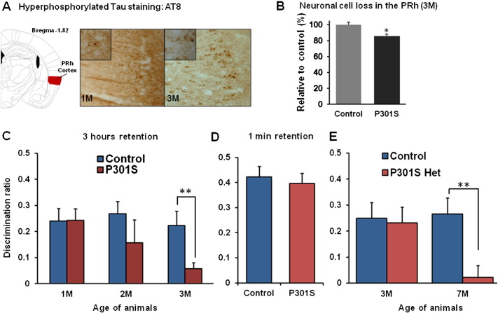
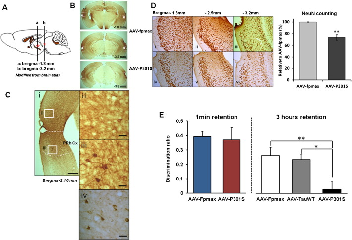
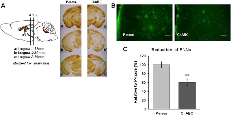
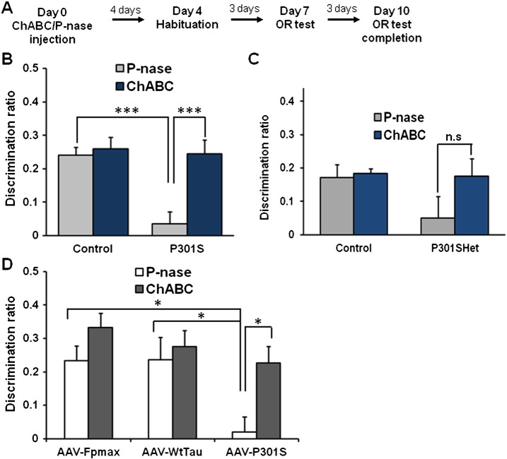
Alzheimer's disease is the most prevalent tauopathy and cause of dementia. We investigate the hypothesis that reactivation of plasticity can restore function in the presence of neuronal damage resulting from tauopathy. We investigated two models with tau hyperphosphorylation, aggregation and neurodegeneration: a transgenic mouse model in which the mutant P301S tau is expressed in neurons (Tg P301S), and a model in which an adeno-associated virus expressing P301S tau (AAV-P301S) was injected in the perirhinal cortex, a region critical for object recognition (OR) memory. Both models show profound loss of OR memory despite only 15% neuronal loss in the Tg P301S and 26% in AAV-P301S-injected mice. Recordings from perirhinal cortex slices of 3month-old P301S transgenic mice showed a diminution in synaptic transmission following temporal stimulation. Chondroitinase ABC (ChABC) can reactivate plasticity and affect memory through actions on perineuronal nets. ChABC was injected into the perirhinal cortex and animals were tested for OR memory 1week later, demonstrating restoration of OR memory to normal levels. Synaptic transmission indicated by fEPSP amplitude was restored to control levels following ChABC treatment. ChABC did not affect the progression of neurodegenerative tauopathy. These findings suggest that increasing plasticity by manipulation of perineuronal nets offers a novel therapeutic approach to the treatment of memory loss in neurodegenerative disorders.
Overview
The claims and statistics of this paper haven’t been checked yet.
Run the check — free, no account needed. It reads the open-access text and checks the sources it cites.
Integrity signals
Registry
Cites
Statistics
Figures
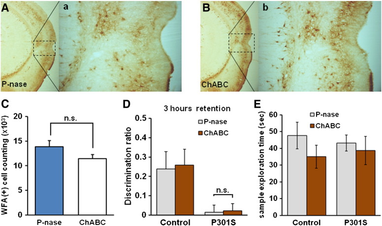
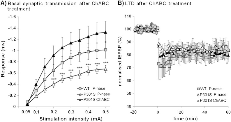
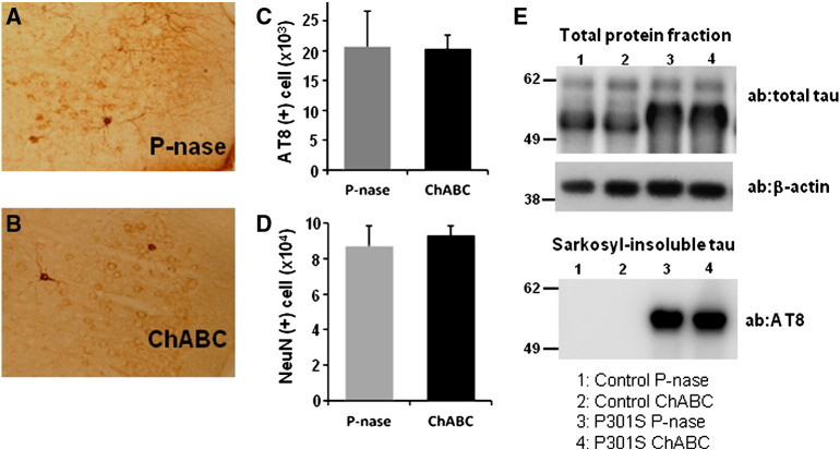
Verification
Claims the paper makes
The claims and statistics of this paper haven’t been checked yet.
Run the check — free, no account needed. It reads the open-access text and checks the sources it cites.
Claims other papers make about this one
Each is checked against this paper’s own text: does it say what the citing paper says it does?
The source backs part of the claim, not all of it — or backs it for a different population, condition or comparison.
In the citing paper
“Although one study by Yang et al. showed no difference in the number of PNNs within the perirhinal cortex of 3-month-old TauP301S (PS19) mouse model of tauopathy compared to age-matched controls (Yang et al. 2015)”
From Perineuronal net deglycosylation associates with tauopathy‐induced gliosis and neurodegeneration (2024) · cites this paper as Yang et al. 2015
Experimental — accuracy not yet measuredThe source backs part of the claim, not all of it — or backs it for a different population, condition or comparison.
In the citing paper
“this investigation was limited to both brain region (perirhinal cortex) and age (3 months).”
From Perineuronal net deglycosylation associates with tauopathy‐induced gliosis and neurodegeneration (2024) · cites this paper as Yang et al. 2015
Experimental — accuracy not yet measuredThe cited source says what the claim attributes to it.
In the citing paper
“Treating these animals with Ch--ABC to the PrC restored object memory to normal levels (Yang et al., 2015)”
From Casting a Wide Net: Role of Perineuronal Nets in Neural Plasticity (2016) · cites this paper as Yang et al., 2015
The check wasn’t confident either way, so it gives no verdict — never disputed.
In the citing paper
“There is some inconsistency on PNN findings in the transgenic mouse model of AD.”
From Disrupted structural connectivity in ArcAβ mouse model of Aβ amyloidosis (2020) · cites this paper as Yang et al., 2015
Experimental — accuracy not yet measuredIn the citing paper
“Remarkably, ChABC injections into perirhinal cortices from transgenic mice with tau pathology restored even the object recognition memory of these animals (Yang et al., 2015).”
From Damaged Neocortical Perineuronal Nets Due to Experimental Focal Cerebral Ischemia in Mice, Rats and Sheep (2017) · cites this paper as Yang et al., 2015
Self-check
Percentages, sums and reported differences re-computed from the paper’s own numbers.
The self-check needs the paper’s full text.
Citations
Cites
Neuropathological stageing of Alzheimer-related changes.
Braak, H., & Braak, E. (1991) · Acta Neuropathologica · doi:10.1007/bf00308809 · Cited by 16,123
The Mouse Brain in Stereotaxic Coordinates
Paxinos, G. (2001) · Cited by 12,859
Shrestha, S., & Offer, S. M. (2016) · Medical Epigenetics · doi:10.1159/000444713 · Cited by 8,951
Chondroitinase ABC promotes functional recovery after spinal cord injury
Bradbury, E. J., Moon, L. D. F., Popat, R. J., King, V. R., Bennett, G. S., Patel, P. N., Fawcett, J. W., & McMahon, S. B. (2002) · Nature · doi:10.1038/416636a · Cited by 2,357
Self-citationSynaptic mechanisms of synchronized gamma oscillations in inhibitory interneuron networks
Bartos, M., Vida, I., & Jonas, P. (2007) · Nature Reviews Neuroscience · doi:10.1038/nrn2044 · Cited by 2,070
Reactivation of Ocular Dominance Plasticity in the Adult Visual Cortex
Pizzorusso, T., Medini, P., Berardi, N., Chierzi, S., Fawcett, J. W., & Maffei, L. (2002) · Science · doi:10.1126/science.1072699 · Cited by 1,679
Self-citationTau pathology and neurodegeneration
Spillantini, M. G., & Goedert, M. (2013) · The Lancet Neurology · doi:10.1016/s1474-4422(13)70090-5 · Cited by 1,133
Self-citationGoedert, M., Spillantini, M. G., Potier, M. C., Ulrich, J., & Crowther, R. A. (1989) · The EMBO Journal · doi:10.1002/j.1460-2075.1989.tb03390.x · Cited by 1,037
Self-citationGoedert, M., Jakes, R., Spillantini, M. G., Hasegawa, M., Smith, M. J., & Crowther, R. A. (1996) · Nature · doi:10.1038/383550a0 · Cited by 1,036
Self-citationAllen, B., Ingram, E., Takao, M., Smith, M. J., Jakes, R., Virdee, K., Yoshida, H., Holzer, M., Craxton, M., Emson, P. C., et al. (2002) · Journal of Neuroscience · doi:10.1523/jneurosci.22-21-09340.2002 · Cited by 763
Self-citation
Cited by
Sorted by citations
Items 101–138 of 138
Dynamic Organization of Neuronal Extracellular Matrix Revealed by HaloTag-HAPLN1
Sterin, I., Niazi, A., Kim, J. S., Park, J., & Park, S. (2024) · Journal of Neuroscience · doi:10.1523/jneurosci.0666-24.2024 · Cited by 10
Motor recovery through perineuronal net modulation in a Parkinson’s disease mouse model
Benacom, D., Chataing, C., Prochiantz, A., & Nardo, A. A. D. (2025) · Brain · doi:10.1093/brain/awaf226 · Cited by 6
Richard, A., & Lu, X. (2019) · Neural Regeneration Research · doi:10.4103/1673-5374.247459 · Cited by 6
Croce, K. R., Ng, C., Pankiv, S., Albarran, E., Langfelder, P., de Jesus, A. R., Duncan, G. M., Wang, N., Basile, A. O., McHugh, C., et al. (2025) · Neuron · doi:10.1016/j.neuron.2025.08.018 · Cited by 4
Next generation tau models in Alzheimer's disease research – virus based gene delivery systems
Cubinkova, V., Valachova, B., Brezováková, V., SZABÓ, R., Zimova, I., Kostecká, Z., & Jadhav, S. (2017) · Acta Virologica · doi:10.4149/av_2017_01_13 · Cited by 6
Visualization of Perineuronal Nets in Central Nervous System Tissue Sections
Souter, L., & Kwok, J. C. F. (2019) · Methods in molecular biology · doi:10.1007/978-1-4939-9698-8_20 · Cited by 6
Regulation of CNS Plasticity Through the Extracellular Matrix
Warren, P. M., Dickens, S., Gigout, S., Fawcett, J. W., & Kwok, J. C. F. (2018) · Oxford University Press eBooks · doi:10.1093/oxfordhb/9780190635374.013.11 · Cited by 5
Self-citationBouisset, G., Tixier, F. J., Dupak, T., Lejards, C., & Verret, L. (2025) · iScience · doi:10.1016/j.isci.2025.113344 · Cited by 4
Yang, S., Gigout, S., Molinaro, A., Naito-Matsui, Y., Hilton, S., Foscarin, S., Nieuwenhuis, B., Verhaagen, J., Pizzorusso, T., Saksida, L. M., et al. (2020) · bioRxiv (Cold Spring Harbor Laboratory) · doi:10.1101/2020.01.03.894188 · Cited by 4
Self-citationIrvine, S. F., Gigout, S., Štěpánková, K., Martínez-Varea, N., Urdzíková, L. M., Jendelová, P., & Kwok, J. C. F. (2023) · bioRxiv (Cold Spring Harbor Laboratory) · doi:10.1101/2023.01.23.525137 · Cited by 4
Perineuronal Nets: Old Structures, New Functions
Tewari, B. P., Chaunsali, L., & Sontheimer, H. W. (2026) · The Neuroscientist · doi:10.1177/10738584261421180 · Cited by 3
Micheau, J., Catheline, G., Barse, E., Hiba, B., Marcilhac, A., Michèle Allard, A., Platt, B., & Riedel, G. (2024) · Brain Research Bulletin · doi:10.1016/j.brainresbull.2024.111161 · Cited by 4
Disrupted structural connectivity in ArcAβ mouse model of Aβ amyloidosis
Al-Amin, M. M., Grandjean, J., Klohs, J., & Kim, J. (2020) · doi:10.1101/2020.04.27.064634 · Cited by 2
“For example, some studies argued that the degradation of PNN restores memory in P301s tau mice (Yang et al., 2015) and reduces plaque burden in APPswe/PS1dE9 mice while others demonstrated unaltered PNN in Tg2576 mice (Morawski et al., 2010b).”
BackgroundResultHuang, Y.-F., Liu, C., Liu, Z., Zhuang, X., Chen, J.-H., Zhang, Z.-N., Shen, C., & Hu, Y. (2026) · Brain · doi:10.1093/brain/awag058 · Cited by 3
Emerging Therapeutic Role of Chondroitinase (ChABC) in Neurological Disorders and Cancer
Kolathur, K. K., Kumar, A., Biswas, A., Bojja, S. L., & Subrahmanyam, V. M. (2022) · Current Drug Therapy · doi:10.2174/1574885517666220331151619 · Cited by 3
Baidoe‐Ansah, D., Carceller, H., Mirzapourdelavar, H., Pérez-Rando, M., Strackeljan, L., Garcia-Vazquez, B., Seidenbecher, C. I., Kaushik, R., Nácher, J., & Dityatev, A. E. (2026) · Cellular and Molecular Neurobiology · doi:10.1007/s10571-026-01762-2 · Cited by 2
Perineuronal nets in motor circuitry regulate the performance of learned vocalizations in songbirds
Wan, X., Wang, A. S., Storch, D.-S., Li, V. Y., & Sakata, J. T. (2025) · Communications Biology · doi:10.1038/s42003-025-07520-2 · Cited by 2
Astrocytic Regulation of Aberrant Perineuronal Net Formation in Mecp2 ‐Null Neocortex
Sinha, A., Kowalchuk, A. M., Khatibi, N., Matthews, R. T., & MacDonald, J. L. (2026) · Journal of Neurochemistry · doi:10.1111/jnc.70503 · Cited by 5
Fell, C. W., Hagelkrüys, A., Cicvaric, A., Horrer, M., Liu, L., Li, J. S. S., Stadlmann, J., Polyansky, A. A., Mereiter, S., Tejada, M. A., et al. (2021) · bioRxiv (Cold Spring Harbor Laboratory) · doi:10.1101/2021.09.09.459581 · Cited by 2
Novel extracellular matrix architecture on excitatory neurons revealed by HaloTag-HAPLN1Preprint
Sterin, I., Niazi, A., Kim, J. S., Park, J., & Park, S. (2024) · bioRxiv (Cold Spring Harbor Laboratory) · doi:10.1101/2024.03.29.587384 · Cited by 2
Reichelt, A. C., Hashad, A. M., Ghosh‐Swaby, O. R., Skirzewski, M., Scott, G. A., Machado, M. M. F., Kramar, C. P., Princz‐Lebel, O., Inoue, W., Bussey, T. J., et al. (2026) · Neuropsychopharmacology · doi:10.1038/s41386-026-02343-z · Cited by 1
Self-citationDeasy, S., Amontree, M., Colon, Z. A., Thorland, E., Modi, K., Hummel, K., Blanco, I., Greco, G. A., Maguire‐Zeiss, K. A., & Conant, K. E. (2025) · bioRxiv (Cold Spring Harbor Laboratory) · doi:10.1101/2025.11.09.687435 · Cited by 1
Identification of novel intracellular mechanisms in developmental spine pruning through Ankyrin B.
Wyatt, E. V. (2019) · Carolina Digital Repository (University of North Carolina at Chapel Hill) · doi:10.17615/nv7d-dp91
Popovich, P. G. (2019) · Faculty Opinions – Post-Publication Peer Review of the Biomedical Literature · doi:10.3410/f.733447321.793557618
Research Progress on the Role of Semaphorin3F in Nervous System and Its Mechanism
潘, 浩. (2022) · Advances in Clinical Medicine · doi:10.12677/acm.2022.12121695
Benacom, D., Chataing, C., Prochiantz, A., & Nardo, A. A. D. (2024) · bioRxiv (Cold Spring Harbor Laboratory) · doi:10.1101/2024.05.31.596849
Zhang, H., Cicvaric, A., Bassett, T. E., Wood, E. M., Petrovic, Z., & Radulović, J. (2024) · Elsevier eBooks · doi:10.1016/b978-0-443-15754-7.00066-3
Nadadhur, A. G., Mason, M. J., Rees, J. S., Sidoryk‐Wȩgrzynowicz, M., Tolkovsky, A. M., & Spillantini, M. G. (2025) · Neural Regeneration Research · doi:10.4103/nrr.nrr-d-24-01598
Self-citationMicroglia: Key Players in Perineuronal Net Dynamics
Garmendia-Berges, M., & Puerta, E. (2025) · IntechOpen eBooks · doi:10.5772/intechopen.1012413 · Cited by 1
Chondroitin 4-sulphate depletion enhances synaptic plasticity and memory in agingPreprint
Burianova, J. S., Svoboda, J., Růžička, J., Štěpánková, K., Klausová, T., Gmiterková, L., Spundova, T., Martínez-Varea, N., Králíková, M., Tureček, R., et al. (2026) · bioRxiv (Cold Spring Harbor Laboratory) · doi:10.64898/2026.01.28.702127
Self-citationCimpean, A., Dubišová, J., Martínez-Varea, N., Mareková, D., Urdzíková, L. M., Fawcett, J. W., Jendelová, P., & Kwok, J. C. F. (2026) · Glia · doi:10.1002/glia.70233
Self-citationShibah, S. R. M. (2026) · SSRN Electronic Journal · doi:10.2139/ssrn.6438100
Douceau, S. (2020) · theses.fr (ABES) · doi:10.70675/6f1007f2z8468z4365zb07dz7d863bf32cd5
4-methylumbelliferone attenuates amyloid pathology and learning deficits in the APP/PS1 mouse model
Amontree, M., O’Leary, J., Wonnenberg, P. M., Nelson, M., & Conant, K. E. (2026) · Scientific Reports · doi:10.64898/2026.05.21.726929
Bhuiyan, M. H., Smith, M. J., Hinkley, S. F. R., & Clarkson, A. N. (2026) · Neuroprotection/Neuroprotection (Chichester, England. Print) · doi:10.1002/nep3.70043
The Role of Meprins on the Brain Extracellular Matrix and Perineuronal Nets
Kreiselmaier, S., Keller, M., Nardi, L., Mueller, C., von Wiegen, N., Bickenbach, K., Gröbner, L. S., Abukhalaf, M., Tholey, A., Mueller, M. M., et al. (2026) · The FASEB Journal · doi:10.1096/fj.202601333r
Metadata
- Authors
- Yang, S., Cacquevel, M., Saksida, L. M., Bussey, T. J., Schneider, B. L., Aebischer, P., Melani, R., Pizzorusso, T., Fawcett, J. W., & Spillantini, M. G.
- Affiliations
- University of Cambridge (GB)
- École Polytechnique Fédérale de Lausanne (CH)
- Neuroscience Institute (IT)
- Corresponding author
- Spillantini, Maria Grazia
- ORCID
- Venue
- Experimental Neurology
- Publisher
- Elsevier BV
- Published
- 2015-03
- Volume
- 265
- Pages
- 48-58
- ISSN
- 0014-4886 (ISSN-L)
- PMC
- PMC4353684
- OpenAlex
- W2156939977
- Cited by
- 138
- References
- 54
- Citations by year
- 2026: 12 · 2025: 11 · 2024: 15 · 2023: 9 · 2022: 13 · 2021: 13 · 2020: 13 · 2019: 15 · 2018: 12 · 2017: 10 · 2016: 9 · 2015: 6
- FWCI
- 5.56
- Type
- journal article
- Language
- en
- Open access
- hybrid
- doi.org
- Licence
- Locations
- Experimental Neurology (publishedVersion, cc-by)
- PubMed Central (publishedVersion, cc-by)
- PubMed (publishedVersion)
- Florence Research (University of Florence) (submittedVersion, other-oa)
- Infoscience (Ecole Polytechnique Fédérale de Lausanne) (submittedVersion)
- Infoscience (Ecole Polytechnique Fédérale de Lausanne) (submittedVersion, cc-by-nc-nd)
- Scholarship@Western (Western University) (submittedVersion)
- Scuola Normale Superiore di Pisa (submittedVersion, cc-by)
- Apollo (University of Cambridge) (submittedVersion, cc-by)
- Funders
- Wellcome Trust
- Christopher and Dana Reeve Foundation
- Medical Research Council
- National Institute for Health Research
- Alzheimer’s Research UK
- Topics
- Life Sciences › Neuroscience › Cellular and Molecular Neuroscience › Alzheimer's disease research and treatments
- Life Sciences › Neuroscience › Cognitive Neuroscience › Memory and Neural Mechanisms
- Life Sciences › Neuroscience › Cellular and Molecular Neuroscience › Neuroscience and Neuropharmacology Research
- Keywords
- perineuronal nets, chondroitinase ABC, tauopathy, Alzheimer's disease, P301S tau, object recognition memory, perirhinal cortex, synaptic plasticity, synaptic transmission, neurodegeneration, memory recovery, tau hyperphosphorylation
- MeSH terms
- Animals, Cerebral Cortex, Humans, Injections, Intraventricular, Male, Memory, Mice, Inbred C57BL, Mice, Transgenic, Nerve Net, Neuronal Plasticity, Organ Culture Techniques, Chondroitin ABC Lyase, Tauopathies, Mice
About these checks
Registry, retraction, Cites and statistics results are computed, not judged, with one exception: a printed reference that no index lists is read once by a model before it counts as a citation error. Claims are extracted by a model and kept only when their quote appears verbatim in the text; each cited source is then checked, with a model’s help, for whether it says what the claim relies on. Those verdicts can be wrong — read the claim’s quote and the cited source before relying on one.
Statistics and arithmetic are recomputed, not judged.
Confidence bands are shown only for fields calibrated on our annotated test set. None is calibrated yet, so no band is shown.
What counts as a claim ›How we measure confidence ›How we check this ›
Are you an author, or do you see a problem with this page? Request a review
