Cited by 47Zebrafish Biomedical Research ApplicationsMemory and Neural MechanismsSpatial Cognition and Navigation

The DOI matches its Crossref record; 4 of the 4 references it cites were found in a registry or an index.

Zebrafish Exploit Visual Cues and Geometric Relationships to Form a Spatial Memory

Open access

Published version of a preprint

Abstract

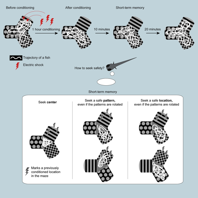
Animals use salient cues to navigate in their environment, but their specific cognitive strategies are largely unknown. We developed a conditioned place avoidance paradigm to discover whether and how zebrafish form spatial memories. In less than an hour, juvenile zebrafish, as young as 3 weeks, learned to avoid the arm of a Y-maze that was cued with a mild electric shock. Interestingly, individual fish solved this task in different ways: by staying in the safe center of the maze or by preference for one, or both, of the safe arms. In experiments in which the learned patterns were swapped, rotated, or replaced, the animals could transfer the association of safety to a different arm or to a different pattern using either visual cues or location as the conditioned stimulus. These findings show that juvenile zebrafish exhibit several complementary spatial learning modes, which generate a flexible repertoire of behavioral strategies.

Overview

The claims and statistics of this paper haven’t been checked yet.

Run the check — free, no account needed. It reads the open-access text and checks the sources it cites.

A few free checks a day · papers already verified open instantly · results are public

Integrity signals

Registry

Crossref · 10.1016/j.isci.2019.07.013

Cites

4 of 4 references verified0 citation errors · 0 not checkable

Statistics

Figures

Graphical abstract
Graphical abstract CC BY-NC-ND · PubMed Central
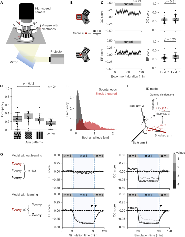
Figure 1 The Conditioned Place Avoidance (CPA) Paradigm (A) Experimental setup. (B) Measures of fish performance in the paradigm, top: arm occupancy (OC); bottom: arm entry frequency (EF); middle: schematic for the calculation of the preference scores for OC/EF. Positive values of the scores correspond to arm preference; negative values correspond to arm avoidance. Red color represents the shocked arm, black and gray represent the safe arms. (C) Left: moving averages of the OC/EF scores in a 2-h control experiment (mean ± SEM). Right: box plots for OC/EF scores in the first and the last 5 min of the control experiments (Mann-Whitney test, n = 24 fish). Box plots show median and quartiles; whiskers show 1.5x interquartile range; dots show values for individual fish; prime symbol stands for minutes. (D) There is no significant preference for any of the visual patterns (ANOVA, n = 24 fish). Box plot annotations are the same as in (C). (E) Distributions of amplitudes for spontaneous (gray) and shock-triggered swim bouts (red). Age of the fish is between 20 and 27 days post fertilization. (F) Pseudo-random 1D walk model used to evaluate CPA measures. Bout amplitude S for simulated movement was drawn from a Gamma distribution, amplitude in the conditioned arm was multiplied by a speed ratio α. (G) Top: model without learning. Bottom: model with learning. Learning rule was implemented by decreasing the probability of entry into the conditioned arm. Middle and right: moving averages of EF and OC scores. Black/gray lines correspond to simulations with different speed ratios α. Dashed vertical blue lines mark the areas where moving average combined information from two sessions. Arrowheads mark the differences in the OC/EF scores between top and bottom. Moving averages are calculated with a 5-min time window and a 30-s time step. See also Table S1.
Figure 1 The Conditioned Place Avoidance (CPA) Paradigm (A) Experimental setup. (B) Measures of fish performance in the paradigm, top: arm occupancy (OC); bottom: arm entry frequency (EF); middle: schematic for the calculation of the preference scores for OC/EF. Positive values of the scores correspond to arm preference; negative values correspond to arm avoidance. Red color represents the shocked arm, black and gray represent the safe arms. (C) Left: moving averages of the OC/EF scores in a 2-h control experiment (mean ± SEM). Right: box plots for OC/EF scores in the first and the last 5 min of the control experiments (Mann-Whitney test, n = 24 fish). Box plots show median and quartiles; whiskers show 1.5x interquartile range; dots show values for individual fish; prime symbol stands for minutes. (D) There is no significant preference for any of the visual patterns (ANOVA, n = 24 fish). Box plot annotations are the same as in (C). (E) Distributions of amplitudes for spontaneous (gray) and shock-triggered swim bouts (red). Age of the fish is between 20 and 27 days post fertilization. (F) Pseudo-random 1D walk model used to evaluate CPA measures. Bout amplitude S for simulated movement was drawn from a Gamma distribution, amplitude in the conditioned arm was multiplied by a speed ratio α. (G) Top: model without learning. Bottom: model with learning. Learning rule was implemented by decreasing the probability of entry into the conditioned arm. Middle and right: moving averages of EF and OC scores. Black/gray lines correspond to simulations with different speed ratios α. Dashed vertical blue lines mark the areas where moving average combined information from two sessions. Arrowheads mark the differences in the OC/EF scores between top and bottom. Moving averages are calculated with a 5-min time window and a 30-s time step. See also Table S1.CC BY-NC-ND · PubMed Central
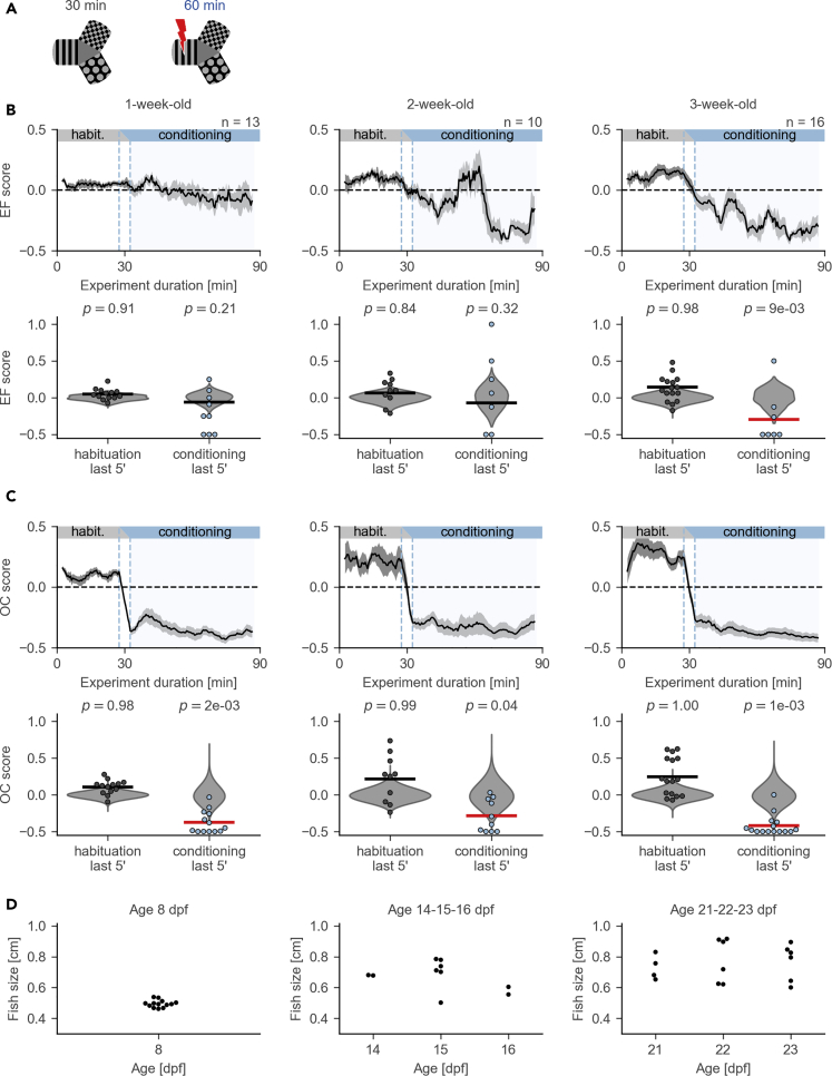
Figure 2 Performance in the CPA Paradigm across Different Age Groups (A) Schematic of the protocol: habituation and conditioning sessions. (B) Changes in EF scores across different age groups. Note that the EF score becomes significantly lower than zero only in 3-week-old fish. Top: moving average (mean ± SEM). Bottom: comparison of the EF scores in the last 5 min of conditioning with the null-distribution (permutation test). Individual values for each fish are shown as dots; null-distributions are shown in gray violin plots. Horizontal lines show the sample means; the line is red if the mean lies to the left of the fifth percentile in the null-distribution. Prime symbol stands for minutes. (C) Changes in OC scores across different age groups. Figure annotations are the same as in (B). (D) Distribution of body sizes across different ages. Average body size and its variability increase with age: 1 week, 4.95 ± 0.23 mm; 2 weeks, 6.74 ± 0.93 mm; 3 weeks, 7.64 ± 1.15 mm, mean ± standard deviation. Dpf, days post fertilization.
Figure 2 Performance in the CPA Paradigm across Different Age Groups (A) Schematic of the protocol: habituation and conditioning sessions. (B) Changes in EF scores across different age groups. Note that the EF score becomes significantly lower than zero only in 3-week-old fish. Top: moving average (mean ± SEM). Bottom: comparison of the EF scores in the last 5 min of conditioning with the null-distribution (permutation test). Individual values for each fish are shown as dots; null-distributions are shown in gray violin plots. Horizontal lines show the sample means; the line is red if the mean lies to the left of the fifth percentile in the null-distribution. Prime symbol stands for minutes. (C) Changes in OC scores across different age groups. Figure annotations are the same as in (B). (D) Distribution of body sizes across different ages. Average body size and its variability increase with age: 1 week, 4.95 ± 0.23 mm; 2 weeks, 6.74 ± 0.93 mm; 3 weeks, 7.64 ± 1.15 mm, mean ± standard deviation. Dpf, days post fertilization.CC BY-NC-ND · PubMed Central
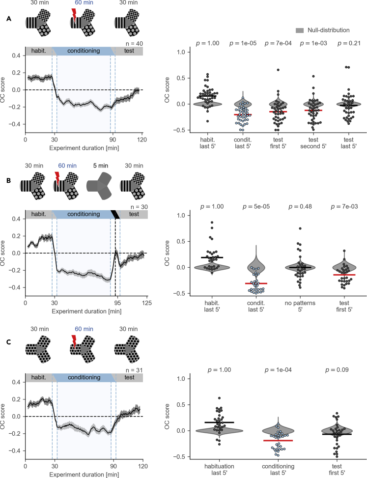
Figure 3 Evaluation of Aversive Memory Formed in the CPA Paradigm (A) Top: schematic of the protocol with habituation, conditioning, and test sessions. Bottom, left: OC score moving average. Black line shows average across individuals, gray ribbon shows SEM. Bottom, right: comparison of the OC scores in the last 5 min of conditioning and in the first, second, and last 5 min of test session with the null-distribution (permutation test, n = 40 fish). Prime symbol indicates minutes. (B) Top: schematic of the protocol with habituation, conditioning, no-pattern (all arms with a gray background), and test sessions. Bottom, left: OC score moving average. No-pattern session is indicated on the top of the plot with a black horizontal bar. Note the deflection in the moving average during the no-pattern session (at around minute 95) with a peak value near zero, the chance level. Bottom, right: comparison of the OC scores in the last 5 min of conditioning, 5 min of no-pattern, and in the first 5 min of test session with the null-distribution (permutation test, n = 30 fish). (C) Top: schematic of the protocol where visual patterns in all arms are identical. Bottom, left: OC score moving average. Bottom, right: comparison of the OC scores in the last 5 min of conditioning and in the first 5 min of test session with the null-distribution (permutation test, n = 31 fish). All moving averages are calculated with a 5-min time window and a 30-s time step. See also Figure S1.
Figure 3 Evaluation of Aversive Memory Formed in the CPA Paradigm (A) Top: schematic of the protocol with habituation, conditioning, and test sessions. Bottom, left: OC score moving average. Black line shows average across individuals, gray ribbon shows SEM. Bottom, right: comparison of the OC scores in the last 5 min of conditioning and in the first, second, and last 5 min of test session with the null-distribution (permutation test, n = 40 fish). Prime symbol indicates minutes. (B) Top: schematic of the protocol with habituation, conditioning, no-pattern (all arms with a gray background), and test sessions. Bottom, left: OC score moving average. No-pattern session is indicated on the top of the plot with a black horizontal bar. Note the deflection in the moving average during the no-pattern session (at around minute 95) with a peak value near zero, the chance level. Bottom, right: comparison of the OC scores in the last 5 min of conditioning, 5 min of no-pattern, and in the first 5 min of test session with the null-distribution (permutation test, n = 30 fish). (C) Top: schematic of the protocol where visual patterns in all arms are identical. Bottom, left: OC score moving average. Bottom, right: comparison of the OC scores in the last 5 min of conditioning and in the first 5 min of test session with the null-distribution (permutation test, n = 31 fish). All moving averages are calculated with a 5-min time window and a 30-s time step. See also Figure S1.CC BY-NC-ND · PubMed Central
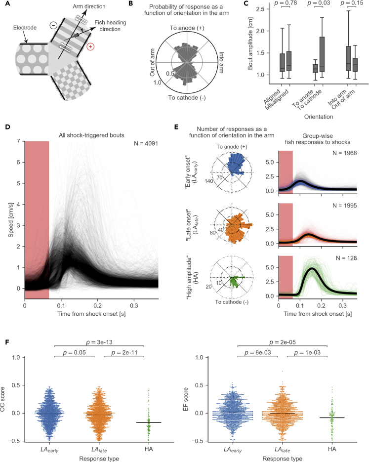
Figure 4 Responsiveness to Electric Shocks (A) Schematic of a fish's orientation in the electric field. The orientation angle is calculated between arm direction and fish heading direction. White arrows show the direction of the electric field. Black lines indicate the positions of the electrodes. (B) A radial histogram of the probabilities of shock-triggered swim bouts (i.e., responses to shocks) plotted against the fish's orientation in the electric field (bin size 9°). (C) Comparison of the bout amplitudes between different orientations in the electric field (2-sample t test). Bout amplitude is calculated as speed integrated over the duration of a bout. “Aligned” bouts include anode- and cathode-facing orientations; “misaligned” bouts include into-arm- and out-of-arm-facing orientations. (D) Variety of amplitudes and onsets of individual shock-triggered swim bouts. Each curve shows the speed during an individual bout (N = 4,091 bouts). (E) Response types identified by hierarchical cluster analysis: low amplitude with early onset (LAearly, N = 1,968 bouts), low amplitude with late onset (LAlate, N = 1,995 bouts), and high-amplitude responses (HA, N = 128 bouts). Left: radial histogram of how many bouts of a certain type occur plotted against the fish's orientation in the electric field (bin size 9°). Right: speed over time after the shock onset. Y axes are the same as in (D). (F) Comparison of OC (left) and EF (right) scores in a 5-min time window after a shock-triggered bout between different response types (Mann-Whitney test). Each dot represents an OC or EF score after an individual shock-triggered bout. Horizontal lines indicate sample means. Bouts were obtained from the experiments with 27 fish.
Figure 4 Responsiveness to Electric Shocks (A) Schematic of a fish's orientation in the electric field. The orientation angle is calculated between arm direction and fish heading direction. White arrows show the direction of the electric field. Black lines indicate the positions of the electrodes. (B) A radial histogram of the probabilities of shock-triggered swim bouts (i.e., responses to shocks) plotted against the fish's orientation in the electric field (bin size 9°). (C) Comparison of the bout amplitudes between different orientations in the electric field (2-sample t test). Bout amplitude is calculated as speed integrated over the duration of a bout. “Aligned” bouts include anode- and cathode-facing orientations; “misaligned” bouts include into-arm- and out-of-arm-facing orientations. (D) Variety of amplitudes and onsets of individual shock-triggered swim bouts. Each curve shows the speed during an individual bout (N = 4,091 bouts). (E) Response types identified by hierarchical cluster analysis: low amplitude with early onset (LAearly, N = 1,968 bouts), low amplitude with late onset (LAlate, N = 1,995 bouts), and high-amplitude responses (HA, N = 128 bouts). Left: radial histogram of how many bouts of a certain type occur plotted against the fish's orientation in the electric field (bin size 9°). Right: speed over time after the shock onset. Y axes are the same as in (D). (F) Comparison of OC (left) and EF (right) scores in a 5-min time window after a shock-triggered bout between different response types (Mann-Whitney test). Each dot represents an OC or EF score after an individual shock-triggered bout. Horizontal lines indicate sample means. Bouts were obtained from the experiments with 27 fish.CC BY-NC-ND · PubMed Central
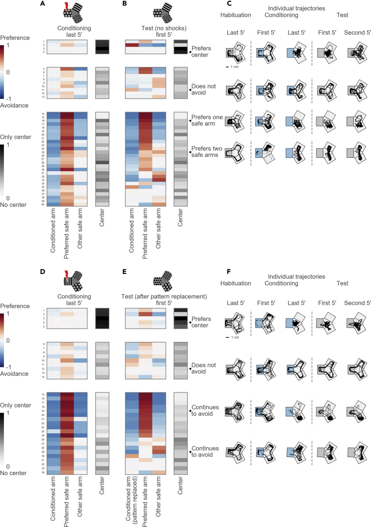
Figure 5 Diverse Strategies Used to Avoid the Conditioned Arm (A) Top: schematic of the maze during the conditioning session. Bottom: hierarchical clustering of arm occupancies in the last 5 min of the conditioning session reveals three groups: 4 fish preferring the center, 9 fish not avoiding the conditioned arm, and 27 avoiding fish. Each group is presented as a table, one row per fish. Columns correspond to maze arms (from left to right: the conditioned arm, the preferred safe arm, the other safe arm) and the center. Each cell shows a color-coded occupancy value in the particular compartment of the maze for a particular fish, with the logarithmic blue-to-red color scheme for the maze arms and the gray color scheme for the maze center. (B) Top: schematic of the maze during the test session. The rows in (A) and (B) correspond to the same fish. Rows within each group are ordered by their similarity to each other in the hierarchical tree in the test session. (C) Example trajectories of individual fish in the last 5 min of habituation, in the first and last 5 min of conditioning, and in the first and second 5 min of test session. Top: a fish uses the central compartment as a “safe haven.” Upper middle: a non-avoiding fish revisits the conditioned arm despite continued shocks. Lower middle: a fish prefers one safe arm. Bottom: a fish swims in both safe arms. The conditioned arm is depicted with gray background for the habituation session, blue for the conditioning session, and again gray for the test session. The orientation of the conditioned arm varied in the experiments and is shown here on the left for clarity. (D) Top: schematic of the maze during the conditioning session in experiments with pattern replacement. Bottom: hierarchical clustering reveals three groups: 5 center-preferring, 9 non-avoiding, and 20 avoiding fish. (E) Top: schematic of the maze during the test session in experiments with pattern replacement. Bottom: occupancies during the test session that correspond to the groups identified in (D). (F) Example trajectories of individual fish before and after the pattern replacement. Top: a fish prefers the center. Upper middle: a non-avoiding fish. Lower middle: a fish swims in one safe arm before and after the replacement of the conditioned pattern. Bottom: a fish swims in both safe arms before and after the pattern replacement. See also Figures S2 and S3.
Figure 5 Diverse Strategies Used to Avoid the Conditioned Arm (A) Top: schematic of the maze during the conditioning session. Bottom: hierarchical clustering of arm occupancies in the last 5 min of the conditioning session reveals three groups: 4 fish preferring the center, 9 fish not avoiding the conditioned arm, and 27 avoiding fish. Each group is presented as a table, one row per fish. Columns correspond to maze arms (from left to right: the conditioned arm, the preferred safe arm, the other safe arm) and the center. Each cell shows a color-coded occupancy value in the particular compartment of the maze for a particular fish, with the logarithmic blue-to-red color scheme for the maze arms and the gray color scheme for the maze center. (B) Top: schematic of the maze during the test session. The rows in (A) and (B) correspond to the same fish. Rows within each group are ordered by their similarity to each other in the hierarchical tree in the test session. (C) Example trajectories of individual fish in the last 5 min of habituation, in the first and last 5 min of conditioning, and in the first and second 5 min of test session. Top: a fish uses the central compartment as a “safe haven.” Upper middle: a non-avoiding fish revisits the conditioned arm despite continued shocks. Lower middle: a fish prefers one safe arm. Bottom: a fish swims in both safe arms. The conditioned arm is depicted with gray background for the habituation session, blue for the conditioning session, and again gray for the test session. The orientation of the conditioned arm varied in the experiments and is shown here on the left for clarity. (D) Top: schematic of the maze during the conditioning session in experiments with pattern replacement. Bottom: hierarchical clustering reveals three groups: 5 center-preferring, 9 non-avoiding, and 20 avoiding fish. (E) Top: schematic of the maze during the test session in experiments with pattern replacement. Bottom: occupancies during the test session that correspond to the groups identified in (D). (F) Example trajectories of individual fish before and after the pattern replacement. Top: a fish prefers the center. Upper middle: a non-avoiding fish. Lower middle: a fish swims in one safe arm before and after the replacement of the conditioned pattern. Bottom: a fish swims in both safe arms before and after the pattern replacement. See also Figures S2 and S3.CC BY-NC-ND · PubMed Central
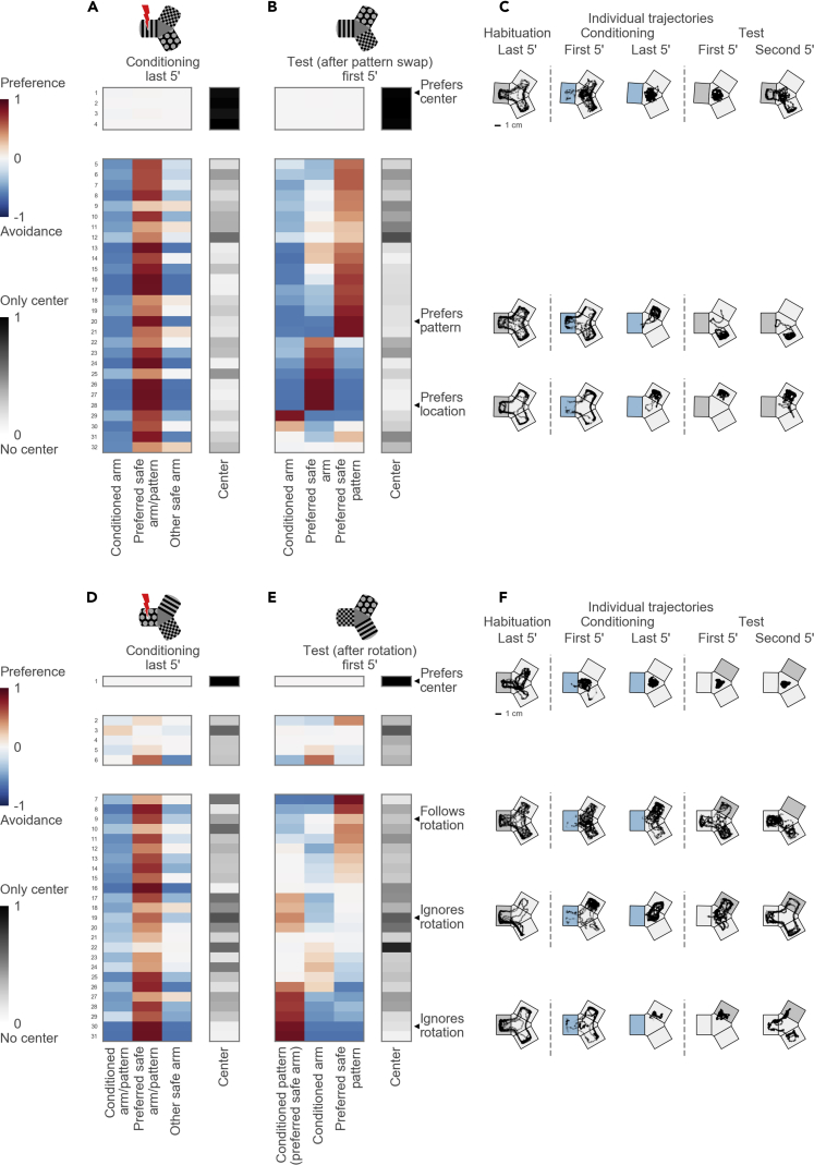
Figure 6 Dissociation of Pattern and Location Preference (A) Top: schematic of the maze during the conditioning session in experiments with pattern swap. Bottom: hierarchical clustering reveals two groups: 4 center-preferring and 28 arm-avoiding fish. (B) Arm occupancies during the test session in experiments with pattern swap. Occupancy groups correspond to the groups identified in (A). (C) Example trajectories of individual fish before and after the pattern swap. Top: a fish prefers the center. Middle: a fish prefers one arm and switches the arm after the patterns are swapped. Bottom: another fish also prefers one arm but stays in the same arm after the pattern swap. (D) Top: schematic of the maze during the conditioning session in experiments with pattern rotation. Bottom: hierarchical clustering reveals three groups: 1 center-preferring, 5 non-avoiding, and 25 avoiding fish. (E) Arm occupancies during the test session in experiments with pattern rotation. The conditioned pattern moves into the preferred safe arm, thus creating a conflict between avoidance and preference cues. The pattern from the preferred arm moves into the non-preferred arm; the pattern from the non-preferred arm moves into the previously conditioned arm. Occupancy groups correspond to the groups identified in (D). (F) Example trajectories of individual fish before and after the pattern rotation. Top: a fish prefers the center. Upper middle: a fish moves its preference following the preferred pattern and starts avoiding its previously preferred arm. Lower middle: a fish ignores the rotation and stays in its preferred arm despite the presence of the conditioned pattern. Bottom: another fish also ignores the rotation and stays in its preferred arm. See also Figure S4.
Figure 6 Dissociation of Pattern and Location Preference (A) Top: schematic of the maze during the conditioning session in experiments with pattern swap. Bottom: hierarchical clustering reveals two groups: 4 center-preferring and 28 arm-avoiding fish. (B) Arm occupancies during the test session in experiments with pattern swap. Occupancy groups correspond to the groups identified in (A). (C) Example trajectories of individual fish before and after the pattern swap. Top: a fish prefers the center. Middle: a fish prefers one arm and switches the arm after the patterns are swapped. Bottom: another fish also prefers one arm but stays in the same arm after the pattern swap. (D) Top: schematic of the maze during the conditioning session in experiments with pattern rotation. Bottom: hierarchical clustering reveals three groups: 1 center-preferring, 5 non-avoiding, and 25 avoiding fish. (E) Arm occupancies during the test session in experiments with pattern rotation. The conditioned pattern moves into the preferred safe arm, thus creating a conflict between avoidance and preference cues. The pattern from the preferred arm moves into the non-preferred arm; the pattern from the non-preferred arm moves into the previously conditioned arm. Occupancy groups correspond to the groups identified in (D). (F) Example trajectories of individual fish before and after the pattern rotation. Top: a fish prefers the center. Upper middle: a fish moves its preference following the preferred pattern and starts avoiding its previously preferred arm. Lower middle: a fish ignores the rotation and stays in its preferred arm despite the presence of the conditioned pattern. Bottom: another fish also ignores the rotation and stays in its preferred arm. See also Figure S4.CC BY-NC-ND · PubMed Central
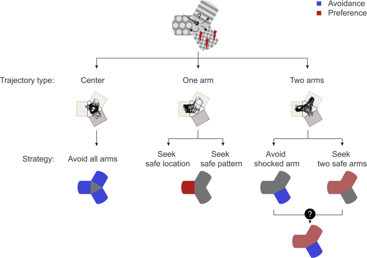
Figure 7 Diverse Strategies for Conditioned Place Avoidance Among Individual Animals Center visitors could avoid all arms. One-arm visitors prefer one arm: either a pattern or a location in the maze. Two-arm visitors avoid the conditioned arm: either by avoiding the conditioned arm or by a more sophisticated strategy, e.g., learning the two safe patterns or the combination of visual and location cues in the maze.
Figure 7 Diverse Strategies for Conditioned Place Avoidance Among Individual Animals Center visitors could avoid all arms. One-arm visitors prefer one arm: either a pattern or a location in the maze. Two-arm visitors avoid the conditioned arm: either by avoiding the conditioned arm or by a more sophisticated strategy, e.g., learning the two safe patterns or the combination of visual and location cues in the maze.CC BY-NC-ND · PubMed Central

Verification

Claims the paper makes

The claims and statistics of this paper haven’t been checked yet.

Run the check — free, no account needed. It reads the open-access text and checks the sources it cites.

A few free checks a day · papers already verified open instantly · results are public

Claims other papers make about this one

Each is checked against this paper’s own text: does it say what the citing paper says it does?

Self-check

Percentages, sums and reported differences re-computed from the paper’s own numbers.

The self-check needs the paper’s full text.

Citations

Cites

Show all 4 references

Research works printed but not linked: 37

  1. Aizenberg, 2011, Cerebellar-dependent learning in larval Zebrafish, J. Neurosci., 31, 8708, 10.1523/JNEUROSCI.6565-10.2011 · DOI
  2. Al-Imari, 2008, Sight of conspecifics as reward in associative learning in zebrafish (Danio rerio), Behav. Brain Res., 189, 216, 10.1016/j.bbr.2007.12.007 · DOI
  3. Aoki, 2015, Y-maze avoidance: an automated and rapid associative learning paradigm in zebrafish, Neurosci. Res., 91, 69, 10.1016/j.neures.2014.10.012 · DOI
  4. Bergmann, 2018, Imaging neuronal activity in the optic tectum of late stage larval Zebrafish, J. Dev. Biol., 6, 6, 10.3390/jdb6010006 · DOI
  5. Braubach, 2009, Olfactory conditioning in the zebrafish (Danio rerio), Behav. Brain Res., 198, 190, 10.1016/j.bbr.2008.10.044 · DOI
  6. Broglio, 2010, Selective involvement of the goldfish lateral pallium in spatial memory, Behav. Brain Res., 210, 191, 10.1016/j.bbr.2010.02.031 · DOI · Paper page
  7. Cheng, 1986, A purely geometric module in the rat’s spatial representation, Cognition, 23, 149, 10.1016/0010-0277(86)90041-7 · DOI
  8. Eichenbaum, 2017, The role of the hippocampus in navigation is memory, J. Neurophysiol., 117, 1785, 10.1152/jn.00005.2017 · DOI · Paper page
  9. Fanselow, 1990, Factors governing one-trial contextual conditioning, Anim. Learn. Behav., 18, 264, 10.3758/BF03205285 · DOI
  10. Franz, 2000, Biomimetic robot navigation, Rob. Auton. Syst., 30, 133, 10.1016/S0921-8890(99)00069-X · DOI · Paper page
  11. Gouteux, 2001, Rhesus monkeys use geometric and nongeometric information during a reorientation task, J. Exp. Psychol. Gen., 130, 505, 10.1037/0096-3445.130.3.505 · DOI
  12. Harmon, 2017, Distinct responses of Purkinje neurons and roles of simple spikes during associative motor learning in larval zebrafish, Elife, 6, 10.7554/eLife.22537 · DOI
  13. Hinz, 2013, Protein synthesis-dependent associative long-term memory in larval Zebrafish, J. Neurosci., 33, 15382, 10.1523/JNEUROSCI.0560-13.2013 · DOI
  14. Kalueff, 2013, Towards a comprehensive catalog of Zebrafish behavior 1.0 and beyond, Zebrafish, 10, 70, 10.1089/zeb.2012.0861 · DOI
  15. Kelly, 1998, Pigeons’ (Columba livia) encoding of geometric and featural properties of a spatial environment, J. Comp. Psychol., 112, 259, 10.1037/0735-7036.112.3.259 · DOI
  16. Kenney, 2017, Contextual fear conditioning in zebrafish, Learn. Mem., 24, 516, 10.1101/lm.045690.117 · DOI
  17. Lal, 2018, Identification of a neuronal population in the telencephalon essential for fear conditioning in zebrafish, BMC Biol., 16, 45, 10.1186/s12915-018-0502-y · DOI
  18. Lee, 2010, The Habenula prevents helpless behavior in larval Zebrafish, Curr. Biol., 20, 2211, 10.1016/j.cub.2010.11.025 · DOI
  19. Lee, 2012, Independent effects of geometry and landmark in a spontaneous reorientation task: a study of two species of fish, Anim. Cogn., 15, 861, 10.1007/s10071-012-0512-z · DOI
  20. Lee, 2013, Navigation by environmental geometry: the use of zebrafish as a model, J. Exp. Biol., 216, 3693
  21. Lee, 2015, Boundary primacy in spatial mapping: evidence from zebrafish (Danio rerio), Behav. Processes, 119, 116, 10.1016/j.beproc.2015.07.012 · DOI
  22. Liu, 2012, Alternative startle motor patterns and behaviors in the larval zebrafish (Danio rerio), J. Comp. Physiol. A, 198, 11, 10.1007/s00359-011-0682-1 · DOI
  23. López, 1999, Multiple spatial learning strategies in goldfish (Carassius auratus), Anim. Cogn., 2, 109, 10.1007/s100710050031 · DOI · Paper page
  24. Matsuda, 2017, Granule cells control recovery from classical conditioned fear responses in the zebrafish cerebellum, Sci. Rep., 7, 11865, 10.1038/s41598-017-10794-0 · DOI
  25. Mueller, 2011, The dorsal pallium in zebrafish, Danio rerio (Cyprinidae, Teleostei), Brain Res., 1381, 95, 10.1016/j.brainres.2010.12.089 · DOI
  26. O'Keefe, 1971, The hippocampus as a spatial map. Preliminary evidence from unit activity in the freely-moving rat, Brain Res., 34, 171, 10.1016/0006-8993(71)90358-1 · DOI · Paper page
  27. Packard, 1996, Inactivation of Hippocampus or caudate nucleus with lidocaine differentially affects expression of place and response learning, Neurobiol. Learn. Mem., 65, 65, 10.1006/nlme.1996.0007 · DOI
  28. Poulos, 2009, Persistence of fear memory across time requires the basolateral amygdala complex, Proc. Natl. Acad. Sci. U S A, 106, 11737, 10.1073/pnas.0905257106 · DOI
  29. Salas, 2006, Neuropsychology of learning and memory in teleost fish, Zebrafish, 3, 157, 10.1089/zeb.2006.3.157 · DOI · Paper page
  30. Sison, 2010, Associative learning in zebrafish (Danio rerio) in the plus maze, Behav. Brain Res., 207, 99, 10.1016/j.bbr.2009.09.043 · DOI
  31. Tabor, 2014, Direct activation of the Mauthner cell by electric field pulses drives ultrarapid escape responses, J. Neurophysiol., 112, 834, 10.1152/jn.00228.2014 · DOI
  32. Temizer, 2015, A visual pathway for looming-evoked escape in larval Zebrafish, Curr. Biol., 25, 1823, 10.1016/j.cub.2015.06.002 · DOI · Paper page
  33. Valente, 2012, Ontogeny of classical and operant learning behaviors in zebrafish, Learn. Mem., 19, 170, 10.1101/lm.025668.112 · DOI
  34. Vallortigara, 1990, Geometric modules in animals’ spatial representations: a test with chicks (Gallus gallus domesticus), J. Comp. Psychol., 104, 248, 10.1037/0735-7036.104.3.248 · DOI
  35. Vargas, 2004, Encoding of geometric and featural spatial information by Goldfish (Carassius auratus), J. Comp. Psychol., 118, 206, 10.1037/0735-7036.118.2.206 · DOI · Paper page
  36. Vendrell-Llopis, 2016, Evolutionary conserved brainstem circuits encode category, concentration and mixtures of taste, Sci. Rep., 5, 17825, 10.1038/srep17825 · DOI
  37. Yang, 2019, Visual contrast modulates operant learning responses in larval Zebrafish, Front. Behav. Neurosci., 13, 4, 10.3389/fnbeh.2019.00004 · DOI

Cited by

Sorted by citations

Metadata

Authors
Yashina, K., Tejero-Cantero, Á., Herz, A., & Baier, H.
Affiliations
  • Max Planck Institute of Neurobiology (DE)
  • Ludwig-Maximilians-Universität München (DE)
Corresponding author
Baier, Herwig
Venue
iScience
Publisher
Elsevier BV
Published
2019-09
Volume
19
Pages
119-134
ISSN
2589-0042 (ISSN-L)
OpenAlex
W2958418972
Cited by
47
References
4
Citations by year
2026: 6 · 2025: 8 · 2024: 5 · 2023: 8 · 2022: 5 · 2021: 8 · 2020: 7
FWCI
2.65
Type
journal article
Language
en
Open access
Locations
  • iScience (publishedVersion, cc-by-nc-nd)
  • bioRxiv (Cold Spring Harbor Laboratory) (acceptedVersion, cc-by-nc-nd)
  • Europe PMC (PubMed Central) (publishedVersion, cc-by-nc-nd)
  • PubMed Central (publishedVersion, cc-by-nc-nd)
  • PubMed (publishedVersion)
  • DOAJ (DOAJ: Directory of Open Access Journals) (submittedVersion, cc-by-sa)
  • MPG.PuRe (Max Planck Society) (submittedVersion, other-oa)
Funders
  • Max-Planck-Gesellschaft
  • Deutsche Forschungsgemeinschaft (RTG 2175)
  • Bundesministerium für Bildung und Forschung
Topics
  • Life Sciences › Biochemistry, Genetics and Molecular Biology › Cell Biology › Zebrafish Biomedical Research Applications
  • Life Sciences › Neuroscience › Cognitive Neuroscience › Memory and Neural Mechanisms
  • Social Sciences › Psychology › Experimental and Cognitive Psychology › Spatial Cognition and Navigation
Keywords
zebrafish, spatial memory, spatial learning, conditioned place avoidance, visual cues, geometric cues, Y-maze, juvenile fish, transfer of learning, behavioral strategies

About these checks

Registry, retraction, Cites and statistics results are computed, not judged, with one exception: a printed reference that no index lists is read once by a model before it counts as a citation error. Claims are extracted by a model and kept only when their quote appears verbatim in the text; each cited source is then checked, with a model’s help, for whether it says what the claim relies on. Those verdicts can be wrong — read the claim’s quote and the cited source before relying on one.

Statistics and arithmetic are recomputed, not judged.

Confidence bands are shown only for fields calibrated on our annotated test set. None is calibrated yet, so no band is shown.

What counts as a claim ›How we measure confidence ›How we check this ›

Are you an author, or do you see a problem with this page? Request a review