Cited by 8Epigenetics and DNA MethylationGenomics and Chromatin DynamicsCancer-related gene regulation

The DOI matches its Crossref record; 1 of the 1 references it cites were found in a registry or an index.

Caenorhabditis elegans MES-3 is a highly divergent ortholog of the canonical PRC2 component SUZ12

Open access

Published version of a preprint

Abstract

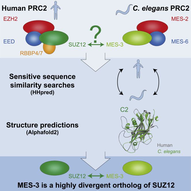
Polycomb Repressive Complex 2 (PRC2) catalyzes the mono-, di-, and trimethylation of histone protein H3 on lysine 27 (H3K27), which is strongly associated with transcriptionally silent chromatin. The functional core of PRC2 is highly conserved in animals and consists of four subunits. One of these, SUZ12, has not been identified in the genetic model Caenorhabditis elegans , whereas C. elegans PRC2 contains the clade-specific MES-3 protein. Through unbiased sensitive sequence similarity searches complemented by high-quality structure predictions of monomers and multimers, we here demonstrate that MES-3 is a highly divergent ortholog of SUZ12. MES-3 shares protein folds and conserved residues of key domains with SUZ12 and is predicted to interact with core PRC2 members similar to SUZ12 in human PRC2. Thus, in agreement with previous genetic and biochemical studies, we provide evidence that C. elegans contains a diverged yet evolutionary conserved core PRC2, like other animals.

Overview

The claims and statistics of this paper haven’t been checked yet.

Run the check — free, no account needed. It reads the open-access text and checks the sources it cites.

A few free checks a day · papers already verified open instantly · results are public

Integrity signals

Registry

Crossref · 10.1016/j.isci.2022.104633

Cites

1 of 1 references verified0 citation errors · 0 not checkable

Statistics

Figures

Graphical abstract
Graphical abstract CC BY · PubMed Central
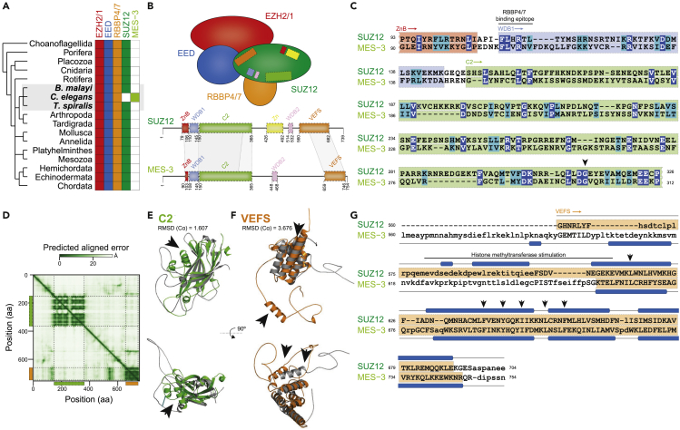
Figure 1 MES-3 is a highly divergent ortholog of the canonical Polycomb Repressive Complex two component SUZ12 (A). The Polycomb Repressive Complex 2 (PRC2) core components EZH2/1, EED, RBBP4/7, and SUZ12 are conserved in a broad range of metazoans; the presence of orthologs is indicated by filled boxes. Notably, based on sequence similarity searches, an ortholog of SUZ12 is absent in the nematode model species Caenorhabditis elegans, but present in other, closely related nematodes (Brugia malayi and Trichinella spiralis). C. elegans encodes the PRC2 core component MES-3 that lacks obvious motifs or sequence similarity to SUZ12 (Ahringer and Gasser, 2018; Bender et al., 2004; Ketel et al., 2005; Paulsen et al., 1995; Xu et al., 2001). (B). Schematic representation of the composition of the core PRC2. The zinc finger binding (ZnB; red), WD-domain binding 1 (WDB1; blue), C2 domain (green), zinc finger (Zn; yellow), WD-domain binding 2 (WDB2; pink), and VEFS (orange) motifs or domains involved in SUZ12 protein-interactions are shown in the schematic as well as along the protein sequence (Chammas et al., 2020; Chen et al., 2018; Kasinath et al., 2018). Schematic representation of the protein sequence of MES-3 is shown, and regions of uncovered sequence (c) and structural (e, f) similarity are highlighted. (C). Protein sequence alignment between the N-terminal region of SUZ12 and MES-3, as identified by sensitive profile-vs-profile sequence similarity searches, covers part of the zinc finger binding (ZnB; red), WD-domain binding 1 (WDB1; blue), and C2 domain (green). The conserved RBBP4/7 binding epitope as well as Gly299 are highlighted (Birve et al., 2001; Rai et al., 2013; Schmitges et al., 2011). Identical amino acids are shown in blue and biochemically similar amino acids are shown in turquoise. (D–F). The predicted aligned error (in Å; based on model 2 ptm) of the MES-3 structure is shown as a heatmap and reveals two separated globular regions in the N- and C-terminus, the former overlaps with the profile-vs-profile match (c) and corresponds to the C2 domain of SUZ12 (e; Figure S1I; RMSD = 1.607), while the latter overlaps with the region that structurally resembles the VEFS domain (f; Figure S1J; RMSD = 3.676). The black arrows (e, f) highlight regions that differ considerably between SUZ12 and MES-3 (Figures S1I and S1J), and the structure predictions of SUZ12 and MES-3 (e, f) are shown in gray as well as green (C2) and orange (VEFS), respectively. (G). Sequence-independent structure alignment of the VEFS regions of SUZ12 and MES-3 reveals significantly structural similarity (Dali Z score = 8.3; TM-score = 0.55), especially along the α helices in the C-terminus; a region previously shown to stimulate histone methyltransferase activity in SUZ12 (Birve et al., 2001) (pos. 580 to 612) is highlighted by a black bar, and individual amino acids important for PRC2 assembly (Birve et al., 2001) are shown by black arrows.
Figure 1 MES-3 is a highly divergent ortholog of the canonical Polycomb Repressive Complex two component SUZ12 (A). The Polycomb Repressive Complex 2 (PRC2) core components EZH2/1, EED, RBBP4/7, and SUZ12 are conserved in a broad range of metazoans; the presence of orthologs is indicated by filled boxes. Notably, based on sequence similarity searches, an ortholog of SUZ12 is absent in the nematode model species Caenorhabditis elegans, but present in other, closely related nematodes (Brugia malayi and Trichinella spiralis). C. elegans encodes the PRC2 core component MES-3 that lacks obvious motifs or sequence similarity to SUZ12 (Ahringer and Gasser, 2018; Bender et al., 2004; Ketel et al., 2005; Paulsen et al., 1995; Xu et al., 2001). (B). Schematic representation of the composition of the core PRC2. The zinc finger binding (ZnB; red), WD-domain binding 1 (WDB1; blue), C2 domain (green), zinc finger (Zn; yellow), WD-domain binding 2 (WDB2; pink), and VEFS (orange) motifs or domains involved in SUZ12 protein-interactions are shown in the schematic as well as along the protein sequence (Chammas et al., 2020; Chen et al., 2018; Kasinath et al., 2018). Schematic representation of the protein sequence of MES-3 is shown, and regions of uncovered sequence (c) and structural (e, f) similarity are highlighted. (C). Protein sequence alignment between the N-terminal region of SUZ12 and MES-3, as identified by sensitive profile-vs-profile sequence similarity searches, covers part of the zinc finger binding (ZnB; red), WD-domain binding 1 (WDB1; blue), and C2 domain (green). The conserved RBBP4/7 binding epitope as well as Gly299 are highlighted (Birve et al., 2001; Rai et al., 2013; Schmitges et al., 2011). Identical amino acids are shown in blue and biochemically similar amino acids are shown in turquoise. (D–F). The predicted aligned error (in Å; based on model 2 ptm) of the MES-3 structure is shown as a heatmap and reveals two separated globular regions in the N- and C-terminus, the former overlaps with the profile-vs-profile match (c) and corresponds to the C2 domain of SUZ12 (e; Figure S1I; RMSD = 1.607), while the latter overlaps with the region that structurally resembles the VEFS domain (f; Figure S1J; RMSD = 3.676). The black arrows (e, f) highlight regions that differ considerably between SUZ12 and MES-3 (Figures S1I and S1J), and the structure predictions of SUZ12 and MES-3 (e, f) are shown in gray as well as green (C2) and orange (VEFS), respectively. (G). Sequence-independent structure alignment of the VEFS regions of SUZ12 and MES-3 reveals significantly structural similarity (Dali Z score = 8.3; TM-score = 0.55), especially along the α helices in the C-terminus; a region previously shown to stimulate histone methyltransferase activity in SUZ12 (Birve et al., 2001) (pos. 580 to 612) is highlighted by a black bar, and individual amino acids important for PRC2 assembly (Birve et al., 2001) are shown by black arrows.CC BY · PubMed Central
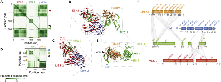
Figure 2 MES-3 provides a structural scaffold for PRC2 in C. elegans (A). The predicted aligned error (in Å) of MES-3 co-folded with MES-2 and MES-6 is shown as a heatmap and is consistent with association of MES-3 with MES-2 and MES-6 in the C-terminal regions of MES-3, which overlaps with the predicted VEFS domain. (B). Experimentally resolved human core PRC2 (rcsbpdb:6WKR (Kasinath et al., 2021)) highlights interactions between SUZ12 and RBBP4 as well as SUZ12 and EZH2 and EED. (C). Predicted C. elegans core PRC2 is formed by MES-2, MES-3, and MES-6. The corresponding region in human PRC2 is highlighted, as well as the position of the MES-3 VEFS domain (green triangle, see a.) and the MES-2 MCSS/SANT2-like region (red triangle, see a.), as well as a short central region of MES-3 (pos. 530-570) that associates with multiple regions in MES-2 and MES-6 (black triangle, see a.). For clarity, only regions of MES-3 interacting with MES-2 and MES-6 are shown (MES-3, pos. 1-530 and 570-640 are hidden). (D). The predicted aligned error (in Å) of MES-3 co-folded with LIN-53 is shown as a heatmap and reveals association between the N-terminal region of MES-3 and LIN-53. (E). Predicted MES-3 with LIN-53 complex. The corresponding region in human PRC2 is highlighted, as well as the MES-3 C2 domain (green triangle, see d.) and regions surrounding the C2 domain that engage in association with LIN-53 (WDB2, black triangle; WDB1, gray triangle; ZnB, light-blue triangle; see d.). For clarity, only regions of MES-3 interacting with LIN-53 are shown (MES-3, pos. 1-80, 365-415, and 470-754 are hidden). (F). Schematic representation of MES-3 and its predicted interactions with MES-2, MES-6, and LIN-53. The positions of protein domains/motifs in MES-2, MES-6, and LIN-53 were inferred via structural alignments of the predicted structures to those of the experimentally determined human EZH2, EED, and RBBP4 (Chammas et al., 2020; Kasinath et al., 2021). MES-3 domains/motifs are indicated as in Figure 1 with the addition of the central MES-2/MES-6 interacting region; domains/motifs shown are WD-domains (WD40) in MES-6 and LIN-53, and MES-6 binding (MES-6b), Swi3, Ada2, N-CoR and TFIIIB DNA-binding domain 1 like (SANT1), Motif connecting SANT1 and SANT2 (MCSS), SANT2, CXC, and the Su(var)3-9 , EZ and Trx domain (SET) in MES-2 (Chammas et al., 2020). We note that the region around the potential MCSS/SANT2 domains in MES-2 is substantially diverged compared with EZH2, yet still displays considerable structural similarity.
Figure 2 MES-3 provides a structural scaffold for PRC2 in C. elegans (A). The predicted aligned error (in Å) of MES-3 co-folded with MES-2 and MES-6 is shown as a heatmap and is consistent with association of MES-3 with MES-2 and MES-6 in the C-terminal regions of MES-3, which overlaps with the predicted VEFS domain. (B). Experimentally resolved human core PRC2 (rcsbpdb:6WKR (Kasinath et al., 2021)) highlights interactions between SUZ12 and RBBP4 as well as SUZ12 and EZH2 and EED. (C). Predicted C. elegans core PRC2 is formed by MES-2, MES-3, and MES-6. The corresponding region in human PRC2 is highlighted, as well as the position of the MES-3 VEFS domain (green triangle, see a.) and the MES-2 MCSS/SANT2-like region (red triangle, see a.), as well as a short central region of MES-3 (pos. 530-570) that associates with multiple regions in MES-2 and MES-6 (black triangle, see a.). For clarity, only regions of MES-3 interacting with MES-2 and MES-6 are shown (MES-3, pos. 1-530 and 570-640 are hidden). (D). The predicted aligned error (in Å) of MES-3 co-folded with LIN-53 is shown as a heatmap and reveals association between the N-terminal region of MES-3 and LIN-53. (E). Predicted MES-3 with LIN-53 complex. The corresponding region in human PRC2 is highlighted, as well as the MES-3 C2 domain (green triangle, see d.) and regions surrounding the C2 domain that engage in association with LIN-53 (WDB2, black triangle; WDB1, gray triangle; ZnB, light-blue triangle; see d.). For clarity, only regions of MES-3 interacting with LIN-53 are shown (MES-3, pos. 1-80, 365-415, and 470-754 are hidden). (F). Schematic representation of MES-3 and its predicted interactions with MES-2, MES-6, and LIN-53. The positions of protein domains/motifs in MES-2, MES-6, and LIN-53 were inferred via structural alignments of the predicted structures to those of the experimentally determined human EZH2, EED, and RBBP4 (Chammas et al., 2020; Kasinath et al., 2021). MES-3 domains/motifs are indicated as in Figure 1 with the addition of the central MES-2/MES-6 interacting region; domains/motifs shown are WD-domains (WD40) in MES-6 and LIN-53, and MES-6 binding (MES-6b), Swi3, Ada2, N-CoR and TFIIIB DNA-binding domain 1 like (SANT1), Motif connecting SANT1 and SANT2 (MCSS), SANT2, CXC, and the Su(var)3-9 , EZ and Trx domain (SET) in MES-2 (Chammas et al., 2020). We note that the region around the potential MCSS/SANT2 domains in MES-2 is substantially diverged compared with EZH2, yet still displays considerable structural similarity.CC BY · PubMed Central

Verification

Claims the paper makes

The claims and statistics of this paper haven’t been checked yet.

Run the check — free, no account needed. It reads the open-access text and checks the sources it cites.

A few free checks a day · papers already verified open instantly · results are public

Self-check

Percentages, sums and reported differences re-computed from the paper’s own numbers.

The self-check needs the paper’s full text.

Citations

Cites

Research works printed but not linked: 41

  1. Ahringer, 2018, Repressive chromatin in Caenorhabditis elegans: establishment, composition, and function, Genetics, 208, 491, 10.1534/genetics.117.300386 · DOI
  2. Bannister, 2011, Regulation of chromatin by histone modifications, Cell Res., 21, 381, 10.1038/cr.2011.22 · DOI · Paper page
  3. Bayly-Jones, 2021, Mining folded proteomes in the era of accurate structure prediction, Biorxiv
  4. Bender, 2004, The MES-2/MES-3/MES-6 complex and regulation of histone H3 methylation in C. elegans, Curr. Biol., 14, 1639, 10.1016/j.cub.2004.08.062 · DOI
  5. Bieluszewski, 2021, PRC2 activity, recruitment, and silencing: a comparative perspective, Trends Plant Sci., 1186, 10.1016/j.tplants.2021.06.006 · DOI
  6. Birve, 2001, Su(z)12 , a novel Drosophila Polycomb group gene that is conserved in vertebrates and plants, Development, 128, 3371, 10.1242/dev.128.17.3371 · DOI
  7. Capowski, 1991, Identification of grandchildless loci whose products are required for normal germ-line development in the nematode Caenorhabditis elegans, Genetics, 129, 1061, 10.1093/genetics/129.4.1061 · DOI
  8. Chammas, 2020, Engaging chromatin: PRC2 structure meets function, Br. J Cancer, 122, 315, 10.1038/s41416-019-0615-2 · DOI
  9. Chen, 2018, Unique structural platforms of Suz12 dictate distinct classes of PRC2 for chromatin binding, Mol. Cell, 69, 840, 10.1016/j.molcel.2018.01.039 · DOI
  10. Cosentino, 2019, SonicParanoid: fast, accurate and easy orthology inference, Bioinformatics, 35, 149, 10.1093/bioinformatics/bty631 · DOI · Paper page
  11. Derelle, 2020, Broccoli: combining phylogenetic and network analyses for orthology assignment, Mol. Biol. Evol., 37, 3389, 10.1093/molbev/msaa159 · DOI
  12. Deutekom, 2020, Benchmarking orthology methods using phylogenetic patterns defined at the base of Eukaryotes, Brief Bioinform., 22, bbaa206, 10.1093/bib/bbaa206 · DOI
  13. Emms, 2015, OrthoFinder: solving fundamental biases in whole genome comparisons dramatically improves orthogroup inference accuracy, Genome Biol., 16, 157, 10.1186/s13059-015-0721-2 · DOI · Paper page
  14. Erdős, 2021, IUPred3: prediction of protein disorder enhanced with unambiguous experimental annotation and visualization of evolutionary conservation, Nucleic Acids Res., 49, W297, 10.1093/nar/gkab408 · DOI
  15. Evans, 2022, Protein complex prediction with AlphaFold-Multimer, Biorxiv
  16. Gabler, 2020, Protein sequence analysis using the MPI Bioinformatics Toolkit, Curr. Protoc. Bioinform, 72, e108, 10.1002/cpbi.108 · DOI · Paper page
  17. Garvin, 1998, The Phenotype of mes-2, mes-3, mes-4 and mes-6, maternal-effect genes required for survival of the germline in Caenorhabditis elegans, is sensitive to chromosome dosage, Genetics, 148, 167, 10.1093/genetics/148.1.167 · DOI
  18. Gaydos, 2014, H3K27me and PRC2 transmit a memory of repression across generations and during development, Science, 345, 1515, 10.1126/science.1255023 · DOI
  19. Glancy, 2021, Structural basis for PRC2 engagement with chromatin, Curr. Opin. Struc. Biol., 67, 135, 10.1016/j.sbi.2020.10.017 · DOI
  20. Grossniklaus, 2014, Transcriptional silencing by Polycomb-Group proteins, Csh Perspect. Biol., 6, a019331
  21. Hauri, 2016, A high-density map for navigating the human Polycomb complexome, Cell Rep., 17, 583, 10.1016/j.celrep.2016.08.096 · DOI
  22. Holm, 1993, Protein structure comparison by alignment of distance matrices, J. Mol. Biol., 233, 123, 10.1006/jmbi.1993.1489 · DOI · Paper page
  23. Huerta-Cepas, 2016, eggNOG 4.5: a hierarchical orthology framework with improved functional annotations for eukaryotic, prokaryotic and viral sequences, Nucleic Acids Res., 44, D286, 10.1093/nar/gkv1248 · DOI
  24. Karakuzu, 2009, MIG-32 and SPAT-3A are PRC1 homologs that control neuronal migration in Caenorhabditis elegans, Development, 136, 943, 10.1242/dev.029363 · DOI
  25. Kasinath, 2021, JARID2 and AEBP2 regulate PRC2 in the presence of H2AK119ub1 and other histone modifications, Science, 371, eabc3393, 10.1126/science.abc3393 · DOI
  26. Kasinath, 2018, Structures of human PRC2 with its cofactors AEBP2 and JARID2, Science, 359, 940, 10.1126/science.aar5700 · DOI
  27. Ketel, 2005, Subunit contributions to histone methyltransferase activities of fly and worm Polycomb Group complexes, Mol. Cell Biol., 25, 6857, 10.1128/MCB.25.16.6857-6868.2005 · DOI
  28. Korf, 1998, The Polycomb group in Caenorhabditis elegans and maternal control of germline development, Development, 125, 2469, 10.1242/dev.125.13.2469 · DOI
  29. Margueron, 2011, The Polycomb complex PRC2 and its mark in life, Nature, 469, 343, 10.1038/nature09784 · DOI · Paper page
  30. Paulsen, 1995, Phenotypic and molecular analysis of mes-3, a maternal-effect gene required for proliferation and viability of the germ line in C. elegans, Genetics, 141, 1383, 10.1093/genetics/141.4.1383 · DOI
  31. Rai, 2013, Elements of the Polycomb repressor SU(Z)12 needed for histone H3-K27 methylation, the interface with E(Z), and in vivo function, Mol. Cell Biol., 33, 4844, 10.1128/MCB.00307-13 · DOI
  32. Rodrigues, 2018, pdb-tools: a swiss army knife for molecular structures, F1000research, 7, 1961, 10.12688/f1000research.17456.1 · DOI
  33. Sanchez-Pulido, 2021, Extending the horizon of homology detection with coevolution-based structure prediction, J. Mol. Biol., 433, 10.1016/j.jmb.2021.167106 · DOI
  34. Schmitges, 2011, Histone methylation by PRC2 is inhibited by active chromatin marks, Mol. Cell, 42, 330, 10.1016/j.molcel.2011.03.025 · DOI
  35. Schuettengruber, 2017, Genome regulation by Polycomb and trithorax: 70 years and counting, Cell, 171, 34, 10.1016/j.cell.2017.08.002 · DOI · Paper page
  36. Sharaf, 2021, Phylogenetic profiling suggests early origin of the core subunits of Polycomb Repressive Complex 2 (PRC2), Biorxiv
  37. Simon, 2009, Mechanisms of Polycomb gene silencing: knowns and unknowns, Nat. Rev. Mol. Cell Bio., 10, 697, 10.1038/nrm2763 · DOI
  38. Szklarczyk, 2012, Iterative orthology prediction uncovers new mitochondrial proteins and identifies C12orf62 as the human ortholog of COX14, a protein involved in the assembly of cytochrome coxidase, Genome Biol., 13, R12
  39. Xu, 2001, The Caenorhabditis elegans maternal-effect sterile proteins, MES-2, MES-3, and MES-6, are associated in a complex in embryos, Proc. Natl. Acad. Sci., 98, 5061, 10.1073/pnas.081016198 · DOI
  40. Yoshida, 2019, A SOSEKI-based coordinate system interprets global polarity cues in Arabidopsis, Nat. Plants, 5, 160, 10.1038/s41477-019-0363-6 · DOI
  41. Zhang, 2005, TM-align: a protein structure alignment algorithm based on the TM-score, Nucleic Acids Res., 33, 2302, 10.1093/nar/gki524 · DOI · Paper page

Metadata

Authors
Snel, B., van den Heuvel, S., & Seidl, M. F.
Affiliations
Utrecht University (NL)
Corresponding author
Seidl, Michael F.
Venue
iScience
Publisher
Elsevier BV
Published
2022-07
Volume
25
Issue
7
Pages
104633
ISSN
2589-0042 (ISSN-L)
OpenAlex
W4283078593
Cited by
8
References
1
Citations by year
2026: 1 · 2025: 1 · 2024: 3 · 2023: 3
FWCI
0.57
Type
journal article
Language
en
Open access
Locations
  • iScience (publishedVersion, cc-by)
  • bioRxiv (Cold Spring Harbor Laboratory) (acceptedVersion, cc-by)
  • PubMed Central (publishedVersion, cc-by)
  • PubMed (publishedVersion)
  • DOAJ (DOAJ: Directory of Open Access Journals) (submittedVersion)
  • Utrecht University Repository (Utrecht University) (submittedVersion, other-oa)
Funders
  • Nederlandse Organisatie voor Wetenschappelijk Onderzoek
  • NWO Earth and Life Sciences (016.160.638, 2019.017)
Topics
  • Life Sciences › Biochemistry, Genetics and Molecular Biology › Molecular Biology › Epigenetics and DNA Methylation
  • Life Sciences › Biochemistry, Genetics and Molecular Biology › Molecular Biology › Genomics and Chromatin Dynamics
  • Life Sciences › Biochemistry, Genetics and Molecular Biology › Molecular Biology › Cancer-related gene regulation
Keywords
SUZ12, polycomb repressive complex 2, H3K27 methylation, H3K27, chromatin silencing, protein structure prediction, protein domain conservation, Caenorhabditis elegans

About these checks

Registry, retraction, Cites and statistics results are computed, not judged, with one exception: a printed reference that no index lists is read once by a model before it counts as a citation error. Claims are extracted by a model and kept only when their quote appears verbatim in the text; each cited source is then checked, with a model’s help, for whether it says what the claim relies on. Those verdicts can be wrong — read the claim’s quote and the cited source before relying on one.

Statistics and arithmetic are recomputed, not judged.

Confidence bands are shown only for fields calibrated on our annotated test set. None is calibrated yet, so no band is shown.

What counts as a claim ›How we measure confidence ›How we check this ›

Are you an author, or do you see a problem with this page? Request a review