

The DOI matches its Crossref record; 1 of the 1 references it cites were found in a registry or an index.
Tumor-derived extracellular vesicle drug delivery system for chemo-photothermal-immune combination cancer treatment
Open accessAbstract
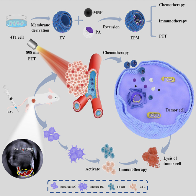
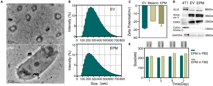
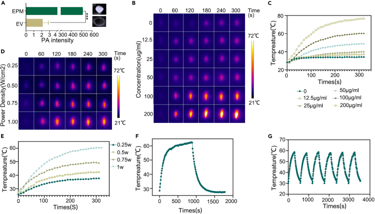
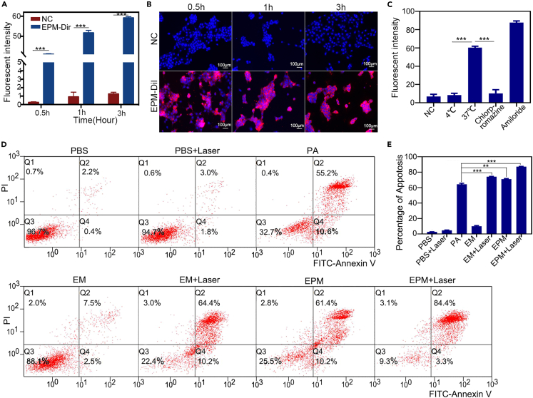
Tumor extracellular vesicles (EVs) demonstrate considerable promise for medication delivery and tumor targeting owing to their natural long-term blood circulation and tissue targeting capabilities. We extracted EVs from mouse breast cancer cell 4T1 using UV stimulation and differential centrifugation. To create a new nano-drug delivery system, the vesicle delivery system (EPM) loaded with melanin and paclitaxel albumin (PA), the collected EVs were repeatedly compressed on a 200 nm porous polycarbonate membrane with melanin and PA. Our findings suggest that EPM is readily absorbed by breast cancer and dendritic cells. EPM generates significant photoacoustic signals and photothermal effects when exposed to near-infrared light and can enhance the infiltration of CD8 + T cells in mouse tumor tissues. EPM is more cytotoxic than PA in in vivo and in vitro investigations. The efficacy of EPM in clinical transformation when paired with chemotherapy/photothermal/immunotherapy treatment is demonstrated in this study.
Overview
The claims and statistics of this paper haven’t been checked yet.
Run the check — free, no account needed. It reads the open-access text and checks the sources it cites.
Integrity signals
Registry
Cites
Statistics
Figures
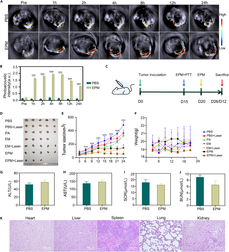
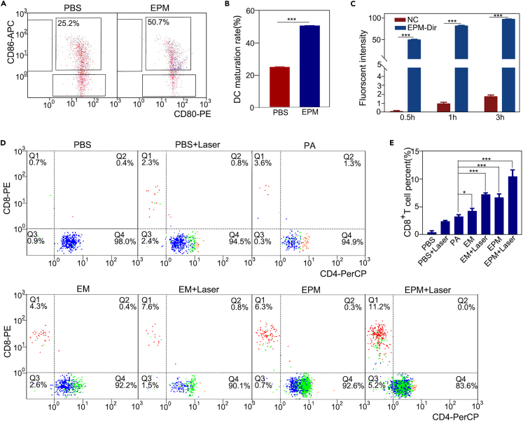
Verification
Claims the paper makes
The claims and statistics of this paper haven’t been checked yet.
Run the check — free, no account needed. It reads the open-access text and checks the sources it cites.
Claims other papers make about this one
Each is checked against this paper’s own text: does it say what the citing paper says it does?
The source backs part of the claim, not all of it — or backs it for a different population, condition or comparison.
In the citing paper
“Small molecule drugs, such as curcumin and paclitaxel, can be co-incubated with parent cells at various conditions for loading into EVs [217, 218].”
From Extracellular vesicles as nanotheranostic platforms for targeted neurological disorder interventions (2024) · cites this paper as [218]
Experimental — accuracy not yet measured
Self-check
Percentages, sums and reported differences re-computed from the paper’s own numbers.
The self-check needs the paper’s full text.
Citations
Cites
Combination strategies to maximize the benefits of cancer immunotherapy
Zhu, S., Zhang, T., Zheng, L., Liu, H., Song, W., Liu, D., Li, Z., & Pan, C.-X. (2021) · Journal of Hematology & Oncology · doi:10.1186/s13045-021-01164-5 · Cited by 654
Research works printed but not linked: 55
- Pasha, 2021, Understanding and overcoming tumor heterogeneity in metastatic breast cancer treatment, Nat. Cancer, 2, 680, 10.1038/s43018-021-00229-1 · DOI
- Riggio, 2021, The lingering mysteries of metastatic recurrence in breast cancer, Br. J. Cancer, 124, 13, 10.1038/s41416-020-01161-4 · DOI
- Sun, 2020, Cancer nanomedicine meets immunotherapy: opportunities and challenges, Acta Pharmacol. Sin., 41, 954, 10.1038/s41401-020-0448-9 · DOI
- Park, 2022, Evolution of drug delivery systems: From 1950 to 2020 and beyond, J. Control. Release, 342, 53, 10.1016/j.jconrel.2021.12.030 · DOI
- Bordeianu, 2023, The Usefulness of Nanotechnology in Improving the Prognosis of Lung Cancer, Biomedicines, 11, 705, 10.3390/biomedicines11030705 · DOI
- Elsharkasy, 2020, Extracellular vesicles as drug delivery systems: Why and how?, Adv. Drug Deliv. Rev., 159, 332, 10.1016/j.addr.2020.04.004 · DOI
- Tkach, 2018, Why the need and how to approach the functional diversity of extracellular vesicles, Philos. Trans. R. Soc. Lond. B Biol. Sci., 373, 20160479, 10.1098/rstb.2016.0479 · DOI
- Walker, 2019, Extracellular vesicle-based drug delivery systems for cancer treatment, Theranostics, 9, 8001, 10.7150/thno.37097 · DOI
- Bose, 2018, Tumor Cell-Derived Extracellular Vesicle-Coated Nanocarriers: An Efficient Theranostic Platform for the Cancer-Specific Delivery of Anti-miR-21 and Imaging Agents, ACS Nano, 12, 10817, 10.1021/acsnano.8b02587 · DOI
- Li, 2021, Cell-derived biomimetic nanocarriers for targeted cancer therapy: cell membranes and extracellular vesicles, Drug Deliv., 28, 1237, 10.1080/10717544.2021.1938757 · DOI
- Xu, 2022, Tumor-Derived Membrane Vesicles: A Promising Tool for Personalized Immunotherapy, Pharmaceuticals, 15, 876, 10.3390/ph15070876 · DOI
- Marar, 2021, Extracellular vesicles in immunomodulation and tumor progression, Nat. Immunol., 22, 560, 10.1038/s41590-021-00899-0 · DOI
- Li, 2021, Tumor-Derived Extracellular Vesicles: Their Role in Immune Cells and Immunotherapy, Int. J. Nanomedicine, 16, 5395, 10.2147/IJN.S313912 · DOI
- Xu, 2022, Cell Membrane-Derived Vesicle: A Novel Vehicle for Cancer Immunotherapy, Front. Immunol., 13, 923598, 10.3389/fimmu.2022.923598 · DOI
- Sun, 2016, Drug combination therapy increases successful drug repositioning, Drug Discov. Today, 21, 1189, 10.1016/j.drudis.2016.05.015 · DOI
- Ayoub, 2021, Editorial: Novel Combination Therapies for the Treatment of Solid Cancers, Front. Oncol., 11, 708943, 10.3389/fonc.2021.708943 · DOI
- Nam, 2018, Chemo-photothermal therapy combination elicits anti-tumor immunity against advanced metastatic cancer, Nat. Commun., 9, 1074, 10.1038/s41467-018-03473-9 · DOI · Paper page
- Li, 2020, Clinical development and potential of photothermal and photodynamic therapies for cancer, Nat. Rev. Clin. Oncol., 17, 657, 10.1038/s41571-020-0410-2 · DOI · Paper page
- Wang, 2021, Near-infrared inorganic nanomaterial-based nanosystems for photothermal therapy, Nanoscale, 13, 8751, 10.1039/D1NR00323B · DOI
- Cordero, 2020, Melanin, Curr. Biol., 30, R142, 10.1016/j.cub.2019.12.042 · DOI
- Liu, 2023, Highly localized, efficient, and rapid photothermal therapy using gold nanobipyramids for liver cancer cells triggered by femtosecond laser, Sci. Rep., 13, 3372, 10.1038/s41598-023-30526-x · DOI
- He, 2022, Thermal-Responsive MXene-DNA Hydrogel for Near-Infrared Light Triggered Localized Photothermal-Chemo Synergistic Cancer Therapy, Small, 18, e2200263, 10.1002/smll.202200263 · DOI
- Chang, 2022, Organic single molecule based nano-platform for NIR-II imaging and chemo-photothermal synergistic treatment of tumor, Biomaterials, 287, 121670, 10.1016/j.biomaterials.2022.121670 · DOI
- Herrmann, 2021, Extracellular vesicles as a next-generation drug delivery platform, Nat. Nanotechnol., 16, 748, 10.1038/s41565-021-00931-2 · DOI · Paper page
- Meng, 2020, Prospects and challenges of extracellular vesicle-based drug delivery system: considering cell source, Drug Deliv., 27, 585, 10.1080/10717544.2020.1748758 · DOI · Paper page
- Igami, 2020, Characterization and function of medium and large extracellular vesicles from plasma and urine by surface antigens and Annexin V, PeerJ Anal. Chem., 2, e4, 10.7717/peerj-achem.4 · DOI
- Zhao, 2021, Apoptotic body–mediated intercellular delivery for enhanced drug penetration and whole tumor destruction, Sci. Adv., 7, eabg0880, 10.1126/sciadv.abg0880 · DOI
- Wang, 2021, Delivering Antisense Oligonucleotides across the Blood-Brain Barrier by Tumor Cell-Derived Small Apoptotic Bodies, Adv. Sci., 8, 2004929, 10.1002/advs.202004929 · DOI
- Yu, 2019, Sequentially responsive biomimetic nanoparticles with optimal size in combination with checkpoint blockade for cascade synergetic treatment of breast cancer and lung metastasis, Biomaterials, 217, 119309, 10.1016/j.biomaterials.2019.119309 · DOI
- Gong, 2020, Macrophage-cancer hybrid membrane-coated nanoparticles for targeting lung metastasis in breast cancer therapy, J. Nanobiotechnology, 18, 92, 10.1186/s12951-020-00649-8 · DOI
- Zhou, 2017, Reprogramming Malignant Cancer Cells toward a Benign Phenotype following Exposure to Human Embryonic Stem Cell Microenvironment, PLoS One, 12, e0169899, 10.1371/journal.pone.0169899 · DOI
- Wortzel, 2019, Exosome-Mediated Metastasis: Communication from a Distance, Dev. Cell, 49, 347, 10.1016/j.devcel.2019.04.011 · DOI
- Wu, 2021, Extracellular vesicles and immunogenic stress in cancer, Cell Death Dis., 12, 894, 10.1038/s41419-021-04171-z · DOI
- Hanahan, 2022, Hallmarks of Cancer: New Dimensions, Cancer Discov., 12, 31, 10.1158/2159-8290.CD-21-1059 · DOI · Paper page
- Forster, 2017, A review of the development of tumor vasculature and its effects on the tumor microenvironment, Hypoxia, 5, 21, 10.2147/HP.S133231 · DOI
- Majidpoor, 2021, Angiogenesis as a hallmark of solid tumors - clinical perspectives, Cell. Oncol., 44, 715, 10.1007/s13402-021-00602-3 · DOI
- Ma, 2016, Reversing drug resistance of soft tumor-repopulating cells by tumor cell-derived chemotherapeutic microparticles, Cell Res., 26, 713, 10.1038/cr.2016.53 · DOI
- Wu, 2019, Surface-Layer Protein-Enhanced Immunotherapy Based on Cell Membrane-Coated Nanoparticles for the Effective Inhibition of Tumor Growth and Metastasis, ACS Appl. Mater. Interfaces, 11, 9850, 10.1021/acsami.9b00294 · DOI
- Wang, 2020, Engineering antigen as photosensitiser nanocarrier to facilitate ROS triggered immune cascade for photodynamic immunotherapy, Biomaterials, 244, 119964, 10.1016/j.biomaterials.2020.119964 · DOI
- Zheng, 2020, In Vivo Monocyte/Macrophage-Hitchhiked Intratumoral Accumulation of Nanomedicines for Enhanced Tumor Therapy, J. Am. Chem. Soc., 142, 382, 10.1021/jacs.9b11046 · DOI
- Barenholz-Cohen, 2020, Lung mechanics modifications facilitating metastasis are mediated in part by breast cancer-derived extracellular vesicles, Int. J. Cancer, 147, 2924, 10.1002/ijc.33229 · DOI
- Mavridi-Printezi, 2023, Functionalization of and through Melanin: Strategies and Bio-Applications, Int. J. Mol. Sci., 24, 9689, 10.3390/ijms24119689 · DOI
- Jiang, 2019, Erythrocyte-cancer hybrid membrane-camouflaged melanin nanoparticles for enhancing photothermal therapy efficacy in tumors, Biomaterials, 192, 292, 10.1016/j.biomaterials.2018.11.021 · DOI
- Jiang, 2017, Red blood cell membrane-camouflaged melanin nanoparticles for enhanced photothermal therapy, Biomaterials, 143, 29, 10.1016/j.biomaterials.2017.07.027 · DOI
- Gujrati, 2019, Bioengineered bacterial vesicles as biological nano-heaters for optoacoustic imaging, Nat. Commun., 10, 1114, 10.1038/s41467-019-09034-y · DOI
- Li, 2023, Cascaded microfluidic circuits for pulsatile filtration of extracellular vesicles from whole blood for early cancer diagnosis, Sci. Adv., 9, eade2819, 10.1126/sciadv.ade2819 · DOI
- Speiser, 2023, CD4+ T cells in cancer, Nat. Cancer, 4, 317, 10.1038/s43018-023-00521-2 · DOI
- Waldman, 2020, A guide to cancer immunotherapy: from T cell basic science to clinical practice, Nat. Rev. Immunol., 20, 651, 10.1038/s41577-020-0306-5 · DOI · Paper page
- Jhunjhunwala, 2021, Antigen presentation in cancer: insights into tumour immunogenicity and immune evasion, Nat. Rev. Cancer, 21, 298, 10.1038/s41568-021-00339-z · DOI
- Zou, 2022, Immune regulation in the tumor microenvironment and its relevance in cancer therapy, Cell. Mol. Immunol., 19, 1, 10.1038/s41423-021-00738-0 · DOI
- Tie, 2022, Immunosuppressive cells in cancer: mechanisms and potential therapeutic targets, J. Hematol. Oncol., 15, 61, 10.1186/s13045-022-01282-8 · DOI · Paper page
- Buzas, 2023, The roles of extracellular vesicles in the immune system, Nat. Rev. Immunol., 23, 236, 10.1038/s41577-022-00763-8 · DOI
- Li, 2022, Antigen Capture and Immune Modulation by Bacterial Outer Membrane Vesicles as In Situ Vaccine for Cancer Immunotherapy Post-Photothermal Therapy, Small, 18, e2107461, 10.1002/smll.202107461 · DOI
- Guo, 2022, Engineered nanomaterials for synergistic photo-immunotherapy, Biomaterials, 282, 121425, 10.1016/j.biomaterials.2022.121425 · DOI
- Shang, 2020, Nanomedicine-based tumor photothermal therapy synergized immunotherapy, Biomater. Sci., 8, 5241, 10.1039/D0BM01158D · DOI
Cited by
Sorted by citations
Extracellular vesicles as nanotheranostic platforms for targeted neurological disorder interventions
Choi, H. K., Chen, M., Goldston, L. L., & Lee, K.-B. (2024) · Nano Convergence · doi:10.1186/s40580-024-00426-5 · Cited by 26
Metadata
- Authors
- Bi, Y., Chen, J., Li, Q., Li, Y., Zhang, L., Zhida, L., Yuan, F., & Zhang, R.
- Affiliations
- Shanxi Medical University (CN)
- Corresponding author
- Zhang, Ruiping
- Venue
- iScience
- Publisher
- Elsevier BV
- Published
- 2024-02
- Volume
- 27
- Issue
- 2
- Pages
- 108833
- ISSN
- 2589-0042 (ISSN-L)
- PMC
- PMC10850737
- OpenAlex
- W4390661715
- Cited by
- 24
- References
- 1
- Citations by year
- 2026: 3 · 2025: 17 · 2024: 4
- FWCI
- 3.27
- Type
- journal article
- Language
- en
- Open access
- gold
- doi.org
- Licence
- Locations
- iScience (publishedVersion, cc-by-nc-nd)
- PubMed Central (publishedVersion, cc-by-nc-nd)
- PubMed (publishedVersion)
- DOAJ (DOAJ: Directory of Open Access Journals) (submittedVersion)
- Funders
- Shaanxi Key Science and Technology Innovation Team Project (202204051001024)
- National Key Research and Development Program of China (202104010910010, 2023YFC3402800, 2021YZ17)
- National Natural Science Foundation of China (82002606, 82071987, 82120108016)
- Topics
- Life Sciences › Biochemistry, Genetics and Molecular Biology › Molecular Biology › Extracellular vesicles in disease
- Physical Sciences › Engineering › Biomedical Engineering › Nanoplatforms for cancer theranostics
- Physical Sciences › Materials Science › Biomaterials › Nanoparticle-Based Drug Delivery
- Keywords
- tumor-derived extracellular vesicles, melanin, photothermal therapy, near-infrared light, photoacoustic imaging, combination cancer therapy, chemotherapy, immunotherapy, CD8+ T cell infiltration, breast cancer, 4T1 cells, dendritic cells
About these checks
Registry, retraction, Cites and statistics results are computed, not judged, with one exception: a printed reference that no index lists is read once by a model before it counts as a citation error. Claims are extracted by a model and kept only when their quote appears verbatim in the text; each cited source is then checked, with a model’s help, for whether it says what the claim relies on. Those verdicts can be wrong — read the claim’s quote and the cited source before relying on one.
Statistics and arithmetic are recomputed, not judged.
Confidence bands are shown only for fields calibrated on our annotated test set. None is calibrated yet, so no band is shown.
What counts as a claim ›How we measure confidence ›How we check this ›
Are you an author, or do you see a problem with this page? Request a review
