

The DOI matches its Crossref record; 137 of the 146 references it cites were found in a registry or an index.
The Liver Tumor Segmentation Benchmark (LiTS)
Open accessAbstract
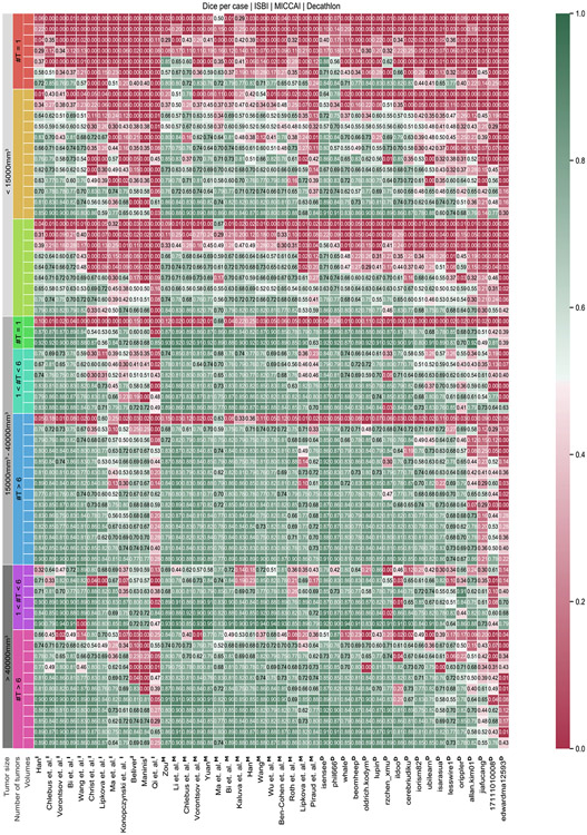
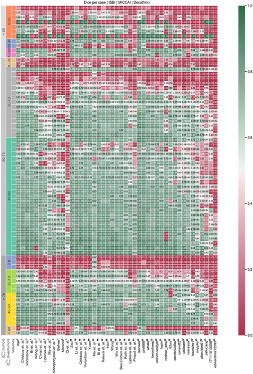
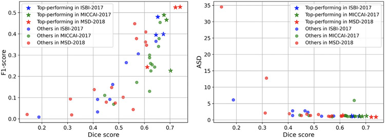
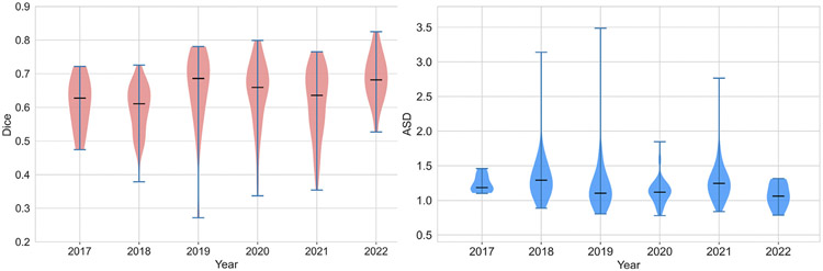
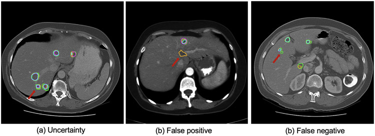
In this work, we report the set-up and results of the Liver Tumor Segmentation Benchmark (LiTS), which was organized in conjunction with the IEEE International Symposium on Biomedical Imaging (ISBI) 2017 and the International Conferences on Medical Image Computing and Computer-Assisted Intervention (MICCAI) 2017 and 2018. The image dataset is diverse and contains primary and secondary tumors with varied sizes and appearances with various lesion-to-background levels (hyper-/hypo-dense), created in collaboration with seven hospitals and research institutions. Seventy-five submitted liver and liver tumor segmentation algorithms were trained on a set of 131 computed tomography (CT) volumes and were tested on 70 unseen test images acquired from different patients. We found that not a single algorithm performed best for both liver and liver tumors in the three events. The best liver segmentation algorithm achieved a Dice score of 0.963, whereas, for tumor segmentation, the best algorithms achieved Dices scores of 0.674 (ISBI 2017), 0.702 (MICCAI 2017), and 0.739 (MICCAI 2018). Retrospectively, we performed additional analysis on liver tumor detection and revealed that not all top-performing segmentation algorithms worked well for tumor detection. The best liver tumor detection method achieved a lesion-wise recall of 0.458 (ISBI 2017), 0.515 (MICCAI 2017), and 0.554 (MICCAI 2018), indicating the need for further research. LiTS remains an active benchmark and resource for research, e.g., contributing the liver-related segmentation tasks in http://medicaldecathlon.com/. In addition, both data and online evaluation are accessible via https://competitions.codalab.org/competitions/17094.
Overview
The claims and statistics of this paper haven’t been checked yet.
Run the check — free, no account needed. It reads the open-access text and checks the sources it cites.
Integrity signals
Registry
Cites
Statistics
Figures
Verification
Claims the paper makes
The claims and statistics of this paper haven’t been checked yet.
Run the check — free, no account needed. It reads the open-access text and checks the sources it cites.
Claims other papers make about this one
Each is checked against this paper’s own text: does it say what the citing paper says it does?
The cited source says what the claim attributes to it.
In the citing paper
“different lesion appearances and annotation targets have been reported across tumor benchmarks [3,8,23]”
From Correction-aware interactive 3D tumor segmentation with sparse and revisable prompts (2026) · cites this paper as [3]
The cited source says what the claim attributes to it.
In the citing paper
“Tumor segmentation is less stable because lesions are not tied to a fixed anatomical shape or location and may be small, heterogeneous, or weakly contrasted [3,8,23].”
From Correction-aware interactive 3D tumor segmentation with sparse and revisable prompts (2026) · cites this paper as [3]
The check wasn’t confident either way, so it gives no verdict — never disputed.
In the citing paper
“The source of the OrganMNIST3D is the same as that of the Organ{A,C,S}MNIST. Instead of 2D images, we directly use the 3D bounding boxes and process the images into 28 × 28 × 28 to perform multi-class classification of 11 body organs.”
From MedMNIST v2 - A large-scale lightweight benchmark for 2D and 3D biomedical image classification (2023) · cites this paper as [30]
Experimental — accuracy not yet measured
Self-check
Percentages, sums and reported differences re-computed from the paper’s own numbers.
The self-check needs the paper’s full text.
Citations
Cites
U-Net: Convolutional Networks for Biomedical Image Segmentation
Ronneberger, O., Fischer, P., & Brox, T. (2015) · Lecture notes in computer science · doi:10.1007/978-3-319-24574-4_28 · Cited by 99,404
New response evaluation criteria in solid tumours: revised RECIST guideline (version 1.1)
Eisenhauer, E. A., Therasse, P., Bogaerts, J., Schwartz, L. H., Sargent, D., Ford, R., Dancey, J., Arbuck, S., Gwyther, S., Mooney, M., et al. (2009) · European Journal of Cancer · doi:10.1016/j.ejca.2008.10.026 · Cited by 29,534
Kass, M., Witkin, A., & Terzopoulos, D. (1988) · International Journal of Computer Vision · doi:10.1007/bf00133570 · Cited by 17,330
A survey on deep learning in medical image analysis
Litjens, G., Kooi, T., Bejnordi, B. E., Setio, A. A. A., Ciompi, F., Ghafoorian, M., van der Laak, J. A. W. M., van Ginneken, B., & Sánchez, C. I. (2017) · Medical Image Analysis · doi:10.1016/j.media.2017.07.005 · Cited by 15,332
Fronts propagating with curvature-dependent speed: Algorithms based on Hamilton-Jacobi formulations
Osher, S., & Sethian, J. A. (1988) · Journal of Computational Physics · doi:10.1016/0021-9991(88)90002-2 · Cited by 14,196
V-Net: Fully Convolutional Neural Networks for Volumetric Medical Image Segmentation
Milletari, F., Navab, N., & Ahmadi, S.-A. (2016) · 2016 Fourth International Conference on 3D Vision (3DV) · doi:10.1109/3dv.2016.79 · Cited by 11,931
Fully convolutional networks for semantic segmentation
Shelhamer, E., Long, J., & Darrell, T. (2017) · IEEE Transactions on Pattern Analysis and Machine Intelligence · doi:10.1109/tpami.2016.2572683 · Cited by 11,898
nnU-Net: a self-configuring method for deep learning-based biomedical image segmentation
Isensee, F., Jaeger, P. F., Kohl, S. A. A., Petersen, J., & Maier-Hein, K. H. (2021) · Nature Methods · doi:10.1038/s41592-020-01008-z · Cited by 10,477
Self-citationYushkevich, P. A., Piven, J., Hazlett, H. C., Smith, R. G., Ho, S., Gee, J. C., & Gerig, G. (2006) · NeuroImage · doi:10.1016/j.neuroimage.2006.01.015 · Cited by 8,868
Active Shape Models-Their Training and Application
Cootes, T. F., Taylor, C. J., Cooper, D. H., & Graham, J. (1995) · Computer Vision and Image Understanding · doi:10.1006/cviu.1995.1004 · Cited by 7,190
Fast approximate energy minimization via graph cuts
Boykov, Y., Veksler, O., & Zabih, R. (2001) · IEEE Transactions on Pattern Analysis and Machine Intelligence · doi:10.1109/34.969114 · Cited by 6,988
The Cancer Imaging Archive (TCIA): Maintaining and Operating a Public Information Repository
Clark, K., Vendt, B., Smith, K., Freymann, J., Kirby, J., Koppel, P., Moore, S., Phillips, S., Maffitt, D., Pringle, M., et al. (2013) · Journal of Digital Imaging · doi:10.1007/s10278-013-9622-7 · Cited by 4,599
Learning Deconvolution Network for Semantic Segmentation
Noh, H., Hong, S., & Han, B. (2015) · 2015 IEEE International Conference on Computer Vision (ICCV) · doi:10.1109/iccv.2015.178 · Cited by 4,422
The future of digital health with federated learning
Rieke, N., Hancox, J., Li, W., Milletarì, F., Roth, H. R., Albarqouni, S., Bakas, S., Galtier, M. N., Landman, B. A., Maier-Hein, K., et al. (2020) · npj Digital Medicine · doi:10.1038/s41746-020-00323-1 · Cited by 4,015 · cited 1×
“Another recent and intriguing use case is the research direction of federated learning, where the multi-institutional nature of the LiTS benchmark dataset could further contribute to federated learning simulations studies and benchmarks (Sheller et al., 2018, 2020; Rieke et al., 2020; Pati et al., 2021).”
BackgroundSelf-citationCodella, N. C. F., Gutman, D., Celebi, M. E., Helba, B., Marchetti, M. A., Dusza, S. W., Kalloo, A., Liopyris, K., Mishra, N., Kittler, H., et al. (2018) · 2018 IEEE 15th International Symposium on Biomedical Imaging (ISBI 2018) · doi:10.1109/isbi.2018.8363547 · Cited by 2,620
H-DenseUNet: Hybrid Densely Connected UNet for Liver and Tumor Segmentation From CT Volumes
Li, X., Chen, H., Qi, X., Dou, Q., Fu, C., & Heng, P. (2018) · IEEE Transactions on Medical Imaging · doi:10.1109/tmi.2018.2845918 · Cited by 2,530
Self-citationInternational Encyclopedia of Statistical Science
Lovric, M. (2011) · Springer eBooks · doi:10.1007/978-3-642-04898-2 · Cited by 1,696
A ‘missing’ family of classical orthogonal polynomials
Vinet, L., & Zhedanov, A. S. (2011) · Journal of Physics A Mathematical and Theoretical · doi:10.1088/1751-8113/44/8/085201 · Cited by 1,649
Sheller, M. J., Edwards, B., Reina, G. A., Martin, J., Pati, S., Kotrotsou, A., Milchenko, M., Xu, W., Marcus, D., Colen, R. R., et al. (2020) · Scientific Reports · doi:10.1038/s41598-020-69250-1 · Cited by 1,646
Self-citationAlbain, K. S., Swann, R. S., Rusch, V. W., Turrisi, A. T., Shepherd, F. A., Smith, C. J., Chen, Y., Livingston, R. B., Feins, R. H., Gandara, D. R., et al. (2009) · The Lancet · doi:10.1016/s0140-6736(09)60737-6 · Cited by 1,472
The Medical Segmentation Decathlon
Antonelli, M., Reinke, A., Bakas, S., Farahani, K., Kopp-Schneider, A., Landman, B. A., Litjens, G., Menze, B., Ronneberger, O., Summers, R. M., et al. (2022) · Nature Communications · doi:10.1038/s41467-022-30695-9 · Cited by 1,384
Self-citationThe burden of liver disease in Europe: A review of available epidemiological data
Blachier, M., Leleu, H., Peck‐Radosavljevic, M., Valla, D. C., & Roudot‐Thoraval, F. (2013) · Journal of Hepatology · doi:10.1016/j.jhep.2012.12.005 · Cited by 1,340
Clemente, C. G., Mihm, M. C., Bufalino, R., Zurrida, S., Collini, P., & Cascinelli, N. (1996) · Cancer · doi:10.1002/(sici)1097-0142(19960401)77:7 · Cited by 1,194
Comparison and evaluation of methods for liver segmentation from CT datasets
Heimann, T., van Ginneken, B., Styner, M. A., Arzhaeva, Y., Aurich, V., Bauer, C., Beck, A., Becker, C., Beichel, R., Bekes, G., et al. (2009) · IEEE Transactions on Medical Imaging · doi:10.1109/tmi.2009.2013851 · Cited by 1,135
The Corsini encyclopedia of psychology
(2010) · Choice Reviews Online · doi:10.5860/choice.47-6008 · Cited by 1,082
Hepatic artery embolization in 120 patients with unresectable hepatoma.
Yamada, R., Sato, M., Kawabata, M., Nakatsuka, H., Nakamura, K., & Takashima, S. (1983) · Radiology · doi:10.1148/radiology.148.2.6306721 · Cited by 994
Livraghi, T., Giorgio, A., Marin, G., Salmi, A., de Sio, I., Bolondi, L., Pompili, M., Brunello, F., Lazzaroni, S., & Torzilli, G. (1995) · Radiology · doi:10.1148/radiology.197.1.7568806 · Cited by 934
CHAOS Challenge - combined (CT-MR) healthy abdominal organ segmentation
Kavur, A. E., Gezer, N. S., BARIŞ, M. M., Aslan, S., Conze, P.-H., Groza, V., Pham, D. D., Chatterjee, S., Ernst, P., Özkan, S., et al. (2020) · Medical Image Analysis · doi:10.1016/j.media.2020.101950 · Cited by 893
Self-citationPercutaneous RF interstitial thermal ablation in the treatment of hepatic cancer.
Rossi, S. M., Stasi, M., Buscarini, E., Quaretti, P., Garbagnati, F., Squassante, L., Paties, C. T., Silverman, D., & Buscarini, L. (1996) · American Journal of Roentgenology · doi:10.2214/ajr.167.3.8751696 · Cited by 759
Kubota, K., Makuuchi, M., Kusaka, K., Kobayashi, T., Miki, K., Hasegawa, K., Harihara, Y., & Takayama, T. (1997) · Hepatology · doi:10.1002/hep.510260514 · Cited by 736
Christ, P. F., Elshaer, M. E. A., Ettlinger, F., Tatavarty, S., Bickel, M., Bilic, P., Rempfler, M., Armbruster, M., Hofmann, F., D’Anastasi, M., et al. (2016) · Lecture Notes in Computer Science · doi:10.1007/978-3-319-46723-8_48 · Cited by 625
Self-citationSheller, M. J., Reina, G. A., Edwards, B., Martin, J., & Bakas, S. (2019) · Lecture Notes in Computer Science · doi:10.1007/978-3-030-11723-8_9 · Cited by 609
Self-citationRey, D., & Neuhäuser, M. (2011) · International Encyclopedia of Statistical Science · doi:10.1007/978-3-642-04898-2_616 · Cited by 452
Zhou, Z., Sodha, V., Pang, J., Gotway, M. B., & Liang, J. (2020) · Medical Image Analysis · doi:10.1016/j.media.2020.101840 · Cited by 375
Causality matters in medical imaging
Castro, D. C., Walker, I., & Glocker, B. (2020) · Nature Communications · doi:10.1038/s41467-020-17478-w · Cited by 364
3D Deeply Supervised Network for Automatic Liver Segmentation from CT Volumes
Dou, Q., Chen, H., Jin, Y., Yu, L., Qin, J., & Heng, P.-A. (2016) · Lecture Notes in Computer Science · doi:10.1007/978-3-319-46723-8_18 · Cited by 344
Self-citationMachine learning analysis of whole mouse brain vasculature
Todorov, M. I., Paetzold, J. C., Schoppe, O., Tetteh, G., Shit, S., Efremov, V., Todorov-Völgyi, K., Düring, M., Dichgans, M., Piraud, M., et al. (2020) · Nature Methods · doi:10.1038/s41592-020-0792-1 · Cited by 338
Self-citationConstruction of an abdominal probabilistic atlas and its application in segmentation
Park, H., Bland, P. H., & Meyer, C. (2003) · IEEE Transactions on Medical Imaging · doi:10.1109/tmi.2003.809139 · Cited by 330
Soler, L., Delingette, H., Malandain, G., Johan Montagnat, A., Ayache, N., Koehl, C., Dourthe, O., Malassagne, B., Smith, M., Mutter, D., et al. (2001) · Computer Aided Surgery · doi:10.1002/igs.1016 · Cited by 322
Self-citationCausality matters in medical imaging
Castro, D. C., Walker, I. D., & Glocker, B. (2020) · RePEc: Research Papers in Economics · Cited by 288
Kuhnigk, J., Dicken, V., Bornemann, L., Bakai, A., Wormanns, D., Krass, S., & Peitgen, H. (2006) · IEEE Transactions on Medical Imaging · doi:10.1109/tmi.2006.871547 · Cited by 287
Automatic 3D liver location and segmentation via convolutional neural network and graph cut
Lu, F., Wu, F., Hu, P., Peng, Z., & Kong, D. (2017) · International Journal of Computer Assisted Radiology and Surgery · doi:10.1007/s11548-016-1467-3 · Cited by 284 · cited 1×
“One approach introduced a 11-layer 3D CNN and graph cut for segmentation refinement[90] and Christ et al.”
MethodChlebus, G., Schenk, A., Moltz, J. H., van Ginneken, B., Hahn, H. K., & Meine, H. (2018) · Scientific Reports · doi:10.1038/s41598-018-33860-7 · Cited by 254
Self-citationFully Convolutional Network for Liver Segmentation and Lesions Detection
Ben-Cohen, A., Diamant, I., Klang, E., Amitai, M. M., & Greenspan, H. K. (2016) · Lecture notes in computer science · doi:10.1007/978-3-319-46976-8_9 · Cited by 241
Self-citationMulti-Organ Segmentation Over Partially Labeled Datasets With Multi-Scale Feature Abstraction
Fang, X., & Yan, P. (2020) · IEEE Transactions on Medical Imaging · doi:10.1109/tmi.2020.3001036 · Cited by 229
3D Segmentation in the Clinic: A Grand Challenge
van Ginneken, B., Heimann, T., & Styner, M. A. (2007) · Cited by 212
Fully convolutional network ensembles for white matter hyperintensities segmentation in MR images
Li, H., Jiang, G., Zhang, J., Wang, R., Wang, Z., Zheng, W., & Menze, B. H. (2018) · NeuroImage · doi:10.1016/j.neuroimage.2018.07.005 · Cited by 209
Self-citationAutomatic 3D liver segmentation based on deep learning and globally optimized surface evolution
Hu, P., Wu, F., Peng, J., Liang, P., & Kong, D.-X. (2016) · Physics in Medicine and Biology · doi:10.1088/1361-6560/61/24/8676 · Cited by 205
Jagannath, S., Velasquez, W. S., Tucker, S. L., Fuller, L. M., McLaughlin, P. W., Manning, J. T., North, L. B., & Cabanillas, F. (1986) · Journal of Clinical Oncology · doi:10.1200/jco.1986.4.6.859 · Cited by 186
Automatic liver segmentation technique for three-dimensional visualization of CT data.
Gao, L., Heath, D. G., Kuszyk, B. S., & Fishman, E. K. (1996) · Radiology · doi:10.1148/radiology.201.2.8888223 · Cited by 177
Efficient Semiautomatic Segmentation of 3D Objects in Medical Images
Schenk, A., Prause, G. P. M., & Peitgen, H. (2000) · Lecture notes in computer science · doi:10.1007/978-3-540-40899-4_19 · Cited by 176
Self-citationShape Constrained Automatic Segmentation of the Liver based on a Heuristic Intensity Model
Kainmüller, D., Lange, T., & Lamecker, H. (2007) · Cited by 173
Massoptier, L., & Casciaro, S. (2008) · European Radiology · doi:10.1007/s00330-008-0924-y · Cited by 173
Jiménez–del–Toro, O. A., Müller, H., Krenn, M., Gruenberg, K., Taha, A. A., Winterstein, M., Eggel, I., Foncubierta–Rodríguez, A., Göksel, O., András, J., et al. (2016) · IEEE Transactions on Medical Imaging · doi:10.1109/tmi.2016.2578680 · Cited by 165
Self-citationChallenges and Methodologies of Fully Automatic Whole Heart Segmentation: A Review
Zhuang, X. (2013) · Journal of Healthcare Engineering · doi:10.1260/2040-2295.4.3.371 · Cited by 159
Shiina, S., Sato, K., Tateishi, R., Shimizu, M., Ohama, H., Hatanaka, T., Takawa, M., Nagamatsu, H., & Imai, Y. (2018) · Canadian Journal of Gastroenterology and Hepatology · doi:10.1155/2018/4756147 · Cited by 157
Moghbel, M., Mashohor, S., Mahmud, R., & Saripan, M. I. (2017) · Artificial Intelligence Review · doi:10.1007/s10462-017-9550-x · Cited by 153
Haghighi, F., Taher, M. R. H., Zhou, Z., Gotway, M. B., & Liang, J. (2021) · IEEE Transactions on Medical Imaging · doi:10.1109/tmi.2021.3060634 · Cited by 151
Moltz, J. H., Bornemann, L., Kuhnigk, J., Dicken, V., Peitgen, E., Meier, S., Bolte, H., Fabel, M., Bauknecht, H.-C., Hittinger, M., et al. (2009) · IEEE Journal of Selected Topics in Signal Processing · doi:10.1109/jstsp.2008.2011107 · Cited by 144
Self-citationAutomatic Liver Segmentation Using a Statistical Shape Model With Optimal Surface Detection
Zhang, X., Tian, J., Deng, K., Wu, Y., & Li, X. (2010) · IEEE Transactions on Biomedical Engineering · doi:10.1109/tbme.2010.2056369 · Cited by 142
Self-citationHierarchical, learning-based automatic liver segmentation
Ling, H., Zhou, S., Zheng, Y., Georgescu, B., Suehling, M., & Comaniciu, D. (2008) · IEEE Computer Society Conference on Computer Vision and Pattern Recognition/Proceedings - IEEE Computer Society Conference on Computer Vision and Pattern Recognition/Proceedings · doi:10.1109/cvpr.2008.4587393 · Cited by 140
Self-citationAutomatic liver segmentation for volume measurement in CT Images
Lim, S., Jeong, Y., & Ho, Y. (2005) · Journal of Visual Communication and Image Representation · doi:10.1016/j.jvcir.2005.07.001 · Cited by 139
Tumor Burden Analysis on Computed Tomography by Automated Liver and Tumor Segmentation
Linguraru, M. G., Richbourg, W. J., Liu, J., Watt, J. M., Pamulapati, V., Wang, S., & Summers, R. M. (2012) · IEEE Transactions on Medical Imaging · doi:10.1109/tmi.2012.2211887 · Cited by 136
BIAS: Transparent reporting of biomedical image analysis challenges
Maier‐Hein, L., Reinke, A., Martel, A. L., Tal, A., Matthias, E., Allan, H., Jannin, P., Henning, M., Sinan, O., Sáez-Rodríguez, J., et al. (2020) · Veřejné služby Informačního systému (Masarykiana Brunensis Universitas) · Cited by 134
The Federated Tumor Segmentation (FeTS) ChallengePreprint
Pati, S., Baid, U., Zenk, M., Edwards, B., Sheller, M., Reina, G. A., Foley, P., Gruzdev, A., Martin, J., Albarqouni, S., et al. (2021) · arXiv (Cornell University) · doi:10.48550/arxiv.2105.05874 · Cited by 132
Self-citationCholangiocarcinoma of the extrahepatic bile ducts
Jarnagin, W. R. (2000) · Seminars in Surgical Oncology · doi:10.1002/1098-2388(200009)19:2 · Cited by 120
Segmentation of the Liver using a 3D Statistical Shape Model
Lamecker, H., Lange, T., & Seebaß, M. (2004) · Cited by 112
Medical Image Computing and Computer-Assisted Intervention – MICCAI 2007
Ayache, N., Sebastien Ourselin, A., & Maeder, A. (2007) · Lecture notes in computer science · doi:10.1007/978-3-540-75759-7 · Cited by 110
Efficient multi-atlas abdominal segmentation on clinically acquired CT with SIMPLE context learning
Xu, Z., Burke, R. P., Lee, C. P., Baucom, R. B., Poulose, B. K., Abramson, R. G., & Landman, B. A. (2015) · Medical Image Analysis · doi:10.1016/j.media.2015.05.009 · Cited by 105
Deep learning-enabled multi-organ segmentation in whole-body mouse scans
Schoppe, O., Pan, C., Coronel, J., Mai, H., Rong, Z., Todorov, M. I., Müskes, A., Navarro, F., Li, H., Ertürk, A., et al. (2020) · Nature Communications · doi:10.1038/s41467-020-19449-7 · Cited by 104
Self-citationHeimann, T., Münzing, S., Meinzer, H., & Ivo Wolf, A. (2007) · Lecture notes in computer science · doi:10.1007/978-3-540-73273-0_1 · Cited by 103
Automatic Liver Segmentation on Volumetric CT Images Using Supervoxel-Based Graph Cuts
Wu, W., Zhou, Z., Wu, S., & Zhang, Y. (2016) · Computational and Mathematical Methods in Medicine · doi:10.1155/2016/9093721 · Cited by 100
Disentangling Human Error from the Ground Truth in Segmentation of Medical Images
Zhang, L., Tanno, R., Xu, M., Jin, C., Jacob, J., Ciccarelli, O., Barkhof, F., & Alexander, D. C. (2020) · Amsterdam UMC (VU Amsterdam) - Institutional Repository · doi:10.48550/arxiv.2007.15963 · Cited by 100
Self-citationYan, K., Cai, J., Zheng, Y., Harrison, A. P., Jin, D., Tang, Y., Tang, Y., Huang, L., Xiao, J. Z., & Lü, L. (2020) · IEEE Transactions on Medical Imaging · doi:10.1109/tmi.2020.3047598 · Cited by 92
Methods and open-source toolkit for analyzing and visualizing challenge results
Wiesenfarth, M., Reinke, A., Landman, B. A., Eisenmann, M., Saiz, L. A., Cardoso, M. J., Maier-Hein, L., & Kopp‐Schneider, A. (2021) · Scientific Reports · doi:10.1038/s41598-021-82017-6 · Cited by 89
Fully Automatic Liver Segmentation through Graph-Cut Technique
Massoptier, L., & Casciaro, S. (2007) · Conference proceedings · doi:10.1109/iembs.2007.4353524 · Cited by 84
Heimann, T., Wolf, I., & Meinzer, H. (2006) · Lecture notes in computer science · doi:10.1007/11866763_6 · Cited by 83
Häme, Y., & Pollari, M. (2011) · Medical Image Analysis · doi:10.1016/j.media.2011.06.006 · Cited by 79
Haghighi, F., Taher, M. R. H., Zhou, Z., Gotway, M. B., & Liang, J. (2020) · Lecture notes in computer science · doi:10.1007/978-3-030-59710-8_14 · Cited by 78
Bauknecht, H.-C., Romano, V. C., Rogalla, P., Klingebiel, R., Wolf, C., Bornemann, L., Hamm, B., & Hein, P. A. (2009) · Investigative Radiology · doi:10.1097/rli.0b013e3181c02ed5 · Cited by 77
Machine Learning with Multi-Site Imaging Data: An Empirical Study on the Impact of Scanner Effects
Glocker, B., Robinson, R., Castro, D. C., Dou, Q., & Konukoglu, E. (2019) · arXiv · doi:10.48550/arxiv.1910.04597 · Cited by 74
Self-citationClinical outcomes of primary arterial embolization in severe hepatic trauma: A systematic review
Virdis, F., Reccia, I., Saverio, S. D., Tugnoli, G., Kwan, S. H., Kumar, J., Atzeni, J., & Podda, M. (2018) · Diagnostic and Interventional Imaging · doi:10.1016/j.diii.2018.10.004 · Cited by 71
Sho, T., Oost, E., Shimizu, A., Watanabe, H., & Nawano, S. (2013) · Medical Image Analysis · doi:10.1016/j.media.2013.10.003 · Cited by 70
Hoogi, A., Subramaniam, A., Veerapaneni, R., & Rubin, D. L. (2016) · IEEE Transactions on Medical Imaging · doi:10.1109/tmi.2016.2628084 · Cited by 69
Segmentation of Liver Metastases in CT Scans by Adaptive Thresholding and Morphological Processing
Moltz, J. H., Bornemann, L., Dicken, V., & Peitgen, H. (2008) · doi:10.54294/msg94u · Cited by 69
Self-citationConze, P.-H., Noblet, V., Rousseau, F., Heitz, F., Blasi, V. D., Memeo, R., & Pessaux, P. (2016) · International Journal of Computer Assisted Radiology and Surgery · doi:10.1007/s11548-016-1493-1 · Cited by 66
Zhou, B., Augenfeld, Z., Chapiro, J., Zhou, S. K., Liu, C., & Duncan, J. S. (2021) · Medical Image Analysis · doi:10.1016/j.media.2021.102041 · Cited by 65
Learning Geodesic Active Contours for Embedding Object Global Information in Segmentation CNNs
Ma, J., He, J., & Yang, X. (2020) · IEEE Transactions on Medical Imaging · doi:10.1109/tmi.2020.3022693 · Cited by 60
Self-citationvan Rikxoort, E. M., Arzhaeva, Y., & van Ginneken, B. (2007) · Cited by 55
Information Processing in Medical Imaging
(2007) · Lecture notes in computer science · doi:10.1007/978-3-540-73273-0 · Cited by 55
Multi-organ Segmentation via Co-training Weight-Averaged Models from Few-Organ Datasets
Huang, R., Zheng, Y., Hu, Z., Zhang, S., & Li, H. (2020) · Lecture notes in computer science · doi:10.1007/978-3-030-59719-1_15 · Cited by 55
Self-citationChapiro, J., Durán, R., Lin, M., Schernthaner, R. E., Wang, Z., Gorodetski, B., & Geschwind, J.-F. H. (2014) · Radiology · doi:10.1148/radiol.14141180 · Cited by 53
The clinical value of tumor burden at diagnosis in Hodgkin lymphoma
Gobbi, P. G., Broglia, C. M., Giulio, G. D., Mantelli, M., Anselmo, P., Merli, F., Zinzani, P. L., Rossi, G., Callea, V., D, E. I. M., et al. (2004) · Cancer · doi:10.1002/cncr.20568 · Cited by 52
Dawant, B. M., Li, R., Lennon, B., & Li, S. (2007) · Cited by 50
Huang, W., Yang, Y., Lin, Z., Huang, G.-B., Zhou, J., Duan, Y., & Xiong, W. (2014) · Annual International Conference of the IEEE Engineering in Medicine and Biology Society (EMBC) · doi:10.1109/embc.2014.6944667 · Cited by 49
Liver segment approximation in CT data for surgical resection planning
Beichel, R., Pock, T., Janko, C., Zotter, R. B., Reitinger, B., Bornik, A., Palágyi, K., Sorantin, E., Werkgartner, G., Bischof, H., et al. (2004) · Proceedings of SPIE, the International Society for Optical Engineering/Proceedings of SPIE · doi:10.1117/12.535514 · Cited by 49
Ensemble segmentation using AdaBoost with application to liver lesion extraction from a CT volume
Shimizu, A., Narihira, T., FURUKAWA, D., Kobatake, H., Nawano, S., & SHINOZAKI, K. (2008) · doi:10.54294/wrtw01 · Cited by 49
Heußel, C. P., Meier, S., Wittelsberger, S., Götte, H., Mildenberger, P. C., & Kauczor, H. (2007) · RöFo - Fortschritte auf dem Gebiet der Röntgenstrahlen und der bildgebenden Verfahren · doi:10.1055/s-2007-963171 · Cited by 47
Zhou, X., Kitagawa, T., Hara, T., Fujita, H., Zhang, X., Yokoyama, R., Kondo, H., Kanematsu, M., & Hoshi, H. (2006) · Lecture notes in computer science · doi:10.1007/11866763_105 · Cited by 46
A Machine Learning Approach for Locating Boundaries of Liver Tumors in CT Images
Li, Y., Hara, S., & Shimura, K. (2006) · Proceedings - International Conference on Pattern Recognition/Proceedings/International Conference on Pattern Recognition · doi:10.1109/icpr.2006.93 · Cited by 43
Automatic liver segmentation on computed tomography using random walkers for treatment planning
Moghbel, M., Mashohor, S., Mahmud, R., & Saripan, M. I. (2016) · EXCLI journal · doi:10.17179/excli2016-473 · Cited by 41
Global-to-Local Shape Matching for Liver Segmentation in CT Imaging
Saddi, K. A., Chefd, C., & Farida Cheriet, A. (2007) · Cited by 39
Neutrosophic Sets and Fuzzy C-Means Clustering for Improving CT Liver Image Segmentation
Anter, A. M., Hassanien, A. E., Elsoud, M. A. A., & Tolba, M. F. (2014) · Advances in intelligent systems and computing · doi:10.1007/978-3-319-08156-4_20 · Cited by 39
OncoTREAT: a software assistant for cancer therapy monitoring
Bornemann, L., Dicken, V., Kuhnigk, J., Wormanns, D., Shin, H., Bauknecht, H.-C., Diehl, V., Fabel, M., Meier, S., Kress, O., et al. (2006) · International Journal of Computer Assisted Radiology and Surgery · doi:10.1007/s11548-006-0059-z · Cited by 39
Metastatic Liver Tumor Segmentation Using Texture-Based Omni-Directional Deformable Surface Models
Vorontsov, E., Abi‐Jaoudeh, N., & Kadoury, S. (2014) · Lecture notes in computer science · doi:10.1007/978-3-319-13692-9_7 · Cited by 38
Self-citationLiver Segmentation Using Sparse 3D Prior Models with Optimal Data Support
Florin, C., Nikos Paragios, A., Funka-Lea, G., & Williams, J. (2007) · Lecture notes in computer science · doi:10.1007/978-3-540-73273-0_4 · Cited by 38
ImplicitAtlas: Learning Deformable Shape Templates in Medical Imaging
Yang, J., Wickramasinghe, U., Ni, B., & Fua, P. (2022) · IEEE Computer Society Conference on Computer Vision and Pattern Recognition/Proceedings - IEEE Computer Society Conference on Computer Vision and Pattern Recognition/Proceedings · doi:10.1109/cvpr52688.2022.01540 · Cited by 38
Segmentation of the Liver Using the Deformable Contour Method on CT Images
Lim, S., Jeong, Y., & Ho, Y. (2005) · Lecture notes in computer science · doi:10.1007/11581772_50 · Cited by 35
Lim, S., Jeong, Y., Lee, C.-W., & Ho, Y. (2004) · Proceedings of SPIE, the International Society for Optical Engineering/Proceedings of SPIE · doi:10.1117/12.533586 · Cited by 35
Liver Tumor Segmentation Using Implicit Surface Evolution
Häme, Y. (2008) · doi:10.54294/lwmcho · Cited by 33
SEMI-automated liver CT segmentation using Laplacian meshes
Chartrand, G., Cresson, T., Chav, R., Gotra, A., Tang, A., & DeGuise, J. (2014) · 2014 IEEE 11th International Symposium on Biomedical Imaging (ISBI) · doi:10.1109/isbi.2014.6867952 · Cited by 32
Self-citationFully automatic liver tumor segmentation from abdominal CT scans
Abdel-massieh, N. H., Hadhoud, M. M., & Amin, K. (2010) · International Conference on Computer Engineering & Systems (ICCES) · doi:10.1109/icces.2010.5674853 · Cited by 32
Atlas based liver segmentation using nonrigid registration with a B-spline transformation model
Slagmolen, P., Elen, A., Seghers, D., Loeckx, D., Maes, F., & Haustermans, K. M. (2007) · Cited by 30
3D liver segmentation in preoperative CT images using a levelsets active surface method
Fernández-de-Manúel, L., Rubio, J. L., Ledesma‐Carbayo, M. J., Pascau, J., Tellado, J. M., Ramon, E., Desco, M., & Santos, A. (2009) · Annual International Conference of the IEEE Engineering in Medicine and Biology Society (EMBC) · doi:10.1109/iembs.2009.5333760 · Cited by 29
Automated Segmentation of 3D CT Images Based on Statistical Atlas and Graph Cuts
Shimizu, A., Nakagomi, K., Narihira, T., Kobatake, H., Nawano, S., Shinozaki, K., Ishizu, K., & Togashi, K. (2011) · Lecture notes in computer science · doi:10.1007/978-3-642-18421-5_21 · Cited by 28
Pairwise learning for medical image segmentation
Wang, R., Cao, S., Ma, K., Zheng, Y., & Meng, D. (2020) · Medical Image Analysis · doi:10.1016/j.media.2020.101876 · Cited by 27
Automatic Liver Segmentation Using Statistical Prior Models and Free-form Deformation
Li, X., Huang, C., Jia, F., Li, Z., Fang, C., & Fan, Y. (2014) · Lecture notes in computer science · doi:10.1007/978-3-319-13972-2_17 · Cited by 26
Self-citationSmeets, D., Stijnen, B., Loeckx, D., Dobbelaer, B. D., & Suetens, P. (2008) · doi:10.54294/dxbugc · Cited by 25
Biomarker Localization From Deep Learning Regression Networks
Cano-Espinosa, C., González, G., Washko, G. R., Cazorla, M., & Estépar, R. S. J. (2020) · IEEE Transactions on Medical Imaging · doi:10.1109/tmi.2020.2965486 · Cited by 24
Prediction of Lymph Node Manifestations in Malignant Lymphoma
Püsken, M., Buerke, B., Gerß, J., Frisch, B., Beyer, F., Weckesser, M., Seifarth, H., Heindel, W. L., & Weßling, J. (2010) · Journal of Computer Assisted Tomography · doi:10.1097/rct.0b013e3181db2901 · Cited by 24
Jiménez‐Carretero, D., Fernández-de-Manúel, L., Pascau, J., Tellado, J. M., Ramon, E., Desco, M., Santos, A., & Ledesma‐Carbayo, M. J. (2011) · Annual International Conference of the IEEE Engineering in Medicine and Biology Society (EMBC) · doi:10.1109/iembs.2011.6090925 · Cited by 24
Neural-network-based boundary detection of liver structure in CT images for 3-D visualization
Tsai, D., & Tanahashi, N. (1994) · IEEE International Conference on Neural Networks/IEEE ... International Conference on Neural Networks · doi:10.1109/icnn.1994.374895 · Cited by 23
Sanromà, G., Benkarim, O. M., Piella, G., Lekadir, K., Hahner, N. M., Eixarch, E., & Ballester, M. Á. G. (2018) · Computerized Medical Imaging and Graphics · doi:10.1016/j.compmedimag.2018.08.007 · Cited by 22
Adaptive Mesh Expansion Model (AMEM) for Liver Segmentation from CT Image
Wang, X., Yang, J., Ai, D., Zheng, Y., Songyuan, T., & Wang, Y. (2015) · PLoS ONE · doi:10.1371/journal.pone.0118064 · Cited by 21
Automatic Segmentation of Single and Multiple Neoplastic Hepatic Lesions in CT Images
Ciecholewski, M., & Ogiela, M. R. (2007) · Lecture notes in computer science · doi:10.1007/978-3-540-73055-2_8 · Cited by 20
32 INVITED New response evaluation criteria in solid tumors: revised RECIST guideline version 1.1
Eisenhauer, E. A., Therasse, P., Bogaerts, J., Schwartz, L. H., Sargent, D. J., Ford, R., Dancey, J. E., Arbuck, S. G., Gwyther, S. J., Mooney, M., et al. (2008) · European Journal of Cancer Supplements · doi:10.1016/s1359-6349(08)71964-5 · Cited by 20
VISCERAL | VISual Concept Extraction challenge in RAdioLogy: ISBI 2014 Challenge Organization
Jim, O. A., Göksel, O., Menze, B. H., Langs, G., Eggel, I., Gruenberg, K., Holzer, M., Kontokotsios, G., Krenn, M., Schaer, R., et al. (2014) · Cited by 20
Self-citationFeature Learning Based Random Walk for Liver Segmentation
Zheng, Y., Ai, D.-N., Zhang, P., Gao, Y., Xia, L., Du, S., Sang, X., & Yang, J. (2016) · PLoS ONE · doi:10.1371/journal.pone.0164098 · Cited by 20
Conquering Data Variations in Resolution: A Slice-Aware Multi-Branch Decoder Network
Wang, S., Cao, S., Chai, Z., Wei, D., Ma, K., Wang, L., & Zheng, Y. (2020) · IEEE Transactions on Medical Imaging · doi:10.1109/tmi.2020.3014433 · Cited by 20
Contrast Enhancement for Liver Tumor Identification
Nugroho, H. A., Ihtatho, D., & Nugroho, H. (2008) · doi:10.54294/1uhwld · Cited by 19
What Makes for Automatic Reconstruction of Pulmonary Segments
Kuang, K., Zhang, L., Li, J., Li, H., Chen, J., Du, B., & Yang, J. (2022) · Lecture notes in computer science · doi:10.1007/978-3-031-16431-6_47 · Cited by 14
Self-citationCutting-edge 3D Medical Image Segmentation Methods in 2020: Are Happy Families All Alike?Preprint
Ma, J. (2021) · arXiv (Cornell University) · doi:10.48550/arxiv.2101.00232 · Cited by 12
Self-citationZhang, F., Dvornek, N. C., Yang, J., Chapiro, J., & Duncan, J. S. (2020) · IEEE Transactions on Medical Imaging · doi:10.1109/tmi.2020.2990625 · Cited by 12
Learning Shape Reconstruction from Sparse Measurements with Neural Implicit Functions
Amiranashvili, T., Lüdke, D., Liu, H. B., Menze, B. H., & Zachow, S. (2022) · Zurich Open Repository and Archive (University of Zurich) · doi:10.5167/uzh-230421 · Cited by 12
Self-citationWen, J., Zhang, X., Xu, Y., Li, Z., & Liu, L. (2009) · International Conference on Biomedical and Pharmaceutical Engineering · doi:10.1109/icbpe.2009.5384087 · Cited by 11
Uncertainty Evaluation Metric for Brain Tumour SegmentationPreprint
Mehta, R., Filos, A., Gal, Y., & Arbel, T. (2020) · arXiv (Cornell University) · doi:10.48550/arxiv.2005.14262 · Cited by 11
Context-aware PolyUNet for Liver and Lesion Segmentation from Abdominal CT ImagesPreprint
Zhang, L., & Yu, S. C. H. (2021) · arXiv (Cornell University) · doi:10.48550/arxiv.2106.11330 · Cited by 5
Self-citation
Research works printed but not linked: 5
- Ben-Dan, 2008, Liver tumor segmentation in CT images using probabilistic methods, vol. 41, 43
- Deng, X., Du, G., 2008. 3D segmentation in the clinic: A grand challenge II-liver tumor segmentation. In: MICCAI Workshop.
- Erickson, 2016, Radiology data from the cancer genome atlas liver hepatocellular carcinoma [TCGA-LIHC] collection, Cancer Imaging Arch.
- Hann, 2000, Diagnostic imaging approaches and relationship to hepatobiliary cancer staging and therapy, vol. 19, 94
- Heussel, 2007, Follow-up CT measurement of liver malignoma according to RECIST and WHO vs. volumetry, RoFo: Fortschr. Geb. Rontgenstr. Nuklearmed., 179, 958
Cited by
Sorted by citations
Sheller, M. J., Edwards, B., Reina, G. A., Martin, J., Pati, S., Kotrotsou, A., Milchenko, M., Xu, W., Marcus, D., Colen, R. R., et al. (2020) · Scientific Reports · doi:10.1038/s41598-020-69250-1 · Cited by 1,646
Self-citationThe Medical Segmentation Decathlon
Antonelli, M., Reinke, A., Bakas, S., Farahani, K., Kopp-Schneider, A., Landman, B. A., Litjens, G., Menze, B., Ronneberger, O., Summers, R. M., et al. (2022) · Nature Communications · doi:10.1038/s41467-022-30695-9 · Cited by 1,384
Self-citationDomain adaptation for medical image analysis: A survey
Guan, H., & Liu, M. (2022) · IEEE Transactions on Biomedical Engineering · doi:10.1109/tbme.2021.3117407 · Cited by 1,057
MedMNIST v2 - A large-scale lightweight benchmark for 2D and 3D biomedical image classification
Yang, J., Shi, R., Wei, D., Liu, Z., Zhao, L., Ke, B., Pfister, H., & Ni, B. (2023) · Scientific Data · doi:10.1038/s41597-022-01721-8 · Cited by 1,043
Medical image segmentation using deep learning: A survey
Wang, R., Lei, T., Cui, R., Zhang, B., Meng, H., & Nandi, A. K. (2022) · IET Image Processing · doi:10.1049/ipr2.12419 · Cited by 1,012
Metadata
- Authors
- Bilic, P., Christ, P., Li, H. B., Vorontsov, E., Ben-Cohen, A., Kaissis, G., Szeskin, A., Jacobs, C., Mamani, G. E. H., Chartrand, G., Lohöfer, F., Holch, J. W., Sommer, W., Hofmann, F., Hostettler, A., Lev-Cohain, N., Drozdzal, M., Amitai, M. M., Vivanti, R., Sosna, J., Ezhov, I., Sekuboyina, A., Navarro, F., Kofler, F., Paetzold, J. C., Shit, S., Hu, X., Lipková, J., Rempfler, M., Piraud, M., Kirschke, J., Wiestler, B., Zhang, Z., Hülsemeyer, C., Beetz, M., Ettlinger, F., Antonelli, M., Bae, W., Bellver, M., Bi, L., Chen, H., Chlebus, G., Dam, E. B., Dou, Q., Fu, C.-W., Georgescu, B., Giró-i-Nieto, X., Gruen, F., Han, X., Heng, P.-A., Hesser, J., Moltz, J. H., Igel, C., Isensee, F., Jäger, P., Jia, F., Kaluva, K. C., Khened, M., Kim, I., Kim, J.-H., Kim, S., Kohl, S., Konopczynski, T., Kori, A., Krishnamurthi, G., Li, F., Li, H., Li, J., Li, X., Lowengrub, J., Ma, J., Maier-Hein, K., Maninis, K.-K., Meine, H., Merhof, D., Pai, A., Perslev, M., Petersen, J., Pont-Tuset, J., Qi, J., Qi, X., Rippel, O., Roth, K., Sarasua, I., Schenk, A., Shen, Z., Torres, J., Wachinger, C., Wang, C., Weninger, L., Wu, J., Xu, D., Yang, X., Yu, S. C.-H., Yuan, Y., Yue, M., Zhang, L., Cardoso, J., Bakas, S., Braren, R., Heinemann, V., Pal, C., Tang, A., Kadoury, S., Soler, L., van Ginneken, B., Greenspan, H., Joskowicz, L., & Menze, B.
- Affiliations
- Technical University of Munich (DE)
- Guangdong University of Foreign Studies (CN)
- Polytechnique Montréal (CA)
- Tel Aviv University (IL)
- TUM Klinikum (DE)
- Hebrew University of Jerusalem (IL)
- Radboud University Medical Center (NL)
- Université de Montréal (CA)
- German Cancer Research Center (DE)
- LMU Klinikum (DE)
- Institut de Recherche contre les Cancers de l’Appareil Digestif (FR)
- Rafael Advanced Defense Systems (Israel) (IL)
- Hadassah Medical Center (IL)
- University of Zurich (CH)
- Helmholtz Munich (DE)
- Chinese University of Hong Kong (HK)
- Brigham and Women's Hospital (US)
- ETH Zurich (CH)
- Nanjing Drum Tower Hospital (CN)
- King's College London (GB)
- Hong Kong University of Science and Technology (HK)
- Barcelona Supercomputing Center (ES)
- The University of Sydney (AU)
- University of Copenhagen (DK)
- Siemens Healthcare (United States) (US)
- Heidelberg University (DE)
- University of North Carolina at Chapel Hill (US)
- Fraunhofer Institute for Digital Medicine (DE)
- Chinese Academy of Sciences (CN)
- Indian Institute of Technology Madras (IN)
- Samsung Medical Center (KR)
- Kakao Brain (South Korea) (KR)
- University of California, Irvine (US)
- University of Bremen (DE)
- RWTH Aachen University (DE)
- University of Hong Kong (HK)
- University of Tübingen (DE)
- Medizinische Hochschule Hannover (DE)
- University of Illinois Urbana-Champaign (US)
- KTH Royal Institute of Technology (SE)
- Nvidia (United States) (US)
- Nanjing University (CN)
- Icahn School of Medicine at Mount Sinai (US)
- Corresponding author
- Li, Hongwei Bran
- Li, Hongchao
- ORCID
- Li, Hongwei Bran
- Vorontsov, Eugene
- Kaissis, Georgios
- Szeskin, Adi
- Jacobs, Colin
- Mamani, Gabriel Efrain Humpire
- Chartrand, Gabriel
- Lohöfer, Fabian
- Holch, Julian Walter
- Hostettler, Alexandre
- Drozdzal, Michal
- Amitai, Michal Marianne
- Sosna, Jacob
- Ezhov, Ivan
- Sekuboyina, Anjany
- Navarro, Fernando
- Kofler, Florian
- Paetzold, Johannes C.
- Shit, Suprosanna
- Hu, Xiaobin
- Lipková, Jana
- Piraud, Marie
- Kirschke, Jan
- Wiestler, Benedikt
- Zhang, Zhiheng
- Beetz, Marcel
- Antonelli, Michela
- Bae, Woong
- Bellver, Míriam
- Chen, Hao
- Chlebus, Grzegorz
- Dam, Erik B.
- Dou, Qi
- Fu, Chi-Wing
- Georgescu, Bogdan
- Giró-i-Nieto, Xavier
- Han, Xu
- Heng, Pheng-Ann
- Hesser, Jürgen
- Moltz, Jan Hendrik
- Igel, Christian
- Isensee, Fabian
- Jäger, Paul
- Jia, Fucang
- Kaluva, Krishna Chaitanya
- Khened, Mahendra
- Kim, Jae-Hun
- Kim, Sungwoong
- Kori, Avinash
- Krishnamurthi, Ganapathy
- Li, Fan
- Li, Hongchao
- Li, Junbo
- Li, Xiaomeng
- Lowengrub, John
- Ma, Jun
- Maier-Hein, Klaus
- Maninis, Kevis-Kokitsi
- Meine, Hans
- Merhof, Dorit
- Pai, Akshay
- Perslev, Mathias
- Petersen, Jens
- Pont-Tuset, Jordi
- Qi, Xiaojuan
- Rippel, Oliver
- Roth, Karsten
- Schenk, Andrea
- Shen, Zengming
- Torres, Jordi
- Wachinger, Christian
- Wang, Chunliang
- Weninger, Leon
- Xu, Daguang
- Yang, Xiaoping
- Yu, Simon Chun-Ho
- Yuan, Yading
- Zhang, Liping
- Bakas, Spyridon
- Braren, Rickmer
- Heinemann, Volker
- Pal, Christopher
- Tang, An
- Kadoury, Samuel
- van Ginneken, Bram
- Greenspan, Hayit
- Menze, Bjoern
- Venue
- Medical Image Analysis
- Publisher
- Elsevier BV
- Published
- 2023-02
- Volume
- 84
- Pages
- 102680
- ISSN
- 1361-8415 (ISSN-L)
- PMC
- PMC10631490
- OpenAlex
- W2910094941
- Cited by
- 1,412
- References
- 146
- Citations by year
- 2026: 259 · 2025: 337 · 2024: 311 · 2023: 195
- FWCI
- 77.21
- Type
- journal article
- Language
- en
- Open access
- hybrid
- doi.org
- Licence
- Locations
- Medical Image Analysis (publishedVersion, cc-by-nc-nd)
- PubMed (publishedVersion)
- Research at the University of Copenhagen (University of Copenhagen) (publishedVersion, cc-by-nc-nd)
- PubMed Central (acceptedVersion, cc-by-nc-nd)
- Zurich Open Repository and Archive (University of Zurich) (acceptedVersion, cc-by-nc-nd)
- arXiv (Cornell University) (submittedVersion, cc-by-nc-nd)
- Rare & Special e-Zone (The Hong Kong University of Science and Technology) (submittedVersion)
- Rare & Special e-Zone (The Hong Kong University of Science and Technology) (submittedVersion, cc-by-nc-sa)
- eScholarship (California Digital Library) (submittedVersion, cc-by-nc-nd)
- Radboud Repository (Radboud University) (submittedVersion, cc-by-nc-sa)
- IRIS eCampus Telematic University (Università degli Studi eCampus) (submittedVersion)
- mediaTUM (Technical University of Munich) (submittedVersion)
- Site cant be reached (submittedVersion, cc-by-nc-nd)
- Fraunhofer-Publica (Fraunhofer-Gesellschaft) (submittedVersion)
- PolyPublie (École Polytechnique de Montréal) (submittedVersion)
- PuSH - Publication Server of Helmholtz Zentrum München (submittedVersion, cc-by-nc-nd)
- UPCommons institutional repository (Universitat Politècnica de Catalunya) (submittedVersion, cc-by-nc-nd)
- Repository for Publications and Research Data (ETH Zurich) (submittedVersion, cc-by-nc-nd)
- Zurich Open Repository and Archive (University of Zurich) (submittedVersion, cc-by-nc-nd)
- Radboud Repository (Radboud University) (submittedVersion, other-oa)
- arXiv (Cornell University)
- ETH Zürich Research Collection
- Zurich Open Repository and Archive (University of Zurich)
- Funders
- Deutsche Forschungsgemeinschaft (SFB824, GSC81, subproject B12)
- Universität Zürich
- Fondation de l'Association des radiologistes du Québec (34939)
- National Institutes of Health (1s10od023495-01)
- Fonds de Recherche du Québec - Santé (34939, Junior 2)
- National Cancer Institute (U01CA242871)
- International Graduate School of Science and Engineering (GSC 81)
- Topics
- Physical Sciences › Computer Science › Computer Vision and Pattern Recognition › Medical Image Segmentation Techniques
- Physical Sciences › Computer Science › Computer Vision and Pattern Recognition › Brain Tumor Detection and Classification
- Health Sciences › Medicine › Radiology, Nuclear Medicine and Imaging › Radiomics and Machine Learning in Medical Imaging
- Keywords
- liver tumor segmentation, liver segmentation, liver cancer, computed tomography, segmentation benchmark, primary liver tumors, secondary liver tumors, lesion detection, medical image segmentation
- MeSH terms
- Algorithms, Humans, Image Processing, Computer-Assisted, Liver, Liver Neoplasms, Retrospective Studies, Benchmarking
About these checks
Registry, retraction, Cites and statistics results are computed, not judged, with one exception: a printed reference that no index lists is read once by a model before it counts as a citation error. Claims are extracted by a model and kept only when their quote appears verbatim in the text; each cited source is then checked, with a model’s help, for whether it says what the claim relies on. Those verdicts can be wrong — read the claim’s quote and the cited source before relying on one.
Statistics and arithmetic are recomputed, not judged.
Confidence bands are shown only for fields calibrated on our annotated test set. None is calibrated yet, so no band is shown.
What counts as a claim ›How we measure confidence ›How we check this ›
Are you an author, or do you see a problem with this page? Request a review
