

The DOI matches its Crossref record; 137 of the 146 references it cites were found in a registry or an index.
The Liver Tumor Segmentation Benchmark (LiTS)
Open accessAbstract
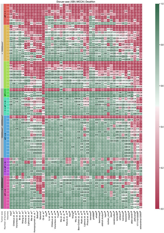
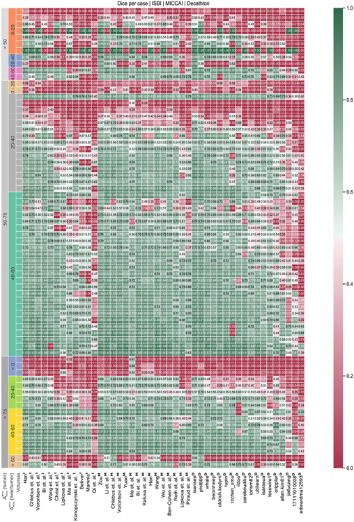
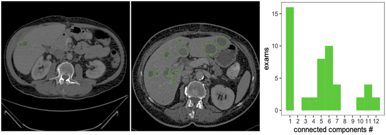
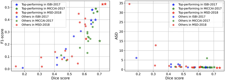
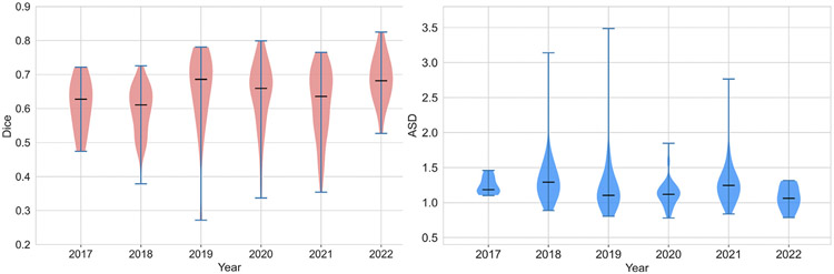
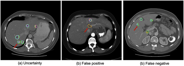
In this work, we report the set-up and results of the Liver Tumor Segmentation Benchmark (LiTS), which was organized in conjunction with the IEEE International Symposium on Biomedical Imaging (ISBI) 2017 and the International Conferences on Medical Image Computing and Computer-Assisted Intervention (MICCAI) 2017 and 2018. The image dataset is diverse and contains primary and secondary tumors with varied sizes and appearances with various lesion-to-background levels (hyper-/hypo-dense), created in collaboration with seven hospitals and research institutions. Seventy-five submitted liver and liver tumor segmentation algorithms were trained on a set of 131 computed tomography (CT) volumes and were tested on 70 unseen test images acquired from different patients. We found that not a single algorithm performed best for both liver and liver tumors in the three events. The best liver segmentation algorithm achieved a Dice score of 0.963, whereas, for tumor segmentation, the best algorithms achieved Dices scores of 0.674 (ISBI 2017), 0.702 (MICCAI 2017), and 0.739 (MICCAI 2018). Retrospectively, we performed additional analysis on liver tumor detection and revealed that not all top-performing segmentation algorithms worked well for tumor detection. The best liver tumor detection method achieved a lesion-wise recall of 0.458 (ISBI 2017), 0.515 (MICCAI 2017), and 0.554 (MICCAI 2018), indicating the need for further research. LiTS remains an active benchmark and resource for research, e.g., contributing the liver-related segmentation tasks in http://medicaldecathlon.com/. In addition, both data and online evaluation are accessible via https://competitions.codalab.org/competitions/17094.
Overview
The claims and statistics of this paper haven’t been checked yet.
Run the check — free, no account needed. It reads the open-access text and checks the sources it cites.
Integrity signals
Registry
Cites
Statistics
Figures
Verification
Claims the paper makes
The claims and statistics of this paper haven’t been checked yet.
Run the check — free, no account needed. It reads the open-access text and checks the sources it cites.
Claims other papers make about this one
Each is checked against this paper’s own text: does it say what the citing paper says it does?
The cited source says what the claim attributes to it.
In the citing paper
“different lesion appearances and annotation targets have been reported across tumor benchmarks [3,8,23]”
From Correction-aware interactive 3D tumor segmentation with sparse and revisable prompts (2026) · cites this paper as [3]
The cited source says what the claim attributes to it.
In the citing paper
“Tumor segmentation is less stable because lesions are not tied to a fixed anatomical shape or location and may be small, heterogeneous, or weakly contrasted [3,8,23].”
From Correction-aware interactive 3D tumor segmentation with sparse and revisable prompts (2026) · cites this paper as [3]
The check wasn’t confident either way, so it gives no verdict — never disputed.
In the citing paper
“The source of the OrganMNIST3D is the same as that of the Organ{A,C,S}MNIST. Instead of 2D images, we directly use the 3D bounding boxes and process the images into 28 × 28 × 28 to perform multi-class classification of 11 body organs.”
From MedMNIST v2 - A large-scale lightweight benchmark for 2D and 3D biomedical image classification (2023) · cites this paper as [30]
Experimental — accuracy not yet measured
Self-check
Percentages, sums and reported differences re-computed from the paper’s own numbers.
The self-check needs the paper’s full text.
Citations
Cites
U-Net: Convolutional Networks for Biomedical Image Segmentation
Ronneberger, O., Fischer, P., & Brox, T. (2015) · Lecture notes in computer science · doi:10.1007/978-3-319-24574-4_28 · Cited by 99,404
New response evaluation criteria in solid tumours: revised RECIST guideline (version 1.1)
Eisenhauer, E. A., Therasse, P., Bogaerts, J., Schwartz, L. H., Sargent, D., Ford, R., Dancey, J., Arbuck, S., Gwyther, S., Mooney, M., et al. (2009) · European Journal of Cancer · doi:10.1016/j.ejca.2008.10.026 · Cited by 29,534
Kass, M., Witkin, A., & Terzopoulos, D. (1988) · International Journal of Computer Vision · doi:10.1007/bf00133570 · Cited by 17,330
A survey on deep learning in medical image analysis
Litjens, G., Kooi, T., Bejnordi, B. E., Setio, A. A. A., Ciompi, F., Ghafoorian, M., van der Laak, J. A. W. M., van Ginneken, B., & Sánchez, C. I. (2017) · Medical Image Analysis · doi:10.1016/j.media.2017.07.005 · Cited by 15,332
Fronts propagating with curvature-dependent speed: Algorithms based on Hamilton-Jacobi formulations
Osher, S., & Sethian, J. A. (1988) · Journal of Computational Physics · doi:10.1016/0021-9991(88)90002-2 · Cited by 14,196
V-Net: Fully Convolutional Neural Networks for Volumetric Medical Image Segmentation
Milletari, F., Navab, N., & Ahmadi, S.-A. (2016) · 2016 Fourth International Conference on 3D Vision (3DV) · doi:10.1109/3dv.2016.79 · Cited by 11,931
Fully convolutional networks for semantic segmentation
Shelhamer, E., Long, J., & Darrell, T. (2017) · IEEE Transactions on Pattern Analysis and Machine Intelligence · doi:10.1109/tpami.2016.2572683 · Cited by 11,898
nnU-Net: a self-configuring method for deep learning-based biomedical image segmentation
Isensee, F., Jaeger, P. F., Kohl, S. A. A., Petersen, J., & Maier-Hein, K. H. (2021) · Nature Methods · doi:10.1038/s41592-020-01008-z · Cited by 10,477
Self-citationYushkevich, P. A., Piven, J., Hazlett, H. C., Smith, R. G., Ho, S., Gee, J. C., & Gerig, G. (2006) · NeuroImage · doi:10.1016/j.neuroimage.2006.01.015 · Cited by 8,868
Active Shape Models-Their Training and Application
Cootes, T. F., Taylor, C. J., Cooper, D. H., & Graham, J. (1995) · Computer Vision and Image Understanding · doi:10.1006/cviu.1995.1004 · Cited by 7,190
Research works printed but not linked: 5
- Ben-Dan, 2008, Liver tumor segmentation in CT images using probabilistic methods, vol. 41, 43
- Deng, X., Du, G., 2008. 3D segmentation in the clinic: A grand challenge II-liver tumor segmentation. In: MICCAI Workshop.
- Erickson, 2016, Radiology data from the cancer genome atlas liver hepatocellular carcinoma [TCGA-LIHC] collection, Cancer Imaging Arch.
- Hann, 2000, Diagnostic imaging approaches and relationship to hepatobiliary cancer staging and therapy, vol. 19, 94
- Heussel, 2007, Follow-up CT measurement of liver malignoma according to RECIST and WHO vs. volumetry, RoFo: Fortschr. Geb. Rontgenstr. Nuklearmed., 179, 958
Cited by
Sorted by citations
Sheller, M. J., Edwards, B., Reina, G. A., Martin, J., Pati, S., Kotrotsou, A., Milchenko, M., Xu, W., Marcus, D., Colen, R. R., et al. (2020) · Scientific Reports · doi:10.1038/s41598-020-69250-1 · Cited by 1,646
Self-citationThe Medical Segmentation Decathlon
Antonelli, M., Reinke, A., Bakas, S., Farahani, K., Kopp-Schneider, A., Landman, B. A., Litjens, G., Menze, B., Ronneberger, O., Summers, R. M., et al. (2022) · Nature Communications · doi:10.1038/s41467-022-30695-9 · Cited by 1,384
Self-citationDomain adaptation for medical image analysis: A survey
Guan, H., & Liu, M. (2022) · IEEE Transactions on Biomedical Engineering · doi:10.1109/tbme.2021.3117407 · Cited by 1,057
MedMNIST v2 - A large-scale lightweight benchmark for 2D and 3D biomedical image classification
Yang, J., Shi, R., Wei, D., Liu, Z., Zhao, L., Ke, B., Pfister, H., & Ni, B. (2023) · Scientific Data · doi:10.1038/s41597-022-01721-8 · Cited by 1,043
Medical image segmentation using deep learning: A survey
Wang, R., Lei, T., Cui, R., Zhang, B., Meng, H., & Nandi, A. K. (2022) · IET Image Processing · doi:10.1049/ipr2.12419 · Cited by 1,012
A Review of Deep-Learning-Based Medical Image Segmentation Methods
Liu, X., Song, L., Liu, S., & Zhang, Y. (2021) · Sustainability · doi:10.3390/su13031224 · Cited by 941
CHAOS Challenge - combined (CT-MR) healthy abdominal organ segmentation
Kavur, A. E., Gezer, N. S., BARIŞ, M. M., Aslan, S., Conze, P.-H., Groza, V., Pham, D. D., Chatterjee, S., Ernst, P., Özkan, S., et al. (2020) · Medical Image Analysis · doi:10.1016/j.media.2020.101950 · Cited by 893
Self-citationSegment anything model for medical image analysis: An experimental study
Mazurowski, M. A., Dong, H., Gu, H., Yang, J., Konz, N., & Zhang, Y. (2023) · Medical Image Analysis · doi:10.1016/j.media.2023.102918 · Cited by 802
“CT-Liver The Liver Tumor Segmentation Benchmark (LiTS) [4] CT 1 Liver 5,501”
BackgroundCoelho, L. P. (2023) · Bioengineering · doi:10.3390/bioengineering10121435 · Cited by 812
Loss odyssey in medical image segmentation
Ma, J., Chen, J., Ng, M., Huang, R., Li, Y., Li, C., Yang, X., & Martel, A. L. (2021) · Medical Image Analysis · doi:10.1016/j.media.2021.102035 · Cited by 662
Self-citationHeller, N. E., Isensee, F., Maier-Hein, K. H., Hou, X., Xie, C., Li, F., Nan, Y., Mu, G., Lin, Z., Han, M., et al. (2020) · Medical Image Analysis · doi:10.1016/j.media.2020.101821 · Cited by 629
Self-citationTransformation-Consistent Self-Ensembling Model for Semisupervised Medical Image Segmentation
Li, X., Yu, L., Chen, H., Fu, C., Xing, L., & Heng, P. (2020) · IEEE Transactions on Neural Networks and Learning Systems · doi:10.1109/tnnls.2020.2995319 · Cited by 553
Self-citationFederated learning for medical image analysis: A survey
Guan, H., Yap, P.-T., Bozoki, A., & Liu, M. (2024) · Pattern Recognition · doi:10.1016/j.patcog.2024.110424 · Cited by 518
INet: Convolutional Networks for Biomedical Image Segmentation
Weng, W., & Zhu, X. (2021) · IEEE Access · doi:10.1109/access.2021.3053408 · Cited by 569
Segment anything model for medical images?
Huang, Y., Yang, X., Liu, L., Zhou, H., Chang, A., Zhou, X., Chen, R., Yu, J., Chen, J., Chen, C., et al. (2024) · Medical Image Analysis · doi:10.1016/j.media.2023.103061 · Cited by 536
“1) DICE coefficient (DICE, %) [85]: A similarity measure to evaluate the overlap between the prediction and GT.”
Self-citationMA-Net: A Multi-Scale Attention Network for Liver and Tumor Segmentation
Fan, T., Wang, G., Li, Y., & Wang, H. (2020) · IEEE Access · doi:10.1109/access.2020.3025372 · Cited by 523
AbdomenCT-1K: Is Abdominal Organ Segmentation a Solved Problem?
Ma, J., Zhang, Y., Gu, S., Zhu, C. G., Ge, C., Zhang, Y., An, X., Wang, C., Wang, Q., Liu, X., et al. (2021) · IEEE Transactions on Pattern Analysis and Machine Intelligence · doi:10.1109/tpami.2021.3100536 · Cited by 429
Self-citationMedMNIST Classification Decathlon: A Lightweight AutoML Benchmark for Medical Image Analysis
Yang, J., Shi, R., & Ni, B. (2021) · 2021 IEEE 18th International Symposium on Biomedical Imaging (ISBI) · doi:10.1109/isbi48211.2021.9434062 · Cited by 414
nnU-Net Revisited: A Call for Rigorous Validation in 3D Medical Image Segmentation
Isensee, F., Wald, T., Ulrich, C., Baumgartner, M., Roy, S., Maier-Hein, K. H., & Jäger, P. F. (2024) · Lecture notes in computer science · doi:10.1007/978-3-031-72114-4_47 · Cited by 408
Self-citationLi, J., Chen, J., Tang, Y., Wang, C., Landman, B. A., & Zhou, S. K. (2023) · Medical Image Analysis · doi:10.1016/j.media.2023.102762 · Cited by 454
Self-citationJiao, R., Zhang, Y., Ding, L., Xue, B., Zhang, J., Cai, R., & Jin, C. (2023) · Computers in Biology and Medicine · doi:10.1016/j.compbiomed.2023.107840 · Cited by 374
CLIP-Driven Universal Model for Organ Segmentation and Tumor Detection
Liu, J., Zhang, Y., Chen, J.-N., Xiao, J., Lu, Y., Landman, B. A., Yuan, Y., Yuille, A., Tang, Y., & Zhou, Z. (2023) · IEEE/CVF International Conference on Computer Vision (ICCV) · doi:10.1109/iccv51070.2023.01934 · Cited by 381
Self-citationDeep learning for medical image segmentation: State-of-the-art advancements and challenges
Rayed, M. E., Islam, S. M. S., Niha, S. I., Jim, J. R., Kabir, M. M., & Mridha, M. F. (2024) · Informatics in Medicine Unlocked · doi:10.1016/j.imu.2024.101504 · Cited by 410
Zhou, Z., Sodha, V., Pang, J., Gotway, M. B., & Liang, J. (2020) · Medical Image Analysis · doi:10.1016/j.media.2020.101840 · Cited by 375
Semantic segmentation using Vision Transformers: A survey
Thisanke, H., Deshan, C., Chamith, K., Seneviratne, S., Vidanaarachchi, R., & Herath, D. (2023) · Engineering Applications of Artificial Intelligence · doi:10.1016/j.engappai.2023.106669 · Cited by 288
Mall, P. K., Singh, P. K., Srivastav, S., Narayan, V., Paprzycki, M., Jaworska, T., & Ganzha, M. (2023) · Healthcare Analytics · doi:10.1016/j.health.2023.100216 · Cited by 310
KiU-Net: Overcomplete Convolutional Architectures for Biomedical Image and Volumetric Segmentation
Valanarasu, J. M. J., Sindagi, V. A., Hacihaliloglu, I., & Patel, V. M. (2021) · IEEE Transactions on Medical Imaging · doi:10.1109/tmi.2021.3130469 · Cited by 295
Deep semi-supervised learning for medical image segmentation: A review
Han, K., Sheng, V. S., Song, Y., Liu, Y., Qiu, C., Ma, S., & Liu, Z. (2024) · Expert Systems with Applications · doi:10.1016/j.eswa.2023.123052 · Cited by 256
Understanding metric-related pitfalls in image analysis validation
Reinke, A., Tizabi, M. D., Baumgartner, M., Eisenmann, M., Heckmann-Nötzel, D., Kavur, A. E., Rädsch, T., Sudre, C. H., Acion, L., Antonelli, M., et al. (2024) · Nature Methods · doi:10.1038/s41592-023-02150-0 · Cited by 241
Self-citationDetection of autism spectrum disorder (ASD) in children and adults using machine learning
Farooq, M. S., Tehseen, R., Sabir, M., & Atal, Z. (2023) · Scientific Reports · doi:10.1038/s41598-023-35910-1 · Cited by 244
Multi-Organ Segmentation Over Partially Labeled Datasets With Multi-Scale Feature Abstraction
Fang, X., & Yan, P. (2020) · IEEE Transactions on Medical Imaging · doi:10.1109/tmi.2020.3001036 · Cited by 229
Efficient Medical Image Segmentation Based on Knowledge Distillation
Qin, D., Bu, J., Liu, Z., Shen, X., Zhou, S., Gu, J., Wang, Z., Wu, L., & Dai, H.-F. (2021) · IEEE Transactions on Medical Imaging · doi:10.1109/tmi.2021.3098703 · Cited by 213
UniverSeg: Universal Medical Image Segmentation
Butoi, V. I., Gonzalez Ortiz, J. J., Ma, T., Sabuncu, M. R., Guttag, J., & Dalca, A. V. (2023) · 2023 IEEE/CVF International Conference on Computer Vision (ICCV) · doi:10.1109/iccv51070.2023.01960 · Cited by 195
DoDNet: Learning to Segment Multi-Organ and Tumors from Multiple Partially Labeled Datasets
Zhang, J., Xie, Y., Xia, Y., & Shen, C. (2021) · IEEE Computer Society Conference on Computer Vision and Pattern Recognition/Proceedings - IEEE Computer Society Conference on Computer Vision and Pattern Recognition/Proceedings · doi:10.1109/cvpr46437.2021.00125 · Cited by 193
Luo, X., Liao, W., Xiao, J., Chen, J., Song, T., Zhang, X., Li, K., Metaxas, D., Wang, G., & Zhang, S. (2022) · Medical Image Analysis · doi:10.1016/j.media.2022.102642 · Cited by 237
Lei, T., Zhang, D., Du, X., Wang, X., Wan, Y., & Nandi, A. K. (2022) · IEEE Transactions on Medical Imaging · doi:10.1109/tmi.2022.3225687 · Cited by 185
Li, D., Yang, J., Kreis, K., Torralba, A. B., & Fidler, S. (2021) · IEEE Computer Society Conference on Computer Vision and Pattern Recognition/Proceedings - IEEE Computer Society Conference on Computer Vision and Pattern Recognition/Proceedings · doi:10.1109/cvpr46437.2021.00820 · Cited by 201
Weakly supervised machine learning
Ren, Z., Wang, S., & Zhang, Y. (2023) · CAAI Transactions on Intelligence Technology · doi:10.1049/cit2.12216 · Cited by 182
Advances in Deep Learning-Based Medical Image Analysis
Liu, X., Gao, K., Liu, B., Pan, C., Liang, K., Yan, L., Ma, J., He, F., Zhang, S., Pan, S., et al. (2021) · Health Data Science · doi:10.34133/2021/8786793 · Cited by 175
Self-citationFast and Low-GPU-memory abdomen CT organ segmentation: The FLARE challenge
Ma, J., Zhang, Y., Gu, S., An, X., Wang, Z., Ge, C., Wang, C., Zhang, F., Wang, Y., Xu, Y., et al. (2022) · Medical Image Analysis · doi:10.1016/j.media.2022.102616 · Cited by 182
Self-citation3DSAM-adapter: Holistic adaptation of SAM from 2D to 3D for promptable tumor segmentation
Gong, S., Zhong, Y., Ma, W., Li, J., Wang, Z., Zhang, J., Heng, P.-A., & Dou, Q. (2024) · Medical Image Analysis · doi:10.1016/j.media.2024.103324 · Cited by 156
Self-citationDíaz, O., Kushibar, K., Osuala, R. O., Linardos, A., Garrucho, L., Igual, L., Radeva, P. I., Prior, F., Gkontra, P., & Lekadir, K. (2021) · Physica Medica · doi:10.1016/j.ejmp.2021.02.007 · Cited by 159
Karimi, D., Warfield, S. K., & Gholipour, A. (2021) · Artificial Intelligence in Medicine · doi:10.1016/j.artmed.2021.102078 · Cited by 181
Medical image super-resolution for smart healthcare applications: A comprehensive survey
Umirzakova, S., Ahmad, S., Khan, L. U., & Whangbo, T. K. (2023) · Information Fusion · doi:10.1016/j.inffus.2023.102075 · Cited by 155
Kushnure, D. T., & Talbar, S. N. (2021) · Computerized Medical Imaging and Graphics · doi:10.1016/j.compmedimag.2021.101885 · Cited by 153
DefED-Net: Deformable Encoder-Decoder Network for Liver and Liver Tumor Segmentation
Lei, T., Wang, R., Zhang, Y., Wan, Y., Liu, C., & Nandi, A. K. (2021) · IEEE Transactions on Radiation and Plasma Medical Sciences · doi:10.1109/trpms.2021.3059780 · Cited by 155
Morshid, A. I., Elsayes, K. M., Khalaf, A., Elmohr, M. M., Yu, J., Kaseb, A. O., Hassan, M. M., Mahvash, A., Wang, Z., Hazle, J. D., et al. (2019) · Radiology Artificial Intelligence · doi:10.1148/ryai.2019180021 · Cited by 148
Advantages of transformer and its application for medical image segmentation: a survey
Pu, Q., Xi, Z., Yin, S., Zhao, Z., & Zhao, L. (2024) · BioMedical Engineering OnLine · doi:10.1186/s12938-024-01212-4 · Cited by 157
CT-ORG, a new dataset for multiple organ segmentation in computed tomography
Rister, B., Yi, D., Shivakumar, K., Nobashi, T. W., & Rubin, D. L. (2020) · Scientific Data · doi:10.1038/s41597-020-00715-8 · Cited by 146
Zhou, Y., Huang, J., Wang, C., Song, L., & Yang, G. (2023) · IEEE/CVF International Conference on Computer Vision (ICCV) · doi:10.1109/iccv51070.2023.01928 · Cited by 145
Self-citationHaghighi, F., Taher, M. R. H., Zhou, Z., Gotway, M. B., & Liang, J. (2021) · IEEE Transactions on Medical Imaging · doi:10.1109/tmi.2021.3060634 · Cited by 151
Interactive Few-Shot Learning: Limited Supervision, Better Medical Image Segmentation
Feng, R., Zheng, X., Gao, T., Chen, J., Wang, W., Chen, D. Z., & Wu, J. (2021) · IEEE Transactions on Medical Imaging · doi:10.1109/tmi.2021.3060551 · Cited by 141
Self-citationRadiomics feature reproducibility under inter-rater variability in segmentations of CT images
Haarburger, C., Müller-Franzes, G., Weninger, L., Kuhl, C., Truhn, D., & Merhof, D. (2020) · Scientific Reports · doi:10.1038/s41598-020-69534-6 · Cited by 139
Self-citationFederated learning for healthcare applications
Chaddad, A., Wu, Y., & Desrosiers, C. (2024) · IEEE Internet of Things Journal · doi:10.1109/jiot.2023.3325822 · Cited by 136
Eres-UNet++: Liver CT image segmentation based on high-efficiency channel attention and Res-UNet++
Li, J., Liu, K., Hu, Y., Zhang, H., Heidari, A. A., Chen, H., Zhang, W., Algarni, A. D., & Elmannai, H. (2023) · Computers in Biology and Medicine · doi:10.1016/j.compbiomed.2022.106501 · Cited by 132
Self-citationFedMix: Mixed Supervised Federated Learning for Medical Image Segmentation
Wicaksana, J., Yan, Z., Zhang, D., Huang, X., Wu, H., Yang, X. Y., & Cheng, K.-T. T. (2022) · IEEE Transactions on Medical Imaging · doi:10.1109/tmi.2022.3233405 · Cited by 142
Self-citationAdapting Visual-Language Models for Generalizable Anomaly Detection in Medical Images
Huang, C., Jiang, A., Feng, J., Zhang, Y., Wang, X., & Wang, Y. (2024) · IEEE Computer Society Conference on Computer Vision and Pattern Recognition/Proceedings - IEEE Computer Society Conference on Computer Vision and Pattern Recognition/Proceedings · doi:10.1109/cvpr52733.2024.01081 · Cited by 143
Recent progress in transformer-based medical image analysis
Liu, Z., Lv, Q., Yang, Z., Li, Y., Lee, C. H., & Shen, L. (2023) · Computers in Biology and Medicine · doi:10.1016/j.compbiomed.2023.107268 · Cited by 133
CLIP in medical imaging: A survey
Zhao, Z., Liu, Y., Wu, H., Wang, M., Li, Y., Wang, S., Teng, L., Liu, D., Cui, Z., Wang, Q., et al. (2025) · Medical Image Analysis · doi:10.1016/j.media.2025.103551 · Cited by 248
A lightweight neural network with multiscale feature enhancement for liver CT segmentation
Ansari, M. Y., Yang, Y., Balakrishnan, S., Abinahed, J. A., Al-Ansari, A. A., Warfa, M., Almokdad, O., Barah, A., Omer, A., Singh, A. V., et al. (2022) · Scientific Reports · doi:10.1038/s41598-022-16828-6 · Cited by 121
Ma, J., Zhang, Y., Gu, S., Ge, C., Mae, S., Young, A., Zhu, C., Yang, X., Meng, K., Huang, Z., et al. (2024) · The Lancet Digital Health · doi:10.1016/s2589-7500(24)00154-7 · Cited by 145
Self-citationReinventing 2D Convolutions for 3D Images
Yang, J., Huang, X., He, Y. F., Xu, J., Yang, C., Xu, G., & Ni, B. (2021) · IEEE Journal of Biomedical and Health Informatics · doi:10.1109/jbhi.2021.3049452 · Cited by 126
Chen, Y., Wang, L., Qin, X., Wang, J., & Xie, X. (2023) · IEEE Transactions on Neural Networks and Learning Systems · doi:10.1109/tnnls.2023.3297103 · Cited by 127
An attempt at beating the 3D U-Net
Isensee, F., & Maier-Hein, K. H. (2019) · Kidney Tumor Segmentation Challenge (KiTS) · doi:10.24926/548719.001 · Cited by 112
Self-citationZheng, R., Wang, Q., Lv, S., Li, C., Wang, C., Chen, W., & Wang, H. (2022) · IEEE Transactions on Medical Imaging · doi:10.1109/tmi.2022.3175461 · Cited by 118
Self-citationXia, K., Yin, H., Qian, P., Jiang, Y., & Wang, S. (2019) · IEEE Access · doi:10.1109/access.2019.2929270 · Cited by 109
Hong, J., Yu, S. C. H., & Chen, W. (2022) · Applied Soft Computing · doi:10.1016/j.asoc.2022.108729 · Cited by 106
Self-citationZhou, H.-Y., Lu, C., Yang, S., Han, X., & Yu, Y. (2021) · IEEE/CVF International Conference on Computer Vision (ICCV) · doi:10.1109/iccv48922.2021.00348 · Cited by 105
Self-citationHaghighi, F., Taher, M., Gotway, M., & Liang, J.-M. (2022) · Computer Vision and Pattern Recognition · doi:10.1109/cvpr52688.2022.02016 · Cited by 97
Label-Free Liver Tumor Segmentation
Hu, Q., Chen, Y., Xiao, J., Sun, S.-W., Chen, J., Yuille, A., & Zhou, Z. (2023) · IEEE Computer Society Conference on Computer Vision and Pattern Recognition/Proceedings - IEEE Computer Society Conference on Computer Vision and Pattern Recognition/Proceedings · doi:10.1109/cvpr52729.2023.00717 · Cited by 106
Medical Image Segmentation With Limited Supervision: A Review of Deep Network Models
Peng, J., & Wang, Y. (2021) · IEEE Access · doi:10.1109/access.2021.3062380 · Cited by 115
A Multiple Layer U-Net, U n -Net, for Liver and Liver Tumor Segmentation in CT
Tran, S.-T., Cheng, C.-H., & Liu, D. (2020) · IEEE Access · doi:10.1109/access.2020.3047861 · Cited by 96
Segment Anything Model (SAM) for Medical Image Segmentation: A Preliminary Review
Zhang, L., Deng, X., & Yu, L. (2023) · IEEE International Conference on Bioinformatics and Biomedicine (BIBM) · doi:10.1109/bibm58861.2023.10386032 · Cited by 103
Self-citationMine yOur owN Anatomy: Revisiting Medical Image Segmentation With Extremely Limited Labels
You, C., Dai, W., Liu, F., Min, Y., Dvornek, N. C., Li, X., Clifton, D. A., Staib, L. H., & Duncan, J. S. (2024) · IEEE Transactions on Pattern Analysis and Machine Intelligence · doi:10.1109/tpami.2024.3461321 · Cited by 109
Self-citationLi, C., Lin, X., Mao, Y., Lin, W. Y., Qi, Q., Ding, X., Huang, Y., Liang, D., & Yu, Y. (2021) · Computers in Biology and Medicine · doi:10.1016/j.compbiomed.2021.105144 · Cited by 98
SAM: Self-Supervised Learning of Pixel-Wise Anatomical Embeddings in Radiological Images
Yan, K., Cai, J., Jin, D., Miao, S., Guo, D., Harrison, A. P., Tang, Y., Xiao, J., Lu, J., & Lu, L. (2022) · IEEE Transactions on Medical Imaging · doi:10.1109/tmi.2022.3169003 · Cited by 95
BMAD: Benchmarks for Medical Anomaly Detection
Bao, J., Sun, H., Deng, H., He, Y., Zhang, Z., & Li, X. (2024) · IEEE/CVF Conference on Computer Vision and Pattern Recognition Workshops (CVPRW) · doi:10.1109/cvprw63382.2024.00408 · Cited by 122
Self-citationA Comprehensive Survey of Foundation Models in Medicine
Khan, W., Leem, S., See, K. B., Wong, J. K., Zhang, S., & Fang, R. (2025) · IEEE Reviews in Biomedical Engineering · doi:10.1109/rbme.2025.3531360 · Cited by 137
Yan, K., Cai, J., Zheng, Y., Harrison, A. P., Jin, D., Tang, Y., Tang, Y., Huang, L., Xiao, J. Z., & Lü, L. (2020) · IEEE Transactions on Medical Imaging · doi:10.1109/tmi.2020.3047598 · Cited by 92
Personalized Federated Learning With Adaptive Batchnorm for Healthcare
Wang, L., Wang, J., Chen, Y., Qin, X., Xu, R., Dimitriadis, D., & Qin, T. (2022) · IEEE Transactions on Big Data · doi:10.1109/tbdata.2022.3177197 · Cited by 101
Dive into the details of self-supervised learning for medical image analysis
Zhang, C., Zheng, H., & Gu, Y. (2023) · Medical Image Anal. · doi:10.1016/j.media.2023.102879 · Cited by 101
Artificial intelligence in precision medicine in hepatology
Su, T., Wu, C., & Kao, J. (2021) · Journal of Gastroenterology and Hepatology · doi:10.1111/jgh.15415 · Cited by 97
Deep learning and level set approach for liver and tumor segmentation from CT scans
Alirr, O. I. (2020) · Journal of Applied Clinical Medical Physics · doi:10.1002/acm2.13003 · Cited by 88
VoCo: A Simple-Yet-Effective Volume Contrastive Learning Framework for 3D Medical Image Analysis
Wu, L., Zhuang, J., & Chen, H. (2024) · IEEE Computer Society Conference on Computer Vision and Pattern Recognition/Proceedings - IEEE Computer Society Conference on Computer Vision and Pattern Recognition/Proceedings · doi:10.1109/cvpr52733.2024.02158 · Cited by 88
Self-citationBousabarah, K., Letzen, B. S., Tefera, J., Savic, L. J., Schobert, I. T., Schlachter, T. R., Staib, L. H., Köcher, M., Chapiro, J., & Lin, M. (2020) · Abdominal Radiology · doi:10.1007/s00261-020-02604-5 · Cited by 86
Data synthesis and adversarial networks: A review and meta-analysis in cancer imaging
Osuala, R. O., Kushibar, K., Garrucho, L., Linardos, A., Szafranowska, Z., Klein, S., Glocker, B., Díaz, O., & Lekadir, K. (2022) · Medical Image Analysis · doi:10.1016/j.media.2022.102704 · Cited by 106
Deep learning techniques for tumor segmentation: a review
Jiang, H., Diao, Z., & Yao, Y. (2021) · The Journal of Supercomputing · doi:10.1007/s11227-021-03901-6 · Cited by 86
Zhou, H.-Y., Lu, C., Chen, C., Yang, S., & Yu, Y. (2023) · IEEE Transactions on Pattern Analysis and Machine Intelligence · doi:10.1109/tpami.2023.3234002 · Cited by 87
Haider, A., Khan, S. I., Iqbal, D. N., Khan, S. U., Haider, S., Mohammad, K., Mustfa, G., Rizwan, M., & Haider, A. (2024) · International Journal of Biological Macromolecules · doi:10.1016/j.ijbiomac.2024.134172 · Cited by 86
MedLSAM: Localize and segment anything model for 3D CT images
Lei, W., Xu, W., Li, K., Zhang, X., & Zhang, S. (2024) · Medical Image Analysis · doi:10.1016/j.media.2024.103370 · Cited by 135
Mai, D. V. C., Drami, I., Pring, E. T., Gould, L. E., Lung, P. F. C., Popuri, K., Chow, V., Beg, M. F., Athanasiou, T., & Jenkins, J. T. (2023) · Journal of Cachexia Sarcopenia and Muscle · doi:10.1002/jcsm.13310 · Cited by 78
TD-Net: A Hybrid End-to-End Network for Automatic Liver Tumor Segmentation From CT Images
Di, S., Zhao, Y., Liao, M., Zhang, F., & Li, X. (2022) · IEEE Journal of Biomedical and Health Informatics · doi:10.1109/jbhi.2022.3181974 · Cited by 74
Self-citationQian, L., Wen, C., Li, Y., Hu, Z., Zhou, X. Y., Xia, X., & Kim, S.-H. (2023) · Computer Methods and Programs in Biomedicine · doi:10.1016/j.cmpb.2023.107885 · Cited by 78
Self-citationTowards Efficient COVID-19 CT Annotation: A Benchmark for Lung and Infection SegmentationPreprint
Ma, J., Wang, Y., An, X., Ge, C., Yu, Z., Chen, J., Zhu, Q., Dong, G., He, J., He, Z., et al. (2020) · arXiv (Cornell University) · Cited by 74
Self-citationRethinking Boundary Detection in Deep Learning Models for Medical Image Segmentation
Lin, Y., Zhang, D., Fang, X., Chen, Y., Cheng, K.-T. T., & Chen, H. (2023) · Lecture notes in computer science · doi:10.1007/978-3-031-34048-2_56 · Cited by 76
Self-citationSMAFormer: Synergistic Multi-Attention Transformer for Medical Image Segmentation
Zheng, F., Chen, X., Liu, W., Li, H., Lei, Y., He, J., Pun, C., & Zhou, S. (2024) · IEEE International Conference on Bioinformatics and Biomedicine (BIBM) · doi:10.1109/bibm62325.2024.10822736 · Cited by 73
Self-citationX-Net: Multi-branch UNet-like network for liver and tumor segmentation from 3D abdominal CT scans
Chi, J., Han, X., Wu, C., Wang, H., & Ji, P. (2021) · Neurocomputing · doi:10.1016/j.neucom.2021.06.021 · Cited by 72
Self-citationICycle-GAN: Improved cycle generative adversarial networks for liver medical image generation
Chen, Y., Lin, H., Zhang, W., Chen, W., Zhou, Z., Heidari, A. A., Chen, H., & Guohui, X. (2024) · Biomedical Signal Processing and Control · doi:10.1016/j.bspc.2024.106100 · Cited by 71
Self-citationTraining on Polar Image Transformations Improves Biomedical Image Segmentation
Benčević, M., Galić, I., Habijan, M., & Babin, D. (2021) · IEEE Access · doi:10.1109/access.2021.3116265 · Cited by 68
Quinton, F., Popoff, R., Presles, B., Leclerc, S., Mériaudeau, F., Nodari, G., Lopez, O., Pellegrinelli, J., Chevallier, O., Dominique Ginhac, B., et al. (2023) · Data · doi:10.3390/data8050079 · Cited by 69
Metadata
- Authors
- Bilic, P., Christ, P., Li, H. B., Vorontsov, E., Ben-Cohen, A., Kaissis, G., Szeskin, A., Jacobs, C., Mamani, G. E. H., Chartrand, G., Lohöfer, F., Holch, J. W., Sommer, W., Hofmann, F., Hostettler, A., Lev-Cohain, N., Drozdzal, M., Amitai, M. M., Vivanti, R., Sosna, J., Ezhov, I., Sekuboyina, A., Navarro, F., Kofler, F., Paetzold, J. C., Shit, S., Hu, X., Lipková, J., Rempfler, M., Piraud, M., Kirschke, J., Wiestler, B., Zhang, Z., Hülsemeyer, C., Beetz, M., Ettlinger, F., Antonelli, M., Bae, W., Bellver, M., Bi, L., Chen, H., Chlebus, G., Dam, E. B., Dou, Q., Fu, C.-W., Georgescu, B., Giró-i-Nieto, X., Gruen, F., Han, X., Heng, P.-A., Hesser, J., Moltz, J. H., Igel, C., Isensee, F., Jäger, P., Jia, F., Kaluva, K. C., Khened, M., Kim, I., Kim, J.-H., Kim, S., Kohl, S., Konopczynski, T., Kori, A., Krishnamurthi, G., Li, F., Li, H., Li, J., Li, X., Lowengrub, J., Ma, J., Maier-Hein, K., Maninis, K.-K., Meine, H., Merhof, D., Pai, A., Perslev, M., Petersen, J., Pont-Tuset, J., Qi, J., Qi, X., Rippel, O., Roth, K., Sarasua, I., Schenk, A., Shen, Z., Torres, J., Wachinger, C., Wang, C., Weninger, L., Wu, J., Xu, D., Yang, X., Yu, S. C.-H., Yuan, Y., Yue, M., Zhang, L., Cardoso, J., Bakas, S., Braren, R., Heinemann, V., Pal, C., Tang, A., Kadoury, S., Soler, L., van Ginneken, B., Greenspan, H., Joskowicz, L., & Menze, B.
- Affiliations
- Technical University of Munich (DE)
- Guangdong University of Foreign Studies (CN)
- Polytechnique Montréal (CA)
- Tel Aviv University (IL)
- TUM Klinikum (DE)
- Hebrew University of Jerusalem (IL)
- Radboud University Medical Center (NL)
- Université de Montréal (CA)
- German Cancer Research Center (DE)
- LMU Klinikum (DE)
- Institut de Recherche contre les Cancers de l’Appareil Digestif (FR)
- Rafael Advanced Defense Systems (Israel) (IL)
- Hadassah Medical Center (IL)
- University of Zurich (CH)
- Helmholtz Munich (DE)
- Chinese University of Hong Kong (HK)
- Brigham and Women's Hospital (US)
- ETH Zurich (CH)
- Nanjing Drum Tower Hospital (CN)
- King's College London (GB)
- Hong Kong University of Science and Technology (HK)
- Barcelona Supercomputing Center (ES)
- The University of Sydney (AU)
- University of Copenhagen (DK)
- Siemens Healthcare (United States) (US)
- Heidelberg University (DE)
- University of North Carolina at Chapel Hill (US)
- Fraunhofer Institute for Digital Medicine (DE)
- Chinese Academy of Sciences (CN)
- Indian Institute of Technology Madras (IN)
- Samsung Medical Center (KR)
- Kakao Brain (South Korea) (KR)
- University of California, Irvine (US)
- University of Bremen (DE)
- RWTH Aachen University (DE)
- University of Hong Kong (HK)
- University of Tübingen (DE)
- Medizinische Hochschule Hannover (DE)
- University of Illinois Urbana-Champaign (US)
- KTH Royal Institute of Technology (SE)
- Nvidia (United States) (US)
- Nanjing University (CN)
- Icahn School of Medicine at Mount Sinai (US)
- Corresponding author
- Li, Hongwei Bran
- Li, Hongchao
- ORCID
- Li, Hongwei Bran
- Vorontsov, Eugene
- Kaissis, Georgios
- Szeskin, Adi
- Jacobs, Colin
- Mamani, Gabriel Efrain Humpire
- Chartrand, Gabriel
- Lohöfer, Fabian
- Holch, Julian Walter
- Hostettler, Alexandre
- Drozdzal, Michal
- Amitai, Michal Marianne
- Sosna, Jacob
- Ezhov, Ivan
- Sekuboyina, Anjany
- Navarro, Fernando
- Kofler, Florian
- Paetzold, Johannes C.
- Shit, Suprosanna
- Hu, Xiaobin
- Lipková, Jana
- Piraud, Marie
- Kirschke, Jan
- Wiestler, Benedikt
- Zhang, Zhiheng
- Beetz, Marcel
- Antonelli, Michela
- Bae, Woong
- Bellver, Míriam
- Chen, Hao
- Chlebus, Grzegorz
- Dam, Erik B.
- Dou, Qi
- Fu, Chi-Wing
- Georgescu, Bogdan
- Giró-i-Nieto, Xavier
- Han, Xu
- Heng, Pheng-Ann
- Hesser, Jürgen
- Moltz, Jan Hendrik
- Igel, Christian
- Isensee, Fabian
- Jäger, Paul
- Jia, Fucang
- Kaluva, Krishna Chaitanya
- Khened, Mahendra
- Kim, Jae-Hun
- Kim, Sungwoong
- Kori, Avinash
- Krishnamurthi, Ganapathy
- Li, Fan
- Li, Hongchao
- Li, Junbo
- Li, Xiaomeng
- Lowengrub, John
- Ma, Jun
- Maier-Hein, Klaus
- Maninis, Kevis-Kokitsi
- Meine, Hans
- Merhof, Dorit
- Pai, Akshay
- Perslev, Mathias
- Petersen, Jens
- Pont-Tuset, Jordi
- Qi, Xiaojuan
- Rippel, Oliver
- Roth, Karsten
- Schenk, Andrea
- Shen, Zengming
- Torres, Jordi
- Wachinger, Christian
- Wang, Chunliang
- Weninger, Leon
- Xu, Daguang
- Yang, Xiaoping
- Yu, Simon Chun-Ho
- Yuan, Yading
- Zhang, Liping
- Bakas, Spyridon
- Braren, Rickmer
- Heinemann, Volker
- Pal, Christopher
- Tang, An
- Kadoury, Samuel
- van Ginneken, Bram
- Greenspan, Hayit
- Menze, Bjoern
- Venue
- Medical Image Analysis
- Publisher
- Elsevier BV
- Published
- 2023-02
- Volume
- 84
- Pages
- 102680
- ISSN
- 1361-8415 (ISSN-L)
- PMC
- PMC10631490
- OpenAlex
- W2910094941
- Cited by
- 1,412
- References
- 146
- Citations by year
- 2026: 259 · 2025: 337 · 2024: 311 · 2023: 195
- FWCI
- 77.21
- Type
- journal article
- Language
- en
- Open access
- hybrid
- doi.org
- Licence
- Locations
- Medical Image Analysis (publishedVersion, cc-by-nc-nd)
- PubMed (publishedVersion)
- Research at the University of Copenhagen (University of Copenhagen) (publishedVersion, cc-by-nc-nd)
- PubMed Central (acceptedVersion, cc-by-nc-nd)
- Zurich Open Repository and Archive (University of Zurich) (acceptedVersion, cc-by-nc-nd)
- arXiv (Cornell University) (submittedVersion, cc-by-nc-nd)
- Rare & Special e-Zone (The Hong Kong University of Science and Technology) (submittedVersion)
- Rare & Special e-Zone (The Hong Kong University of Science and Technology) (submittedVersion, cc-by-nc-sa)
- eScholarship (California Digital Library) (submittedVersion, cc-by-nc-nd)
- Radboud Repository (Radboud University) (submittedVersion, cc-by-nc-sa)
- IRIS eCampus Telematic University (Università degli Studi eCampus) (submittedVersion)
- mediaTUM (Technical University of Munich) (submittedVersion)
- Site cant be reached (submittedVersion, cc-by-nc-nd)
- Fraunhofer-Publica (Fraunhofer-Gesellschaft) (submittedVersion)
- PolyPublie (École Polytechnique de Montréal) (submittedVersion)
- PuSH - Publication Server of Helmholtz Zentrum München (submittedVersion, cc-by-nc-nd)
- UPCommons institutional repository (Universitat Politècnica de Catalunya) (submittedVersion, cc-by-nc-nd)
- Repository for Publications and Research Data (ETH Zurich) (submittedVersion, cc-by-nc-nd)
- Zurich Open Repository and Archive (University of Zurich) (submittedVersion, cc-by-nc-nd)
- Radboud Repository (Radboud University) (submittedVersion, other-oa)
- arXiv (Cornell University)
- ETH Zürich Research Collection
- Zurich Open Repository and Archive (University of Zurich)
- Funders
- Deutsche Forschungsgemeinschaft (SFB824, GSC81, subproject B12)
- Universität Zürich
- Fondation de l'Association des radiologistes du Québec (34939)
- National Institutes of Health (1s10od023495-01)
- Fonds de Recherche du Québec - Santé (34939, Junior 2)
- National Cancer Institute (U01CA242871)
- International Graduate School of Science and Engineering (GSC 81)
- Topics
- Physical Sciences › Computer Science › Computer Vision and Pattern Recognition › Medical Image Segmentation Techniques
- Physical Sciences › Computer Science › Computer Vision and Pattern Recognition › Brain Tumor Detection and Classification
- Health Sciences › Medicine › Radiology, Nuclear Medicine and Imaging › Radiomics and Machine Learning in Medical Imaging
- Keywords
- liver tumor segmentation, liver segmentation, liver cancer, computed tomography, segmentation benchmark, primary liver tumors, secondary liver tumors, lesion detection, medical image segmentation
- MeSH terms
- Algorithms, Humans, Image Processing, Computer-Assisted, Liver, Liver Neoplasms, Retrospective Studies, Benchmarking
About these checks
Registry, retraction, Cites and statistics results are computed, not judged, with one exception: a printed reference that no index lists is read once by a model before it counts as a citation error. Claims are extracted by a model and kept only when their quote appears verbatim in the text; each cited source is then checked, with a model’s help, for whether it says what the claim relies on. Those verdicts can be wrong — read the claim’s quote and the cited source before relying on one.
Statistics and arithmetic are recomputed, not judged.
Confidence bands are shown only for fields calibrated on our annotated test set. None is calibrated yet, so no band is shown.
What counts as a claim ›How we measure confidence ›How we check this ›
Are you an author, or do you see a problem with this page? Request a review
