

The DOI matches its Crossref record; 78 of the 81 references it cites were found in a registry or an index.
The role of skeletal muscle Akt in the regulation of muscle mass and glucose homeostasis
Open accessAbstract
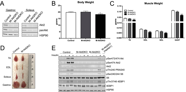
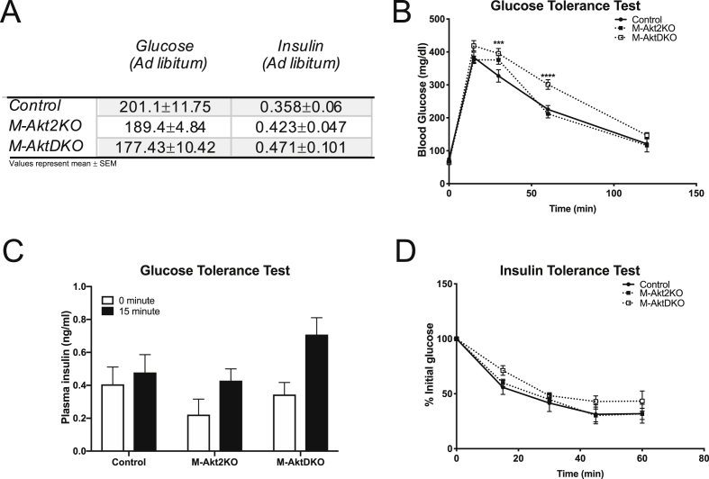
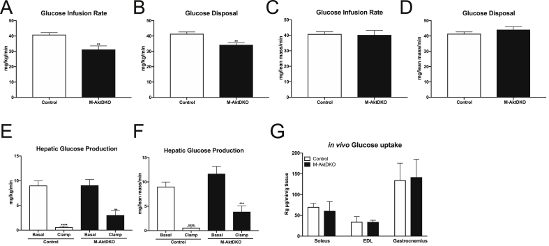
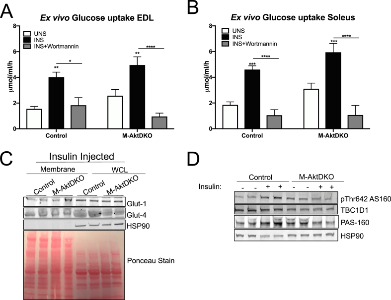
OBJECTIVE: Skeletal muscle insulin signaling is a major determinant of muscle growth and glucose homeostasis. Protein kinase B/Akt plays a prominent role in mediating many of the metabolic effects of insulin. Mice and humans harboring systemic loss-of-function mutations in Akt2, the most abundant Akt isoform in metabolic tissues, are glucose intolerant and insulin resistant. Since the skeletal muscle accounts for a significant amount of postprandial glucose disposal, a popular hypothesis in the diabetes field suggests that a reduction in Akt, specifically in skeletal muscle, leads to systemic glucose intolerance and insulin resistance. Despite this common belief, the specific role of skeletal muscle Akt in muscle growth and insulin sensitivity remains undefined. METHODS: We generated multiple mouse models of skeletal muscle Akt deficiency to evaluate the role of muscle Akt signaling in vivo. The effects of these genetic perturbations on muscle mass, glucose homeostasis and insulin sensitivity were assessed using both in vivo and ex vivo assays. RESULTS: Surprisingly, mice lacking Akt2 alone in skeletal muscle displayed normal skeletal muscle insulin signaling, glucose tolerance, and insulin sensitivity despite a dramatic reduction in phosphorylated Akt. In contrast, deletion of both Akt isoforms (M-AktDKO) prevented downstream signaling and resulted in muscle atrophy. Despite the absence of Akt signaling, in vivo and ex vivo insulin-stimulated glucose uptake were normal in M-AktDKO mice. Similar effects on insulin sensitivity were observed in mice with prolonged deletion (4 weeks) of both skeletal muscle Akt isoforms selectively in adulthood. Conversely, short term deletion (2 weeks) of skeletal muscle specific Akt in adult muscles impaired insulin tolerance paralleling the effect observed by acute pharmacological inhibition of Akt in vitro. Mechanistically, chronic ablation of Akt induced mitochondrial dysfunction and activation of AMPK, which was required for insulin-stimulated glucose uptake in the absence of Akt. CONCLUSIONS: Together, these data indicate that chronic reduction in Akt activity alone in skeletal muscle is not sufficient to induce insulin resistance or prevent glucose uptake in all conditions. Therefore, since insulin-stimulated glucose disposal in skeletal muscle is markedly impaired in insulin-resistant states, we hypothesize that alterations in signaling molecules in addition to skeletal muscle Akt are necessary to perturb glucose tolerance and insulin sensitivity in vivo.
Overview
The claims and statistics of this paper haven’t been checked yet.
Run the check — free, no account needed. It reads the open-access text and checks the sources it cites.
Integrity signals
Registry
Cites
Statistics
Figures
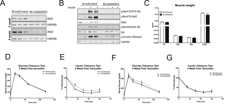
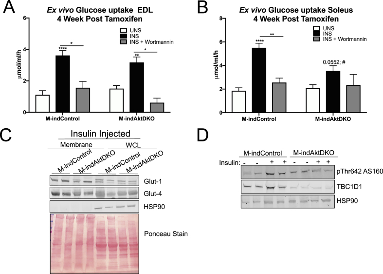
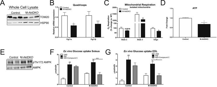
Verification
Claims the paper makes
The claims and statistics of this paper haven’t been checked yet.
Run the check — free, no account needed. It reads the open-access text and checks the sources it cites.
Claims other papers make about this one
Each is checked against this paper’s own text: does it say what the citing paper says it does?
In the citing paper
“recent studies utilizing skeletal muscle-specific knockouts of both Akt1 and Akt2 demonstrated that deletion of both isoforms reduces both muscle size and muscle protein synthesis, indicating that only minimal amounts of Akt in muscle are needed to transmit downstream signaling and preserve muscle mass in vivo [19,20].”
From Antibody blockade of activin type II receptors preserves skeletal muscle mass and enhances fat loss during GLP-1 receptor agonism (2024) · cites this paper as [19]
In the citing paper
“Skeletal muscle Akt signaling is both required and sufficient for muscle hypertrophy, as genetic loss of Akt isoforms in the muscle leads to decreased muscle size in adult mice [19,20,22].”
From Antibody blockade of activin type II receptors preserves skeletal muscle mass and enhances fat loss during GLP-1 receptor agonism (2024) · cites this paper as [19]
In the citing paper
“As described in prior studies, these mice display significant deficits in muscle growth and protein synthesis, consistent with a major anabolic role for Akt in muscle [19,20].”
From Antibody blockade of activin type II receptors preserves skeletal muscle mass and enhances fat loss during GLP-1 receptor agonism (2024) · cites this paper as [19]
In the citing paper
“Residual Akt1 expression in whole muscle tissue is due to the expression of Akt1 in non-muscle cells [19].”
From Antibody blockade of activin type II receptors preserves skeletal muscle mass and enhances fat loss during GLP-1 receptor agonism (2024) · cites this paper as [19]
In the citing paper
“Furthermore, the AKT1 rs1130214 polymorphism affects the AKT pathway, a critical component of the mTOR signaling cascade, influencing MPS rates [59].”
From Beyond Calories: Individual Metabolic and Hormonal Adaptations Driving Variability in Weight Management—A State-of-the-Art Narrative Review (2024) · cites this paper as [59]
Self-check
Percentages, sums and reported differences re-computed from the paper’s own numbers.
The self-check needs the paper’s full text.
Citations
Cites
Watterson, K. R., Bestow, D., Gallagher, J. R., Hamilton, D. L., Ashford, F. B., Meakin, P. J., & Ashford, M. L. J. (2012) · Neurosignals · doi:10.1159/000334144 · Cited by 3,395
The selectivity of protein kinase inhibitors: a further update
Bain, J., Plater, L., Elliott, M., Shpiro, N., Hastie, C. J., McLauchlan, H., Klevernic, I. V., Arthur, J. S. C., Alessi, D. R., & Cohen, P. (2007) · Biochemical Journal · doi:10.1042/bj20070797 · Cited by 2,522
Skeletal Muscle Insulin Resistance Is the Primary Defect in Type 2 Diabetes
DeFronzo, R. A., & Tripathy, D. (2009) · Diabetes Care · doi:10.2337/dc09-s302 · Cited by 2,102
Cho, H., Mu, J., Kim, J. K., Thorvaldsen, J. L., Chu, Q., Crenshaw, E. B., Kaestner, K. H., Bartolomei, M. S., Shulman, G. I., & Birnbaum, M. J. (2001) · Science · doi:10.1126/science.292.5522.1728 · Cited by 1,860
DeFronzo, R. A., Jacot, E., Jequier, E., Maeder, E., Wahren, J., & Felber, J. P. (1981) · Diabetes · doi:10.2337/diab.30.12.1000 · Cited by 1,770
The pathogenesis of insulin resistance: integrating signaling pathways and substrate flux
Samuel, V. T., & Shulman, G. I. (2016) · Journal of Clinical Investigation · doi:10.1172/jci77812 · Cited by 1,484
Pathogenesis of type 2 diabetes mellitus
DeFronzo, R. A. (2004) · Medical Clinics of North America · doi:10.1016/j.mcna.2004.04.013 · Cited by 1,342
Cho, H., Thorvaldsen, J. L., Chu, Q., Feng, F., & Birnbaum, M. J. (2001) · Journal of Biological Chemistry · doi:10.1074/jbc.c100462200 · Cited by 959
Mu, J., Brozinick, J. T., Valladares, O., Bucan, M., & Birnbaum, M. J. (2001) · Molecular Cell · doi:10.1016/s1097-2765(01)00251-9 · Cited by 924
Peng, X.-D., Xu, P.-Z., Chen, M.-L., Hahn-Windgassen, A., Skeen, J. E., Jacobs, J., Sundararajan, D., Chen, W. S., Crawford, S. E., Coleman, K. G., et al. (2003) · Genes & Development · doi:10.1101/gad.1089403 · Cited by 836
Research works printed but not linked: 1
- Moller, 2019, The role of p-21 activated kinases (PAKs) in glucose homeostasis and skeletal muscle glucose uptake, BioRxiv
Cited by
Sorted by citations
The aetiology and molecular landscape of insulin resistance
James, D. E., Stöckli, J., & Birnbaum, M. J. (2021) · Nature Reviews Molecular Cell Biology · doi:10.1038/s41580-021-00390-6 · Cited by 736
Insulin: The master regulator of glucose metabolism
Norton, L., Shannon, C. E., Gastaldelli, A., & DeFronzo, R. A. (2022) · Metabolism · doi:10.1016/j.metabol.2022.155142 · Cited by 367
Mechanisms of muscle insulin resistance and the cross‐talk with liver and adipose tissue
Rosa, S. C. D. S., Nayak, N., Caymo, A. M., & Gordon, J. W. (2020) · Physiological Reports · doi:10.14814/phy2.14607 · Cited by 271
Insulin signalling and GLUT4 trafficking in insulin resistance
van Gerwen, J., Shun-Shion, A. S., & Fazakerley, D. J. (2023) · Biochemical Society Transactions · doi:10.1042/bst20221066 · Cited by 277
Mechanisms of Estrogen Influence on Skeletal Muscle: Mass, Regeneration, and Mitochondrial Function
Pellegrino, A., Tiidus, P. M., & Vandenboom, R. (2022) · Sports Medicine · doi:10.1007/s40279-022-01733-9 · Cited by 178
Targeting PI3K-AKT/mTOR signaling in the prevention of autism
Sharma, A., & Mehan, S. (2021) · Neurochemistry International · doi:10.1016/j.neuint.2021.105067 · Cited by 134
Nunn, E., Jaiswal, N., Gavin, M., Uehara, K., Stefkovich, M., Drareni, K., Calhoun, R., Lee, M., Holman, C. D., Baur, J. A., et al. (2024) · Molecular Metabolism · doi:10.1016/j.molmet.2024.101880 · Cited by 126
Self-citationExcess Accumulation of Lipid Impairs Insulin Sensitivity in Skeletal Muscle
Park, S. S., & Seo, Y. (2020) · International Journal of Molecular Sciences · doi:10.3390/ijms21061949 · Cited by 111
Giromini, C., & Givens, D. I. (2022) · Foods · doi:10.3390/foods11142063 · Cited by 104
Jaiswal, N., Gavin, M., Loro, E., Sostre‐Colón, J., Roberson, P. A., Uehara, K., Rivera‐Fuentes, N., Neinast, M., Arany, Z., Kimball, S. R., et al. (2022) · Journal of Cachexia, Sarcopenia and Muscle · doi:10.1002/jcsm.12846 · Cited by 79
Self-citationYan, Y., Li, M., Lin, J., Ji, Y., Wang, K., Yan, D., Shen, Y., Wang, W., Huang, Z., Jiang, H., et al. (2022) · Frontiers in Pharmacology · doi:10.3389/fphar.2022.947387 · Cited by 84
Ultra-processed diet, systemic oxidative stress, and breach of immunologic tolerance
Martínez Leo, E. E., Peñafiel, A. M., Hernández Escalante, V. M., & Cabrera Araujo, Z. M. (2021) · Nutrition · doi:10.1016/j.nut.2021.111419 · Cited by 49
Kumar, A., Xie, L., Ta, C. M., Hinton, A. O., Jr., Gunasekar, S. K., Minerath, R. A., Shen, K. C., Maurer, J. M., Grueter, C. E., Abel, E. D., et al. (2020) · eLife · doi:10.7554/elife.58941 · Cited by 69
Xia, B., Liu, Q., Sun, D., Wang, Y., Wang, W., & Liu, D. (2023) · Foods · doi:10.3390/foods12030588 · Cited by 64
Richter, E. A., Bilan, P. J., & Klip, A. (2025) · Physiological Reviews · doi:10.1152/physrev.00033.2024 · Cited by 64
AKT ISOFORMS-AS160-GLUT4: The defining axis of insulin resistance
Sharma, M., & Dey, C. S. (2021) · Reviews in Endocrine and Metabolic Disorders · doi:10.1007/s11154-021-09652-2 · Cited by 81
Mishra, S., Cosentino, C., Tamta, A. K., Khan, D., Srinivasan, S., Ravi, V., Abbotto, E., Arathi, B. P., Kumar, S. A., Jain, A., et al. (2022) · Nature Communications · doi:10.1038/s41467-022-32905-w · Cited by 58
Is Arsenic Exposure a Risk Factor for Metabolic Syndrome? A Review of the Potential Mechanisms
Pánico, P., Velasco, M., Salazar, A. M., Picones, A., Ortiz-Huidobro, R. I., Guerrero‐Palomo, G., Salgado-Bernabé, M. E., Ostrosky‐Wegman, P., & Hiriart, M. (2022) · Frontiers in Endocrinology · doi:10.3389/fendo.2022.878280 · Cited by 55
Roy, S., Ghosh, A., Majie, A., Karmakar, V., Das, S., Dinda, S. C., Bose, A., & Gorain, B. (2024) · Phytomedicine · doi:10.1016/j.phymed.2024.155638 · Cited by 58
Theodorakis, N., Kreouzi, M., Pappas, A., & Nikolaou, M. (2024) · International Journal of Molecular Sciences · doi:10.3390/ijms252413438 · Cited by 43
The Therapeutic Effects of Magnesium in Insulin Secretion and Insulin Resistance
Dastgerdi, A. H., Rad, M. G., & Soltani, N. (2022) · Advanced Biomedical Research · doi:10.4103/abr.abr_366_21 · Cited by 41
Rho Family GTPases and Rho GEFs in Glucose Homeostasis
Machin, P. A., Tsonou, E., Hornigold, D. C., & Welch, H. C. E. (2021) · Cells · doi:10.3390/cells10040915 · Cited by 42
Insulin-Stimulated Muscle Glucose Uptake and Insulin Signaling in Lean and Obese Humans
Ramos, P. A., Lytle, K. A., Delivanis, D. A., Nielsen, S., LeBrasseur, N. K., & Jensen, M. D. (2020) · The Journal of Clinical Endocrinology & Metabolism · doi:10.1210/clinem/dgaa919 · Cited by 40
The PPARβ/δ-AMPK Connection in the Treatment of Insulin Resistance
Aguilar-Recarte, D., Palomer, X., Wahli, W., & Vázquez‐Carrera, M. (2021) · International Journal of Molecular Sciences · doi:10.3390/ijms22168555 · Cited by 40
Corylifol A from Psoralea corylifolia L. Enhances Myogenesis and Alleviates Muscle Atrophy
Han, Y., Lee, H., Li, H., & Ryu, J.-H. (2020) · International Journal of Molecular Sciences · doi:10.3390/ijms21051571 · Cited by 34
Feng, S.-Y., Wu, S.-J., Chang, Y., Ng, L., & Chang, S. (2022) · Metabolites · doi:10.3390/metabo12090856 · Cited by 34
Khan, M. N., Choudhary, D., Mehan, S., Khan, Z., Gupta, G. D., & Narula, A. S. (2025) · Neuropeptides · doi:10.1016/j.npep.2025.102516 · Cited by 34
Vav2 catalysis-dependent pathways contribute to skeletal muscle growth and metabolic homeostasis
Rodríguez‐Fdez, S., Lorenzo‐Martín, L. F., Fernández‐Pisonero, I., Porteiro, B., Veyrat‐Durebex, C., Beiroa, D., Al–Massadi, O., Abad, A., Diéguez, C., Coppari, R., et al. (2020) · Nature Communications · doi:10.1038/s41467-020-19489-z · Cited by 33
The insulin signalling network
Burchfield, J. G., Díaz‐Vegas, A., & James, D. E. (2025) · Nature Metabolism · doi:10.1038/s42255-025-01349-z · Cited by 39
Jiang, Y., Feng, C., Shi, Y., Kou, X., & Le, G. (2022) · Frontiers in Nutrition · doi:10.3389/fnut.2022.1039753 · Cited by 33
Chen, K., Chen, X., Lang, C., Yuan, X., Huang, J., Li, Z., Xu, M., Wu, K., Zhou, C., Li, Q., et al. (2023) · Experimental & Molecular Medicine · doi:10.1038/s12276-023-01085-y · Cited by 32
Fukada, S., & Ito, N. (2021) · Experimental Cell Research · doi:10.1016/j.yexcr.2021.112907 · Cited by 32
Insulin action and resistance are dependent on a GSK3β-FBXW7-ERRα transcriptional axis
Xia, H., Scholtes, C., Dufour, C. R., Ouellet, C., Ghahremani, M., & Giguère, V. (2022) · Nature Communications · doi:10.1038/s41467-022-29722-6 · Cited by 33
Deconstructing the Role of PKC Epsilon in Glucose Homeostasis
Schmitz‐Peiffer, C. (2020) · Trends in Endocrinology and Metabolism · doi:10.1016/j.tem.2020.01.016 · Cited by 30
Salucci, S., Bartoletti‐Stella, A., Bavelloni, A., Aramini, B., Blalock, W. L., Fabbri, F., Vannini, I., Sambri, V., Stella, F., & Faenza, I. (2022) · Nutrients · doi:10.3390/nu14173567 · Cited by 30
Gene-Environmental Interactions as Metabolic Drivers of Nonalcoholic Steatohepatitis
Albhaisi, S. A. M., & Sanyal, A. J. (2021) · Frontiers in Endocrinology · doi:10.3389/fendo.2021.665987 · Cited by 28
Arregui, C. D. L. C., Plata-Gómez, A. B., Deleyto-Seldas, N., García, F., Ortega-Molina, A., Abril-Garrido, J., Rodriguez, E., Nemazanyy, I., Tribouillard, L., Martino, A. D., et al. (2021) · Nature Communications · doi:10.1038/s41467-021-23857-8 · Cited by 31
Zhang, L., Shannon, C. E., Bakewell, T. M., Abdul‐Ghani, M. A., Fourcaudot, M. J., & Norton, L. (2020) · American Journal of Physiology-Endocrinology and Metabolism · doi:10.1152/ajpendo.00339.2019 · Cited by 27
Sostre‐Colón, J., Uehara, K., Whitlock, A. E. G., Gavin, M. J., Ishibashi, J., Potthoff, M. J., Seale, P., & Titchenell, P. M. (2021) · Cell Reports · doi:10.1016/j.celrep.2021.109128 · Cited by 26
Self-citationYu, S., Liu, L., Bu, T., Zheng, J., Wang, W., Wu, J., & Liu, D. (2022) · Food & Function · doi:10.1039/d1fo03941e · Cited by 25
AKT2 regulates development and metabolic homeostasis via AMPK-depedent pathway in skeletal muscle
Chen, M., Ji, C., Yang, Q., Gao, S., Peng, Y., Li, Z., Gao, X., Li, Y., Jiang, N., Zhang, Y., et al. (2020) · Clinical Science · doi:10.1042/cs20191320 · Cited by 23
Exercise—A Panacea of Metabolic Dysregulation in Cancer: Physiological and Molecular Insights
Raun, S. H., Buch‐Larsen, K., Schwarz, P., & Sylow, L. (2021) · International Journal of Molecular Sciences · doi:10.3390/ijms22073469 · Cited by 20
Rajabian, N., Choudhury, D., Ikhapoh, I. A., Saha, S., Kalyankar, A. S., Mehrotra, P., Shahini, A., Breed, K., & Andreadis, S. T. (2023) · Aging Cell · doi:10.1111/acel.13764 · Cited by 17
Mohamed, A. I., Erukainure, O. L., Salau, V. F., & Islam, M. S. (2024) · Diabetes & Metabolic Syndrome Clinical Research & Reviews · doi:10.1016/j.dsx.2024.103075 · Cited by 23
Cheng, J., Tan, Y., Zhou, J., Xiao, L., Johnson, M. S., & Qu, X. (2020) · Clinical Science · doi:10.1042/cs20200107 · Cited by 18
Chronic Elevation of Skeletal Muscle [Ca2+]i Impairs Glucose Uptake. An in Vivo and in Vitro Study
Uryash, A., Mijares, A., Lopez, C. E., Adams, J. A., & Lopez, J. R. (2022) · Frontiers in Physiology · doi:10.3389/fphys.2022.872624 · Cited by 17
Gu, Y., Lin, Y., Li, M., Zong, C., Sun, H., Shen, Y., & Zhu, J. (2022) · Annals of Translational Medicine · doi:10.21037/atm-21-6512 · Cited by 17
Liu, L., Zheng, J., Zhou, M., Li, S., He, G., & Wu, J. (2021) · Journal of Agricultural and Food Chemistry · doi:10.1021/acs.jafc.1c00587 · Cited by 17
Martin, D., Nay, K., Robin, F., Rébillard, A., Orfila, L., Martin, B., Leroyer, P., Guggenbuhl, P., Dufresne, S., Noirez, P., et al. (2022) · Journal of Cachexia Sarcopenia and Muscle · doi:10.1002/jcsm.12897 · Cited by 17
AMPK/mTOR balance during exercise: implications for insulin resistance in aging muscle
Mingzheng, X., & You, W. (2025) · Molecular and Cellular Biochemistry · doi:10.1007/s11010-025-05362-4 · Cited by 22
VALLE, M. S., RUSSO, C., CASABONA, A., CRIMI, N., CRIMI, C., COLAIANNI, V., CIONI, M., & MALAGUARNERA, L. (2022) · Minerva Medica · doi:10.23736/s0026-4806.22.07879-x · Cited by 16
Zhao, M., Danneskiold‐Samsøe, N. B., Uličná, L., Nguyen, Q., Coassolo, L., Lee, D. E., White, J. P., Jiang, Z., Cuthbert, N., Paramasivam, S., et al. (2022) · eLife · doi:10.7554/elife.80014 · Cited by 17
CORP: Using transgenic mice to study skeletal muscle physiology
Mobley, C. B., Vechetti, I. J., Valentino, T. R., & McCarthy, J. J. (2020) · Journal of Applied Physiology · doi:10.1152/japplphysiol.00021.2020 · Cited by 16
Drobiova, H., Alhamar, G., Ahmad, R., Al‐Mulla, F., & Madhoun, A. S. A. (2025) · International Journal of Molecular Sciences · doi:10.3390/ijms26157568 · Cited by 26
Diabetes mellitus: Complications, emerging therapeutic targets, and evolving treatment approaches
Mohd, S., Kumar, L. L., Harish, V., Kumar, R., Chaudhary, A., & Sharma, V. (2025) · Obesity Medicine · doi:10.1016/j.obmed.2025.100652 · Cited by 20
Tang, P., Tang, Y., Liu, Y., He, B., Shen, X., Zhang, Z., Qin, D., & Tian, J. (2022) · Biological and Pharmaceutical Bulletin · doi:10.1248/bpb.b22-00597 · Cited by 15
Vieira‐Lara, M. A., & Bakker, B. M. (2024) · Biochimica et Biophysica Acta (BBA) - Molecular Basis of Disease · doi:10.1016/j.bbadis.2024.167172 · Cited by 16
Jin, H., Oh, H., Kim, J., Lee, K., Han, X., Lee, O., & Lee, B.-Y. (2021) · Journal of Functional Foods · doi:10.1016/j.jff.2021.104511 · Cited by 15
Brunetta, H. S., Petrick, H. L., Vachon, B., Nunes, E. A., & Holloway, G. P. (2021) · Biochemical Journal · doi:10.1042/bcj20210264 · Cited by 15
Martínez-Gayo, A., Félix‐Soriano, E., Sáinź, N., González‐Muniesa, P., & Moreno-Aliaga, M. J. (2022) · Nutrients · doi:10.3390/nu14204240 · Cited by 17
Li, W., Chen, S., Lang, J., Luo, J., Chen, J., Zhang, L., Sun, Z., & Dong, D. (2024) · Acta Pharmaceutica Sinica B · doi:10.1016/j.apsb.2024.03.031 · Cited by 15
Low potassium activation of proximal mTOR/AKT signaling is mediated by Kir4.2
Zhang, Y., Bock, F. M., Ferdaus, M. Z., Arroyo, J. P., Rose, K. L., Patel, P. R., Denton, J. S., Delpire, E. J., Weinstein, A. M., Zhang, M., et al. (2024) · Nature Communications · doi:10.1038/s41467-024-49562-w · Cited by 14
Sammut, M. J., Dotzert, M. S., & Melling, C. W. J. (2024) · Journal of Cellular Physiology · doi:10.1002/jcp.31419 · Cited by 19
UBXN9 governs GLUT4-mediated spatial confinement of RIG-I-like receptors and signaling
Harrison, A. G., Yang, D., Cahoon, J. G., Geng, T., Cao, Z., Karginov, T. A., Hu, Y., Li, X., Chiari, C. C., Qyang, Y., et al. (2024) · Nature Immunology · doi:10.1038/s41590-024-02004-7 · Cited by 14
Neural stimulation suppresses mTORC1-mediated protein synthesis in skeletal muscle
Dumitras, A. G., Piccoli, G., Tellkamp, F., Keufgens, L., Baraldo, M., Zorzato, S., Cussonneau, L., Nogara, L., Krüger, M., & Blaauw, B. (2025) · Science Advances · doi:10.1126/sciadv.adt4955 · Cited by 15
Overexpression of Dnmt3a ameliorates diabetic muscle atrophy by modulating the Pten/Akt pathway
Wang, M., Wu, X., Gan, L., Teng, Z., Zhang, H., & Zhang, Y. (2020) · Experimental Physiology · doi:10.1113/ep088894 · Cited by 14
Mann, G., Riddell, M. C., & Adegoke, O. A. J. (2022) · Diabetology · doi:10.3390/diabetology3030032 · Cited by 16
Diabetes-induced muscle wasting: molecular mechanisms and promising therapeutic targets
Qiao, R., Guo, J., Zhang, C., Wang, S., Fang, J., Geng, R., Kang, S., Huang, K., & Tong, T. (2024) · Critical Reviews in Food Science and Nutrition · doi:10.1080/10408398.2024.2382348 · Cited by 13
Faridvand, Y., Haddadi, P., Nejabati, H. R., Ghaffari, S., Zamani-Gharehchamani, E., Nozari, S., Nouri, M., & Jodati, A. (2020) · Molecular Biology Reports · doi:10.1007/s11033-020-05875-9 · Cited by 13
Tang, J.-Y., Chuang, Y.-T., Shiau, J., Yang, K., Chang, F., Hou, M., Farooqı, A. A., & Chang, H. (2022) · Cells · doi:10.3390/cells11192940 · Cited by 13
Bogan, J. S. (2022) · Frontiers in Endocrinology · doi:10.3389/fendo.2022.1019405 · Cited by 12
Mthembu, S. X. H., Mazibuko-Mbeje, S. E., Ziqubu, K., Muvhulawa, N., Marcheggiani, F., Cirilli, I., Nkambule, B. B., Muller, C. J. F., Basson, A. K., Tiano, L., et al. (2023) · Pflügers Archiv - European Journal of Physiology · doi:10.1007/s00424-023-02890-0 · Cited by 13
Gu, L., Wang, Z., Zhang, Y., Zhu, N., Li, J., Yang, M. G., Wang, X., & Rong, S. (2022) · Cell Proliferation · doi:10.1111/cpr.13181 · Cited by 12
Illumination of the Endogenous Insulin-Regulated TBC1D4 Interactome in Human Skeletal Muscle
Larsen, J. K., Larsen, M. R., Birk, J. B., Steenberg, D. E., Hingst, J. R., Højlund, K., Chadt, A., Al‐Hasani, H., Deshmukh, A. S., Wojtaszewski, J. F. P., et al. (2022) · Diabetes · doi:10.2337/db21-0855 · Cited by 12
Effects of Whey Peptide Supplementation on Sarcopenic Obesity in High-Fat Diet-Fed Mice
Lim, G., & Lim, Y. (2022) · Nutrients · doi:10.3390/nu14204402 · Cited by 12
Tuohongerbieke, A., Liu, L., Li, J., Xin, X., & Aisa, H. A. (2023) · Bioorganic Chemistry · doi:10.1016/j.bioorg.2023.106348 · Cited by 11
Dunlop, K., Sarr, O., Stachura, N., Zhao, L., Nygard, K. L., Thompson, J. A., Hadway, J. A., Richardson, B. S., Bureau, Y., Borradaile, N. M., et al. (2021) · Nutrients · doi:10.3390/nu13124315 · Cited by 10
Hepatic sialic acid synthesis modulates glucose homeostasis in both liver and skeletal muscle
Peng, J., Yu, L., Huang, L., Paschoal, V. A., Chu, H., de Souza, C. O., Varre, J. V., Oh, D. Y., Kohler, J. J., Xiao, X., et al. (2023) · Molecular Metabolism · doi:10.1016/j.molmet.2023.101812 · Cited by 10
O’Bryan, S. M., Lavin, K. M., Graham, Z., Drummer, D. J., Tuggle, S. C., Keuren‐Jensen, K. R. V., Reiman, R. A., Alsop, E. B., Kadakia, M., Craig, M. P., et al. (2024) · Journal of Applied Physiology · doi:10.1152/japplphysiol.00680.2023 · Cited by 11
Role of ARHGEF3 as a GEF and mTORC2 Regulator
Khaliq, S. A., Umair, Z., & Yoon, M. (2022) · Frontiers in Cell and Developmental Biology · doi:10.3389/fcell.2021.806258 · Cited by 11
Omega-3 polyunsaturated fatty acids promote SNAREs mediated GLUT4 vesicle docking and fusion
Zhang, Y., He, L., Chen, X., Shentu, P., Xu, Y., & Jiao, J. (2021) · The Journal of Nutritional Biochemistry · doi:10.1016/j.jnutbio.2021.108912 · Cited by 10
The effect of curcumin on PI3K/Akt and AMPK pathways in insulin resistance induced by fructose
Kiren, G., Severcan, Ç., SEVERCAN, S. M., & Paşaoğlu, H. (2024) · Turkish Journal of Biochemistry · doi:10.1515/tjb-2024-0027 · Cited by 9
LaPoint, A., Singer, J., Ferguson, D., Shew, T. M., Renkemeyer, M. K., Palacios, H. H., Field, R. L., Yerrathota, S., Kumari, R., Shankaran, M., et al. (2024) · Journal of Clinical Investigation · doi:10.1172/jci169722 · Cited by 9
Elnageeb, M. E., Elfaki, I., Adam, M. I. E., Adam, K. M., Alameen, A. O., Elfadl, A. K., Albalawi, I. A., Almasoudi, K. S., Almotairi, R., Alsaedi, B. S. O., et al. (2023) · Current Issues in Molecular Biology · doi:10.3390/cimb45090471 · Cited by 9
Tian, L., Smit, D. J., Popova, N. V., Horn, S., Velásquez, L. N., Huber, S., & Jücker, M. (2024) · International Journal of Molecular Sciences · doi:10.3390/ijms25042168 · Cited by 9
Prajapati, P., Kumar, A., Singh, J., Saraf, S. A., & Kushwaha, S. (2023) · Archives of Gerontology and Geriatrics · doi:10.1016/j.archger.2023.105025 · Cited by 9
Kokaji, T., Eto, M., Hatano, A., Yugi, K., Morita, K., Ohno, S., Fujii, M., Hironaka, K., Ito, Y., Egami, R., et al. (2022) · Scientific Reports · doi:10.1038/s41598-022-17964-9 · Cited by 7
Vorotnikov, A. V., Popov, D. V., & Makhnovskii, P. A. (2022) · Biochemistry (Moscow) · doi:10.1134/s0006297922090139 · Cited by 9
Liu, Y., Xie, F., Lu, C., Zhou, Z., Li, S., Zhong, J., Li, Q., & Shao, X. (2023) · General Physiology and Biophysics · doi:10.4149/gpb_2023027 · Cited by 11
Gutiérrez, J., González, D., Escalona‐Rivano, R., Takahashi, C., & Brandan, E. (2021) · The FASEB Journal · doi:10.1096/fj.202001646rr · Cited by 8
Sostre‐Colón, J., Gavin, M. J., Santoleri, D., & Titchenell, P. M. (2022) · Endocrinology · doi:10.1210/endocr/bqac035 · Cited by 7
Self-citationAlterations in cellular metabolisms after Imatinib therapy: a review
Kumar, V., Singh, P., Gupta, S. K., Ali, V., Jyotirmayee, & Verma, M. (2022) · Medical Oncology · doi:10.1007/s12032-022-01699-8 · Cited by 8
Fricke, A. L., Mühlhäuser, W. W. D., Reimann, L., Zimmermann, J. P., Reichenbach, C., Knapp, B. L., Peikert, C. D., Heberle, A. M., Faessler, E., Schäuble, S., et al. (2023) · Journal of Proteome Research · doi:10.1021/acs.jproteome.2c00505 · Cited by 8
Kwon, I. (2023) · Physical Activity and Nutrition · doi:10.20463/pan.2023.0010 · Cited by 8
Jain, A., Behera, M., Ravi, V., Mishra, S., Sundaresan, N. R., & Chatterjee, K. (2020) · Nanomedicine Nanotechnology Biology and Medicine · doi:10.1016/j.nano.2020.102341 · Cited by 8
Park, S., Kim, H., Shin, S., Kim, Y., Kang, Y., Cha, H.-N., Park, S., Park, S. K., & Oh, C. (2025) · Experimental & Molecular Medicine · doi:10.1038/s12276-025-01460-x · Cited by 7
Choi, M., Oh, Y. D., Chung, J. S., Kim, J., & Lim, H. K. (2023) · Aquaculture · doi:10.1016/j.aquaculture.2023.739992 · Cited by 7
Akieda‐Asai, S., Ma, H., Han, W., Nagata, J., Yamaguchi, F., & Date, Y. (2024) · Scientific Reports · doi:10.1038/s41598-024-57727-2 · Cited by 7
IGF-1 knockdown inhibits phosphorylation of Akt and ERK in chicken embryonic myotubes
Saneyasu, T., Nakamura, T., Honda, K., & Kamisoyama, H. (2022) · Growth Hormone & IGF Research · doi:10.1016/j.ghir.2022.101478 · Cited by 7
Identification of XAF1 as an endogenous AKT inhibitor
Chen, M., Wang, K., Han, Y., Yan, S., Yuan, H., Liu, Q., Li, L., Li, N., Zhu, H., Lu, D., et al. (2023) · Cell Reports · doi:10.1016/j.celrep.2023.112690 · Cited by 7
Metadata
- Authors
- Jaiswal, N., Gavin, M. G., Quinn, W. J., Luongo, T. S., Gelfer, R. G., Baur, J. A., & Titchenell, P. M.
- Affiliations
- University of Pennsylvania (US)
- Corresponding author
- Titchenell, P.M.
- Venue
- Molecular Metabolism
- Publisher
- Elsevier BV
- Published
- 2019-10
- Volume
- 28
- Pages
- 1-13
- ISSN
- 2212-8778 (ISSN-L)
- PMC
- PMC6822261
- OpenAlex
- W2964701486
- Cited by
- 167
- References
- 81
- Citations by year
- 2026: 10 · 2025: 28 · 2024: 27 · 2023: 20 · 2022: 40 · 2021: 23 · 2020: 18 · 2019: 1
- FWCI
- 6.78
- Type
- journal article
- Language
- en
- Open access
- gold
- doi.org
- Licence
- Locations
- Molecular Metabolism (publishedVersion, cc-by-nc-nd)
- Europe PMC (PubMed Central) (publishedVersion, cc-by-nc-nd)
- PubMed Central (publishedVersion, cc-by-nc-nd)
- PubMed (publishedVersion)
- DOAJ (DOAJ: Directory of Open Access Journals) (submittedVersion, cc-by-sa)
- Funders
- University of Pennsylvania
- National Institutes of Health (P30 19525, K01 111715)
- Topics
- Life Sciences › Biochemistry, Genetics and Molecular Biology › Molecular Biology › Metabolism, Diabetes, and Cancer
- Life Sciences › Biochemistry, Genetics and Molecular Biology › Molecular Biology › Muscle Physiology and Disorders
- Health Sciences › Nursing › Nutrition and Dietetics › Muscle metabolism and nutrition
- Keywords
- AKT2, Akt isoforms, muscle mass, muscle atrophy, glucose homeostasis, insulin resistance, insulin sensitivity, insulin-stimulated glucose uptake, mitochondrial dysfunction, AMPK, skeletal muscle insulin signaling
- MeSH terms
- Animals, Glucose, Homeostasis, Humans, Male, Weight Gain, Muscle, Skeletal, Proto-Oncogene Proteins c-akt, Mice
About these checks
Registry, retraction, Cites and statistics results are computed, not judged, with one exception: a printed reference that no index lists is read once by a model before it counts as a citation error. Claims are extracted by a model and kept only when their quote appears verbatim in the text; each cited source is then checked, with a model’s help, for whether it says what the claim relies on. Those verdicts can be wrong — read the claim’s quote and the cited source before relying on one.
Statistics and arithmetic are recomputed, not judged.
Confidence bands are shown only for fields calibrated on our annotated test set. None is calibrated yet, so no band is shown.
What counts as a claim ›How we measure confidence ›How we check this ›
Are you an author, or do you see a problem with this page? Request a review
