Student or educator? Sign up with your school email and get 6 months of Pro free.
Cited by 909

The DOI matches its Crossref record; 6 of the 6 references it cites were found in a registry or an index.

ALS/FTD Mutation-Induced Phase Transition of FUS Liquid Droplets and Reversible Hydrogels into Irreversible Hydrogels Impairs RNP Granule Function

Murakami, Tetsuro, Qamar, Seema, Lin, Julie Qiaojin, Schierle, Gabriele S. Kaminski, Rees, Eric, Miyashita, Akinori, Costa, Ana R., Dodd, Roger B., Chan, Fiona T.S., Michel, Claire H., Kronenberg-Versteeg, Deborah, Li, Yi, Yang, Seung-Pil, Wakutani, Yosuke, Meadows, William, Ferry, Rodylyn Rose, Dong, Liang, Tartaglia, Gian Gaetano, Favrin, Giorgio, Lin, Wen-Lang, Dickson, Dennis W., Zhen, Mei, Ron, David, Schmitt-Ulms, Gerold, Fraser, Paul E., Shneider, Neil A., Holt, Christine, Vendruscolo, Michele, Kaminski, Clemens F., St George-Hyslop, Peter·2015·Neuron

Overview

The claims and statistics of this paper haven’t been checked yet.

Run the check — free, no account needed. It reads the open-access text and checks the sources it cites.

A few free checks a day · papers already verified open instantly · results are public

Integrity signals

Registry

Crossref · 10.1016/j.neuron.2015.10.030

Cites

6 of 6 references verified0 citation errors · 0 not checkable

Statistics

Figures

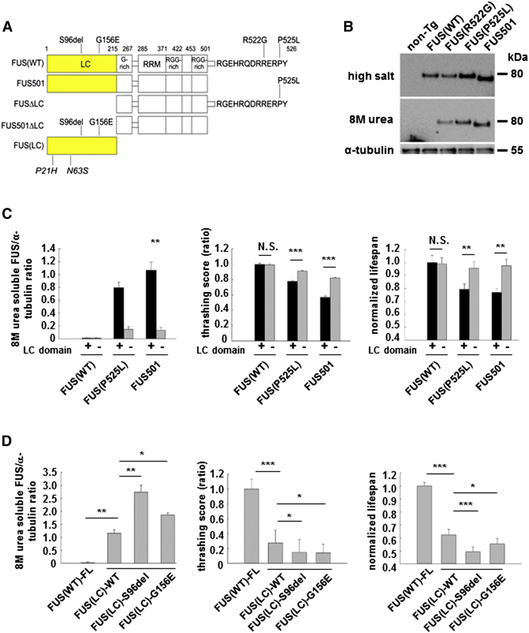
Figure 1 FUS Constructs, Cellular Localization, and Neurotoxicity of Mutant FUS (A) FUS constructs and mutations. Full-length FUS constructs include wild-type FUS(WT) and ALS/FTD mutations (R522G, R524S, P525L, and R495X), or FUS (FUS501), which mimics several ALS/FTD truncation mutants. LC motif at residues 2–214 was deleted in FUS(WT)ΔLC, FUS(P525L)ΔLC, and FUS501ΔLC. FUS(LC)-only constructs include wild-type FUS(LC), pathogenic S96del or G156E mutants, or clinically benign P21H and N63S sequences. LC, low-complexity motif; G-Rich, glycine-rich region; RRM, RNA recognition motif; RGG-rich, arginine-glycine-glycine-rich region. (B) FUS(WT) is located in neuronal nuclei; mutant FUS accumulates as 8 M urea-soluble assemblies in neuronal cytoplasm. Western blot of high-salt buffer extracted whole-worm lysates (top), 8 M urea extracted lysates (middle), and α-tubulin (bottom). (C) FUS LC domain is necessary and sufficient for FUS assembly and neurotoxicity. Deletion of LC domain from FUS(P525L) and FUS 501 reduces formation of 8 M urea-soluble FUS (left panel), improves motor function (middle panel), and lifespan in mutant FUS lines (right panel), but has no impact on FUS(WT) animals. N.S., not significant. ∗∗p < 0.01; ∗∗∗p < 0.001. (D) FUS LC domain is necessary and sufficient for FUS assembly and neurotoxicity. Overexpression of wild-type FUS(LC) alone is associated with increased 8 M urea-soluble assemblies (left panel), impaired motor function (middle panel), and lifespan (right panel). The ALS/FTD FUS(LC)-mutations (S96del and G156E) further augment neurotoxicity. ∗p < 0.05, ∗∗p < 0.01, and ∗∗∗p < 0.001. Error bars are SEM.
Figure 1 FUS Constructs, Cellular Localization, and Neurotoxicity of Mutant FUS (A) FUS constructs and mutations. Full-length FUS constructs include wild-type FUS(WT) and ALS/FTD mutations (R522G, R524S, P525L, and R495X), or FUS (FUS501), which mimics several ALS/FTD truncation mutants. LC motif at residues 2–214 was deleted in FUS(WT)ΔLC, FUS(P525L)ΔLC, and FUS501ΔLC. FUS(LC)-only constructs include wild-type FUS(LC), pathogenic S96del or G156E mutants, or clinically benign P21H and N63S sequences. LC, low-complexity motif; G-Rich, glycine-rich region; RRM, RNA recognition motif; RGG-rich, arginine-glycine-glycine-rich region. (B) FUS(WT) is located in neuronal nuclei; mutant FUS accumulates as 8 M urea-soluble assemblies in neuronal cytoplasm. Western blot of high-salt buffer extracted whole-worm lysates (top), 8 M urea extracted lysates (middle), and α-tubulin (bottom). (C) FUS LC domain is necessary and sufficient for FUS assembly and neurotoxicity. Deletion of LC domain from FUS(P525L) and FUS 501 reduces formation of 8 M urea-soluble FUS (left panel), improves motor function (middle panel), and lifespan in mutant FUS lines (right panel), but has no impact on FUS(WT) animals. N.S., not significant. ∗∗p < 0.01; ∗∗∗p < 0.001. (D) FUS LC domain is necessary and sufficient for FUS assembly and neurotoxicity. Overexpression of wild-type FUS(LC) alone is associated with increased 8 M urea-soluble assemblies (left panel), impaired motor function (middle panel), and lifespan (right panel). The ALS/FTD FUS(LC)-mutations (S96del and G156E) further augment neurotoxicity. ∗p < 0.05, ∗∗p < 0.01, and ∗∗∗p < 0.001. Error bars are SEM.CC BY · PubMed Central
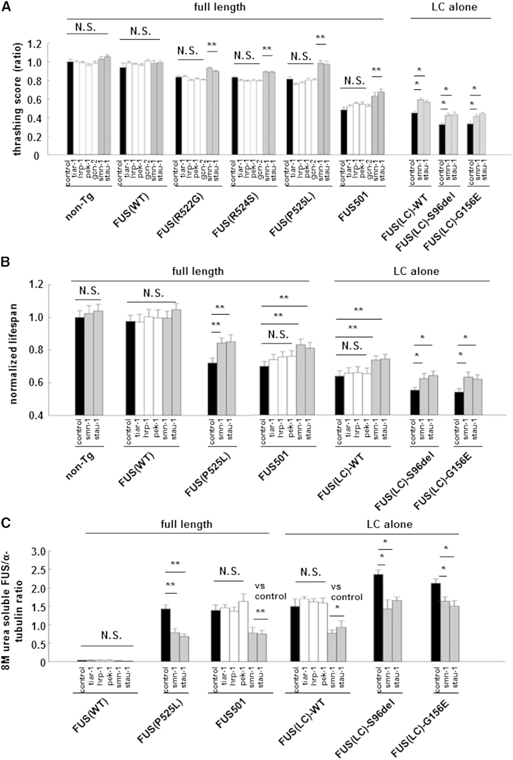
Figure 2 Selected RNA Binding Proteins Co-Localized with FUS in Mutant FUS Assemblies and Their RNAi Knockdown Partially Rescued Neurotoxicity (A) A directed RNAi screen of selected LC-domain containing RBPs revealed that RNAi knockdown of SMN (smn-1) and STAU-1 (stau-1) (gray bars) partially rescued motor function, whereas control RNAi knockdown (black bars) and RNAi knockdown of TIA-1 (tiar-1), hnRNP (hrp-1), EIF2AK (pek-1), and EIF2AK3 (gcn-2) (white bars) had no effect (N.S, not significant; ∗p < 0.05 and ∗∗p < 0.01). (B) RNAi knockdown of SMN and STAU-1 (gray bars) rescued lifespan, whereas RNAi of the other RNPs (white bars) and control RNAi (black bars) had no effect. N.S., not significant. ∗p < 0.05 and ∗∗p < 0.01. (C) RNAi knockdown of SMN and STAU-1 (gray bars) reduced 8 M urea-soluble FUS assemblies whereas control RNAi (black bars) and RNAi of the other RNPs (white bars) had no effect. N.S: not significant. ∗p < 0.05; ∗∗p < 0.01. Error bars are SEM.
Figure 2 Selected RNA Binding Proteins Co-Localized with FUS in Mutant FUS Assemblies and Their RNAi Knockdown Partially Rescued Neurotoxicity (A) A directed RNAi screen of selected LC-domain containing RBPs revealed that RNAi knockdown of SMN (smn-1) and STAU-1 (stau-1) (gray bars) partially rescued motor function, whereas control RNAi knockdown (black bars) and RNAi knockdown of TIA-1 (tiar-1), hnRNP (hrp-1), EIF2AK (pek-1), and EIF2AK3 (gcn-2) (white bars) had no effect (N.S, not significant; ∗p < 0.05 and ∗∗p < 0.01). (B) RNAi knockdown of SMN and STAU-1 (gray bars) rescued lifespan, whereas RNAi of the other RNPs (white bars) and control RNAi (black bars) had no effect. N.S., not significant. ∗p < 0.05 and ∗∗p < 0.01. (C) RNAi knockdown of SMN and STAU-1 (gray bars) reduced 8 M urea-soluble FUS assemblies whereas control RNAi (black bars) and RNAi of the other RNPs (white bars) had no effect. N.S: not significant. ∗p < 0.05; ∗∗p < 0.01. Error bars are SEM.CC BY · PubMed Central
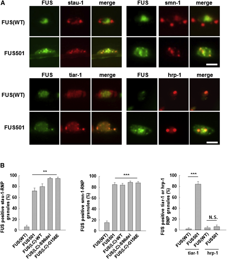
Figure 3 STAU-1, SMN, and TIAR-1 but Not hnRNP1 Colocalize with Mutant FUS in Cytoplasmic RNP Granules STAU-1, SMN, and TIAR-1 (red) colocalized with pathological intraneuronal cytoplasmic and axonal assemblies of mutant full-length FUS501, wild-type FUC-LC domain and mutant FUS(LC) domain (green) but did not colocalize with nuclear FUS(WT) (green). ∗∗∗p < 0.001. Scale bars, 2.0 μm. Additional data are shown in Figures S3B–S3D. STAU-1 colocalization: FUS(WT) = 6.0% ± 3.1%; FUS501 = 72.0 ± 5.3%; FUS(LC) = 80.0 ± 5.2%; FUS(LC)-S96del = 95.0 ± 1.7%; FUS(LC)-G156E = 95.0 ± 2.2; n = 20 neurons; p < 0.001; SMN colocalization: (FUS(WT) = 15.0 ± 3.1%; FUS 501 = 85.0 ± 3.4%; FUS(LC) = 84.0 ± 3.1%; FUS(LC)-S96del = 89.0 ± 2.3%; FUS(LC)-G156E = 88.0 ± 2.5%; n = 20 neurons; p < 0.001). Scale bars, 2.0 μm. Error bars are SEM.
Figure 3 STAU-1, SMN, and TIAR-1 but Not hnRNP1 Colocalize with Mutant FUS in Cytoplasmic RNP Granules STAU-1, SMN, and TIAR-1 (red) colocalized with pathological intraneuronal cytoplasmic and axonal assemblies of mutant full-length FUS501, wild-type FUC-LC domain and mutant FUS(LC) domain (green) but did not colocalize with nuclear FUS(WT) (green). ∗∗∗p < 0.001. Scale bars, 2.0 μm. Additional data are shown in Figures S3B–S3D. STAU-1 colocalization: FUS(WT) = 6.0% ± 3.1%; FUS501 = 72.0 ± 5.3%; FUS(LC) = 80.0 ± 5.2%; FUS(LC)-S96del = 95.0 ± 1.7%; FUS(LC)-G156E = 95.0 ± 2.2; n = 20 neurons; p < 0.001; SMN colocalization: (FUS(WT) = 15.0 ± 3.1%; FUS 501 = 85.0 ± 3.4%; FUS(LC) = 84.0 ± 3.1%; FUS(LC)-S96del = 89.0 ± 2.3%; FUS(LC)-G156E = 88.0 ± 2.5%; n = 20 neurons; p < 0.001). Scale bars, 2.0 μm. Error bars are SEM.CC BY · PubMed Central
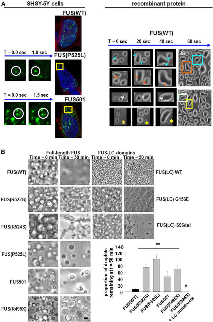
Figure 4 Recombinant FUS Generates Liquid Droplet-Like Assemblies, the Reversibility of which Is Reduced by ALS/FTD Mutants (A) (Left panel) SHSY5Y cells transiently transfected with YFP-tagged wild-type FUS show static intranuclear spherical structures. (Top) In the same cell type, mutant FUS localizes in the cytoplasm as mobile spherical structures (“droplets”) that move, fuse, and disappear (middle and bottom—time series of two fusion events). (Right panel) Cooling of 1–5 μM solutions of either full-length FUS or FUS(LC) domain, induces spontaneous phase transition into liquid droplets, which spontaneously fuse (∗), split over seconds, and redisperse into solute over ∼50 min. (B) Full-length FUS(WT) liquid droplets are more labile than full-length mutant FUS or FUS(LC) droplets. Both mutant and wild-type FUS proteins form multiple bright spherical liquid droplets after cooling (left column). FUS(WT) droplets almost completely disperse by 50 min at 23°C (right column). FUS proteins containing ALS/FTD mutants formed more stable droplets, many of which were still visible at 50 min at 23°C. The residual mutant assemblies had darkly refractile properties, angular edges, and/or spiculated appearances. We interpret these structures to represent further phase transitions to gel-like or solid assemblies. Spiculated assemblies were especially prevalent with the FUS(LC) constructs, where virtually all of the residual FUS(LC) material had this appearance (# in the graph). Error bars are SEM.
Figure 4 Recombinant FUS Generates Liquid Droplet-Like Assemblies, the Reversibility of which Is Reduced by ALS/FTD Mutants (A) (Left panel) SHSY5Y cells transiently transfected with YFP-tagged wild-type FUS show static intranuclear spherical structures. (Top) In the same cell type, mutant FUS localizes in the cytoplasm as mobile spherical structures (“droplets”) that move, fuse, and disappear (middle and bottom—time series of two fusion events). (Right panel) Cooling of 1–5 μM solutions of either full-length FUS or FUS(LC) domain, induces spontaneous phase transition into liquid droplets, which spontaneously fuse (∗), split over seconds, and redisperse into solute over ∼50 min. (B) Full-length FUS(WT) liquid droplets are more labile than full-length mutant FUS or FUS(LC) droplets. Both mutant and wild-type FUS proteins form multiple bright spherical liquid droplets after cooling (left column). FUS(WT) droplets almost completely disperse by 50 min at 23°C (right column). FUS proteins containing ALS/FTD mutants formed more stable droplets, many of which were still visible at 50 min at 23°C. The residual mutant assemblies had darkly refractile properties, angular edges, and/or spiculated appearances. We interpret these structures to represent further phase transitions to gel-like or solid assemblies. Spiculated assemblies were especially prevalent with the FUS(LC) constructs, where virtually all of the residual FUS(LC) material had this appearance (# in the graph). Error bars are SEM.CC BY · PubMed Central
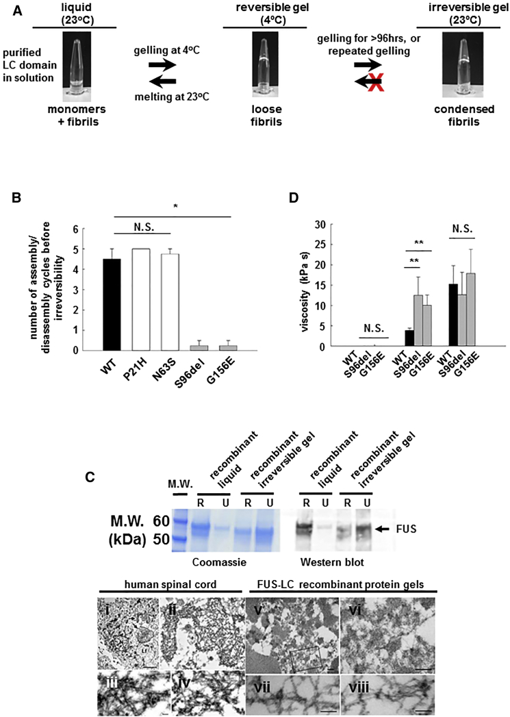
Figure 5 Recombinant FUS Generates Gel-Like Assemblies, the Reversibility of which Is Reduced by ALS/FTD Mutants (A) Purified recombinant FUS(LC) forms clear solution at 23°C (Top left panel) but forms reversible gel-like structures at 4°C (Top center panel). Rewarming to 23°C reverts FUS(LC) to a liquid state (top left panel). Repeated cycling of wild-type FUS(LC) eventually results in the formation of irreversible gels (top right panel). (B) ∼4 to 5 assembly/disassembly cycles can be achieved by wild-type FUS(LC) (black bar) and by FUS(LC) containing benign polymorphic variants (P21H and N63S, white bars). ALS/FTD mutations (gray bars) dramatically reduce the number of cycles to ≤1 before irreversibility (∗∗p < 0.01). (C) Top panel: Coomassie gel (left) and western blot (right) of RIPA- and 8 M urea solubilisation of FUS(LC) reveal that liquid state FUS(LC) is fully soluble in RIPA buffer. Irreversible FUS(LC) is partly soluble in RIPA, but fully soluble in 8 M urea. Bottom panel: Transmission electron microscopy of human FUS inclusions ([Ci]–[Civ]) and irreversible recombinant protein gels ([Cv]–[Cviii]). Both are composed of loose networks of ∼14 nm fibrils. Scale bars are 500 nm ([Cv] and [Cvi]) and 100 nm ([Cvii] and [Cviii]). (D) Analysis of liquid, reversible gel, and irreversible gel using fluorescent particle tracking revealed that liquid (left cluster) and reversible gels (black bars, middle cluster) have viscosities similar to those of P-granules. Mutant reversible gels (gray bars, middle cluster) were more viscous than wild-type reversible gels. Irreversible gels, regardless of whether made from wild-type or ALS/FTD mutant FUS exhibit very high viscosities. N.S., not significant; ∗∗p < 0.01. Error bars are SEM.
Figure 5 Recombinant FUS Generates Gel-Like Assemblies, the Reversibility of which Is Reduced by ALS/FTD Mutants (A) Purified recombinant FUS(LC) forms clear solution at 23°C (Top left panel) but forms reversible gel-like structures at 4°C (Top center panel). Rewarming to 23°C reverts FUS(LC) to a liquid state (top left panel). Repeated cycling of wild-type FUS(LC) eventually results in the formation of irreversible gels (top right panel). (B) ∼4 to 5 assembly/disassembly cycles can be achieved by wild-type FUS(LC) (black bar) and by FUS(LC) containing benign polymorphic variants (P21H and N63S, white bars). ALS/FTD mutations (gray bars) dramatically reduce the number of cycles to ≤1 before irreversibility (∗∗p < 0.01). (C) Top panel: Coomassie gel (left) and western blot (right) of RIPA- and 8 M urea solubilisation of FUS(LC) reveal that liquid state FUS(LC) is fully soluble in RIPA buffer. Irreversible FUS(LC) is partly soluble in RIPA, but fully soluble in 8 M urea. Bottom panel: Transmission electron microscopy of human FUS inclusions ([Ci]–[Civ]) and irreversible recombinant protein gels ([Cv]–[Cviii]). Both are composed of loose networks of ∼14 nm fibrils. Scale bars are 500 nm ([Cv] and [Cvi]) and 100 nm ([Cvii] and [Cviii]). (D) Analysis of liquid, reversible gel, and irreversible gel using fluorescent particle tracking revealed that liquid (left cluster) and reversible gels (black bars, middle cluster) have viscosities similar to those of P-granules. Mutant reversible gels (gray bars, middle cluster) were more viscous than wild-type reversible gels. Irreversible gels, regardless of whether made from wild-type or ALS/FTD mutant FUS exhibit very high viscosities. N.S., not significant; ∗∗p < 0.01. Error bars are SEM.CC BY · PubMed Central
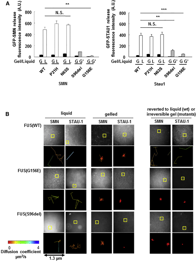
Figure 6 Mutant FUS Forms Poorly Reversible Gels, which Fail to Release Cargo such as SMN and STAU-1 (A) When recombinant GFP-tagged SMN (or STAU-1) protein is mixed with FUS(LC) protein and gel assembly is induced by cooling, little SMN (or STAU-1) is released into the buffer (as measured by fluorescence) regardless of whether the FUS gel is derived from wild-type or ALS/FTD mutant FUS(LC) (black bars). When the gel is reversed by warming to 23°C (white bars), significant SMN (or STAU-1) protein is released into the buffer from FUS(WT) hydrogels. Very little SMN (or STAU-1) is released by the irreversible ALS/FTD mutant FUS(LC) gels (gray bars;∗∗p < 0.001). (B) Fluorescent molecule tracking in liquid, reversible gel, and irreversible gel assemblies reveals that in liquid state, SMN and STAU-1 diffuse rapidly at 1–4 μm/s in both mutant and wild-type samples (left two columns). On formation of a gel (center two columns), movement of SMN and STAU-1 is constrained (<1 μm2/s). On rewarming, reversible FUS(WT) gels disassemble and release SMN and STAU-1. The irreversible mutant gels continue to constrain movement of SMN and STAU-1. Image stacks are captured at 29 Hz; pixel width corresponds to 160 nm on the specimen. Representative areas are boxed and visualized in a 1.3 μm2 plot, color coded to the diffusion coefficient fitted to each track. Error bars are SEM.
Figure 6 Mutant FUS Forms Poorly Reversible Gels, which Fail to Release Cargo such as SMN and STAU-1 (A) When recombinant GFP-tagged SMN (or STAU-1) protein is mixed with FUS(LC) protein and gel assembly is induced by cooling, little SMN (or STAU-1) is released into the buffer (as measured by fluorescence) regardless of whether the FUS gel is derived from wild-type or ALS/FTD mutant FUS(LC) (black bars). When the gel is reversed by warming to 23°C (white bars), significant SMN (or STAU-1) protein is released into the buffer from FUS(WT) hydrogels. Very little SMN (or STAU-1) is released by the irreversible ALS/FTD mutant FUS(LC) gels (gray bars;∗∗p < 0.001). (B) Fluorescent molecule tracking in liquid, reversible gel, and irreversible gel assemblies reveals that in liquid state, SMN and STAU-1 diffuse rapidly at 1–4 μm/s in both mutant and wild-type samples (left two columns). On formation of a gel (center two columns), movement of SMN and STAU-1 is constrained (<1 μm2/s). On rewarming, reversible FUS(WT) gels disassemble and release SMN and STAU-1. The irreversible mutant gels continue to constrain movement of SMN and STAU-1. Image stacks are captured at 29 Hz; pixel width corresponds to 160 nm on the specimen. Representative areas are boxed and visualized in a 1.3 μm2 plot, color coded to the diffusion coefficient fitted to each track. Error bars are SEM.CC BY · PubMed Central
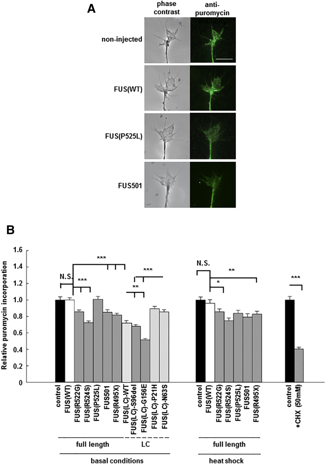
Figure 7 Mutant FUS and FUS(LC) Domain, but Not Full-Length Wild-Type FUS, Cause Reduced New Protein Synthesis When Expressed in Cultured Neurons (A) New protein synthesis was detected by puromycylation in axon terminals of cultured Xenopus retinal ganglion neurons (left panel: phase contrast; right panel: anti-puromycin fluorescent images) expressing equivalent quantities of full-length FUS(WT) or various mutant and truncated human FUS proteins. Scale bar, 10 μm. (B) Under basal conditions, de novo protein synthesis is equivalent in non-injected neurons, and in neurons expressing FUS(WT). However, de novo protein synthesis was significantly reduced in neurons expressing ALS/FTD mutants including: FUS(R522G), FUS(R524S), FUS501, FUS(R495X), and in gelling-prone FUS(LC)(WT), FUS(LC)(S96del), FUS(LC)(G156E), FUS(LC)(P21H), and FUS(LC)(N63S). In good agreement with the in vivo studies in C. elegans, heat shock causes significant reduction of new protein synthesis in neurons expressing mutant FUS, but minimal impact on non-transfected and FUS(WT) expressing neurons (right panel). N.S., not significant; ∗p < 0.05, ∗∗p < 0.01, and ∗∗∗p < 0.001. Error bars are SEM.
Figure 7 Mutant FUS and FUS(LC) Domain, but Not Full-Length Wild-Type FUS, Cause Reduced New Protein Synthesis When Expressed in Cultured Neurons (A) New protein synthesis was detected by puromycylation in axon terminals of cultured Xenopus retinal ganglion neurons (left panel: phase contrast; right panel: anti-puromycin fluorescent images) expressing equivalent quantities of full-length FUS(WT) or various mutant and truncated human FUS proteins. Scale bar, 10 μm. (B) Under basal conditions, de novo protein synthesis is equivalent in non-injected neurons, and in neurons expressing FUS(WT). However, de novo protein synthesis was significantly reduced in neurons expressing ALS/FTD mutants including: FUS(R522G), FUS(R524S), FUS501, FUS(R495X), and in gelling-prone FUS(LC)(WT), FUS(LC)(S96del), FUS(LC)(G156E), FUS(LC)(P21H), and FUS(LC)(N63S). In good agreement with the in vivo studies in C. elegans, heat shock causes significant reduction of new protein synthesis in neurons expressing mutant FUS, but minimal impact on non-transfected and FUS(WT) expressing neurons (right panel). N.S., not significant; ∗p < 0.05, ∗∗p < 0.01, and ∗∗∗p < 0.001. Error bars are SEM.CC BY · PubMed Central
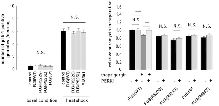
Figure 8 Neurotoxicity of Mutant FUS in C. elegans and Reduced New Protein Synthesis in Xenopus Neurons Are Unlikely to be Due to Activation of the UPR due to Accumulation of Misfolded FUS (A) In vivo stress granule density measured by counting pab-l-positive granules in C. elegans under basal conditions and after heat shock were not different between control (empty vector); FUS(WT) and mutant FUS animals, indicating that mutant FUS animals were not under greater unfolded protein stress. (B) Thapsigargin treatment induced UPR and reduced new protein synthesis in Xenopus neurons expressing FUS(WT). PERK1 inhibitors rescued reduced protein synthesis in thapsigargin-treated neurons expressing FUS(WT), but not mutant FUS-expressing neurons. Error bars are SEM.
Figure 8 Neurotoxicity of Mutant FUS in C. elegans and Reduced New Protein Synthesis in Xenopus Neurons Are Unlikely to be Due to Activation of the UPR due to Accumulation of Misfolded FUS (A) In vivo stress granule density measured by counting pab-l-positive granules in C. elegans under basal conditions and after heat shock were not different between control (empty vector); FUS(WT) and mutant FUS animals, indicating that mutant FUS animals were not under greater unfolded protein stress. (B) Thapsigargin treatment induced UPR and reduced new protein synthesis in Xenopus neurons expressing FUS(WT). PERK1 inhibitors rescued reduced protein synthesis in thapsigargin-treated neurons expressing FUS(WT), but not mutant FUS-expressing neurons. Error bars are SEM.CC BY · PubMed Central

Verification

Claims the paper makes

The claims and statistics of this paper haven’t been checked yet.

Run the check — free, no account needed. It reads the open-access text and checks the sources it cites.

A few free checks a day · papers already verified open instantly · results are public

Claims other papers make about this one

Each is checked against this paper’s own text: does it say what the citing paper says it does?

Self-check

Percentages, sums and reported differences re-computed from the paper’s own numbers.

The self-check needs the paper’s full text.

Citations

Cites

Research works printed but not linked: 43

  1. Akbalik, 2014, Molecular biology. mRNA, live and unmasked, Science, 343, 375, 10.1126/science.1249623 · DOI
  2. Alami, 2014, Axonal transport of TDP-43 mRNA granules is impaired by ALS-causing mutations, Neuron, 81, 536, 10.1016/j.neuron.2013.12.018 · DOI · Paper page
  3. Anderson, 2002, Visibly stressed: the role of eIF2, TIA-1, and stress granules in protein translation, Cell Stress Chaperones, 7, 213, 10.1379/1466-1268(2002)007<0213:VSTROE>2.0.CO;2 · DOI
  4. Axten, 2012, Discovery of 7-methyl-5-(1-[3-(trifluoromethyl)phenyl]acetyl-2,3-dihydro-1H-indol-5-yl)-7H-pyrrolo[2,3-d]pyrimidin-4-amine (GSK2606414), a potent and selective first-in-class inhibitor of protein kinase R (PKR)-like endoplasmic reticulum kinase (PERK), J. Med. Chem., 55, 7193, 10.1021/jm300713s · DOI
  5. Bosco, 2010, Mutant FUS proteins that cause amyotrophic lateral sclerosis incorporate into stress granules, Hum. Mol. Genet., 19, 4160, 10.1093/hmg/ddq335 · DOI
  6. Brangwynne, 2009, Germline P granules are liquid droplets that localize by controlled dissolution/condensation, Science, 324, 1729, 10.1126/science.1172046 · DOI
  7. Couthouis, 2011, Feature Article: A yeast functional screen predicts new candidate ALS disease genes, Proc. Natl. Acad. Sci. USA, 108, 20881, 10.1073/pnas.1109434108 · DOI
  8. Crocker, 1996, J. Colloid Interface Sci., 179, 298, 10.1006/jcis.1996.0217 · DOI
  9. Decker, 2012, P-bodies and stress granules: possible roles in the control of translation and mRNA degradation, Cold Spring Harb. Perspect. Biol., 4, a012286, 10.1101/cshperspect.a012286 · DOI
  10. DeJesus-Hernandez, 2010, De novo truncating FUS gene mutation as a cause of sporadic amyotrophic lateral sclerosis, Hum. Mutat., 31, E1377, 10.1002/humu.21241 · DOI
  11. Dixon, 2006, FGF negatively regulates muscle membrane extension in Caenorhabditis elegans, Development, 133, 1263, 10.1242/dev.02300 · DOI
  12. Dormann, 2009, Proteolytic processing of TAR DNA binding protein-43 by caspases produces C-terminal fragments with disease defining properties independent of progranulin, J. Neurochem., 110, 1082, 10.1111/j.1471-4159.2009.06211.x · DOI
  13. Fang, 2014, Full-length TDP-43 forms toxic amyloid oligomers that are present in frontotemporal lobar dementia-TDP patients, Nat. Commun., 5, 4824, 10.1038/ncomms5824 · DOI
  14. Gendron, 2010, Review: transactive response DNA-binding protein 43 (TDP-43): mechanisms of neurodegeneration, Neuropathol. Appl. Neurobiol., 36, 97, 10.1111/j.1365-2990.2010.01060.x · DOI
  15. Han, 2012, Cell-free formation of RNA granules: bound RNAs identify features and components of cellular assemblies, Cell, 149, 768, 10.1016/j.cell.2012.04.016 · DOI
  16. Hara, 2012, Lower motor neuron disease caused by a novel FUS/TLS gene frameshift mutation, J. Neurol., 259, 2237
  17. Jung, 2014, Remote control of gene function by local translation, Cell, 157, 26, 10.1016/j.cell.2014.03.005 · DOI
  18. Kamath, 2003, Systematic functional analysis of the Caenorhabditis elegans genome using RNAi, Nature, 421, 231, 10.1038/nature01278 · DOI
  19. Kaminski Schierle, 2011, A FRET sensor for non-invasive imaging of amyloid formation in vivo, ChemPhysChem, 12, 673, 10.1002/cphc.201000996 · DOI
  20. Kent, 2014, Autosomal dominant inheritance of rapidly progressive amyotrophic lateral sclerosis due to a truncation mutation in the fused in sarcoma (FUS) gene, Amyotroph. Lateral Scler. Frontotemporal Degener., 15, 557, 10.3109/21678421.2014.920033 · DOI
  21. Li, 2013, Stress granules as crucibles of ALS pathogenesis, J. Cell Biol., 201, 361, 10.1083/jcb.201302044 · DOI
  22. Lin, 2009, Cytoplasmic polyadenylation and cytoplasmic polyadenylation element-dependent mRNA regulation are involved in Xenopus retinal axon development, Neural Dev., 4, 8, 10.1186/1749-8104-4-8 · DOI
  23. Liu-Yesucevitz, 2014, ALS-linked mutations enlarge TDP-43-enriched neuronal RNA granules in the dendritic arbor, J. Neurosci., 34, 4167, 10.1523/JNEUROSCI.2350-13.2014 · DOI
  24. Mao, 2010, Protein misfolding induces hypoxic preconditioning via a subset of the unfolded protein response machinery, Mol. Cell. Biol., 30, 5033, 10.1128/MCB.00922-10 · DOI
  25. Mitchell, 2014, Principles and properties of eukaryotic mRNPs, Mol. Cell, 54, 547, 10.1016/j.molcel.2014.04.033 · DOI
  26. Morita, 2009, Effects of non-steroidal anti-inflammatory drugs on Abeta deposition in Abeta(1-42) transgenic C. elegans, Brain Res., 1295, 186, 10.1016/j.brainres.2009.08.002 · DOI
  27. Murakami, 2012, ALS mutations in FUS cause neuronal dysfunction and death in Caenorhabditis elegans by a dominant gain-of-function mechanism, Hum. Mol. Genet., 21, 1, 10.1093/hmg/ddr417 · DOI
  28. Neumann, 2007, TDP-43 proteinopathy in frontotemporal lobar degeneration and amyotrophic lateral sclerosis: protein misfolding diseases without amyloidosis, Arch. Neurol., 64, 1388, 10.1001/archneur.64.10.1388 · DOI
  29. Nishitoh, 2002, ASK1 is essential for endoplasmic reticulum stress-induced neuronal cell death triggered by expanded polyglutamine repeats, Genes Dev., 16, 1345, 10.1101/gad.992302 · DOI
  30. Rademakers, 2012, Advances in understanding the molecular basis of frontotemporal dementia, Nat. Rev. Neurol., 8, 423, 10.1038/nrneurol.2012.117 · DOI
  31. Ramaswami, 2013, Altered ribostasis: RNA-protein granules in degenerative disorders, Cell, 154, 727, 10.1016/j.cell.2013.07.038 · DOI
  32. Rees, 2013, The construction of power in family medicine bedside teaching: a video observation study, Med. Educ., 47, 154, 10.1111/medu.12055 · DOI
  33. Robinson, 2013, TDP-43 skeins show properties of amyloid in a subset of ALS cases, Acta Neuropathol., 125, 121, 10.1007/s00401-012-1055-8 · DOI
  34. Scheuner, 2001, Translational control is required for the unfolded protein response and in vivo glucose homeostasis, Mol. Cell, 7, 1165, 10.1016/S1097-2765(01)00265-9 · DOI
  35. Schmidt, 2009, SUnSET, a nonradioactive method to monitor protein synthesis, Nat. Methods, 6, 275, 10.1038/nmeth.1314 · DOI · Paper page
  36. Schwartz, 2013, RNA seeds higher-order assembly of FUS protein, Cell Rep., 5, 918, 10.1016/j.celrep.2013.11.017 · DOI
  37. Tartaglia, 2008, The Zyggregator method for predicting protein aggregation propensities, Chem. Soc. Rev., 37, 1395, 10.1039/b706784b · DOI
  38. Ticozzi, 2010, Protein aggregation and defective RNA metabolism as mechanisms for motor neuron damage, CNS Neurol. Disord. Drug Targets, 9, 285, 10.2174/187152710791292585 · DOI
  39. tom Dieck, 2015, Direct visualization of newly synthesized target proteins in situ, Nat. Methods, 12, 411, 10.1038/nmeth.3319 · DOI
  40. Uchikado, 2006, Heterogeneous inclusions in neurofilament inclusion disease, Neuropathology, 26, 417, 10.1111/j.1440-1789.2006.00709.x · DOI
  41. van der Zee, 2014, Dementia in 2013: frontotemporal lobar degeneration-building on breakthroughs, Nat. Rev. Neurol., 10, 70, 10.1038/nrneurol.2013.270 · DOI
  42. Wolozin, 2012, Regulated protein aggregation: stress granules and neurodegeneration, Mol. Neurodegener., 7, 56, 10.1186/1750-1326-7-56 · DOI
  43. Yan, 2010, Frameshift and novel mutations in FUS in familial amyotrophic lateral sclerosis and ALS/dementia, Neurology, 75, 807, 10.1212/WNL.0b013e3181f07e0c · DOI

Cited by

See all 909

Metadata

Authors
Murakami, T., Qamar, S., Lin, J. Q., Schierle, G. S. K., Rees, E., Miyashita, A., Costa, A. R., Dodd, R. B., Chan, F. T. S., Michel, C. H., Kronenberg-Versteeg, D., Li, Y., Yang, S.-P., Wakutani, Y., Meadows, W., Ferry, R. R., Dong, L., Tartaglia, G. G., Favrin, G., Lin, W.-L., Dickson, D. W., Zhen, M., Ron, D., Schmitt-Ulms, G., Fraser, P. E., Shneider, N. A., Holt, C., Vendruscolo, M., Kaminski, C. F., & St George-Hyslop, P.
Venue
Neuron
Publisher
Elsevier BV
Published
2015-11
Volume
88
Issue
4
Pages
678-690
ISSN
0896-6273
Cited by
909
References
6
Language
en

About these checks

Registry, retraction, Cites and statistics results are computed, not judged, with one exception: a printed reference that no index lists is read once by a model before it counts as a citation error. Claims are extracted by a model and kept only when their quote appears verbatim in the text; each cited source is then checked, with a model’s help, for whether it says what the claim relies on. Those verdicts can be wrong — read the claim’s quote and the cited source before relying on one.

Statistics and arithmetic are recomputed, not judged.

Confidence bands are shown only for fields calibrated on our annotated test set. None is calibrated yet, so no band is shown.

What counts as a claim ›How we measure confidence ›How we check this ›

Are you an author, or do you see a problem with this page? Request a review

Data sources

Bibliographic data: Semantic Scholar, OpenAlex, Crossref.

Contains information from the Semantic Scholar Academic Graph Datasets, made available under the ODC Attribution License (ODC-BY 1.0).

Semantic Scholar’s data is described in: Kinney, R., et al. (2023). The Semantic Scholar Open Data Platform. arXiv:2301.10140.