Cited by 133Neural and Behavioral Psychology StudiesNeurotransmitter Receptor Influence on BehaviorDeep Brain Stimulation and Movement Disorders

The DOI matches its Crossref record; 79 of the 79 references it cites were found in a registry or an index.

A supramodal role of the basal ganglia in memory and motor inhibition: Meta-analytic evidence

Open access

Abstract

The ability to stop actions and thoughts is essential for goal-directed behaviour. Neuroimaging research has revealed that stopping actions and thoughts engage similar cortical mechanisms, including the ventro- and dorso-lateral prefrontal cortex. However, whether and how these abilities require similar subcortical mechanisms remains unexplored. Specifically of interest are the basal ganglia, subcortical structures long-known for their motor functions, but less so for their role in cognition. To investigate the potential common mechanisms in the basal ganglia underlying action and thought stopping, we conducted meta-analyses using fMRI data from the Go/No-Go, Stop-signal, and Think/No-Think tasks. All three tasks require active stopping of prepotent actions or thoughts. To localise basal ganglia activations, we performed high-resolution manual segmentations of striatal subregions. We found that all three tasks recovered clusters in the basal ganglia, although the specific localisation of these clusters differed. Although the Go/No-Go and Stop-signal tasks are often interchangeably used for measuring action stopping, their cluster locations in the basal ganglia did not significantly overlap. These different localised clusters suggest that the Go/No-Go and Stop-signal tasks may recruit distinct basal ganglia stopping processes, and therefore should not be treated equivalently. More importantly, the basal ganglia cluster recovered from the Think/No-Think task largely co-localised with that from the Stop-signal task, but not the Go/No-Go task, possibly indicating that the Think/No-Think and Stop-signal tasks share a common striatal circuitry involved in the cancellation of unwanted thoughts and actions. The greater similarity of the Think/No-Think task to the Stop-Signal rather than Go/No-Go task also was echoed at the cortical level, which revealed highly overlapping and largely right lateralized set of regions including the anterior DLPFC, VLPFC, Pre-SMA and ACC. Overall, we provide novel evidence suggesting not only that the basal ganglia are critical for thought stopping, but also that they are involved in specific stopping subprocesses that can be engaged by tasks in different domains. These findings raise the possibility that the basal ganglia may be part of a supramodal network responsible for stopping unwanted processes more broadly.

Overview

The claims and statistics of this paper haven’t been checked yet.

Run the check — free, no account needed. It reads the open-access text and checks the sources it cites.

A few free checks a day · papers already verified open instantly · results are public

Integrity signals

Registry

Crossref · 10.1016/j.neuropsychologia.2017.11.033

Cites

79 of 79 references verified0 citation errors · 0 not checkable

Statistics

Figures

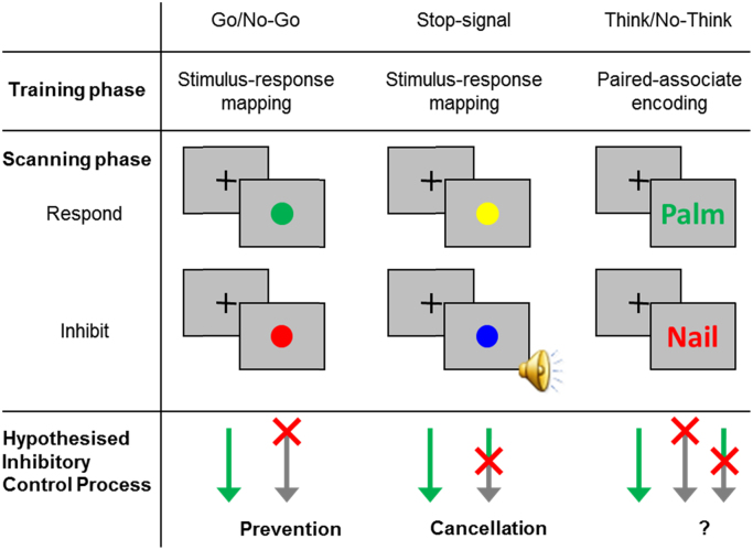
Fig. 1.1 Typical Go/No-Go, Stop-signal, and Think/No-Think Paradigms and the Hypothesised Inhibitory Control Processes. In the hypothesised inhibitory control process panel, the arrows denote the time-flow within a single trial. The colour green represents the respond processes, the red “X” represents when inhibitory control is putatively engaged in the trial, and the grey represents the inhibited processes. On a Go or Think trial, participants would carry out the motor response or memory retrieval, respectively. On an inhibit trial, if prevention processes are engaged, inhibitory control should be effective from the very beginning of the trial, before the corresponding response is even started. If cancellation processes are engaged, inhibitory control would be recruited only to terminate an initiated response. In the lower right, the uncertain positioning of the “X” indicates that we do not know whether prevention or cancellation may be more important for the Think/No-Think task.
Fig. 1.1 Typical Go/No-Go, Stop-signal, and Think/No-Think Paradigms and the Hypothesised Inhibitory Control Processes. In the hypothesised inhibitory control process panel, the arrows denote the time-flow within a single trial. The colour green represents the respond processes, the red “X” represents when inhibitory control is putatively engaged in the trial, and the grey represents the inhibited processes. On a Go or Think trial, participants would carry out the motor response or memory retrieval, respectively. On an inhibit trial, if prevention processes are engaged, inhibitory control should be effective from the very beginning of the trial, before the corresponding response is even started. If cancellation processes are engaged, inhibitory control would be recruited only to terminate an initiated response. In the lower right, the uncertain positioning of the “X” indicates that we do not know whether prevention or cancellation may be more important for the Think/No-Think task.CC BY · PubMed Central
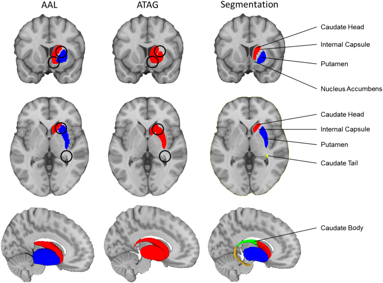
Fig. 2.1 Segmentation of the striatal subregions. The three columns compare the AAL and ATAG atlases with our manual segmentation. The top row shows the coronal section, the middle row shows the axial section, and the bottom row shows the 3D rending of the structures in the sagittal plane. The relevant structures are labelled, and the differences are marked with black circles. Anatomical underlay and subcortical renders are displayed in MNI space.
Fig. 2.1 Segmentation of the striatal subregions. The three columns compare the AAL and ATAG atlases with our manual segmentation. The top row shows the coronal section, the middle row shows the axial section, and the bottom row shows the 3D rending of the structures in the sagittal plane. The relevant structures are labelled, and the differences are marked with black circles. Anatomical underlay and subcortical renders are displayed in MNI space.CC BY · PubMed Central
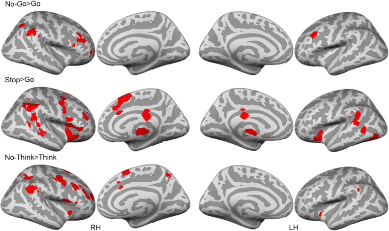
Fig. 3.1 Cortical activations from the task-specific meta-analyses. All clusters are thresholded using cluster-level inference (p<.05, uncorrected p<.001, threshold permutations=1000).
Fig. 3.1 Cortical activations from the task-specific meta-analyses. All clusters are thresholded using cluster-level inference (p<.05, uncorrected p<.001, threshold permutations=1000).CC BY · PubMed Central
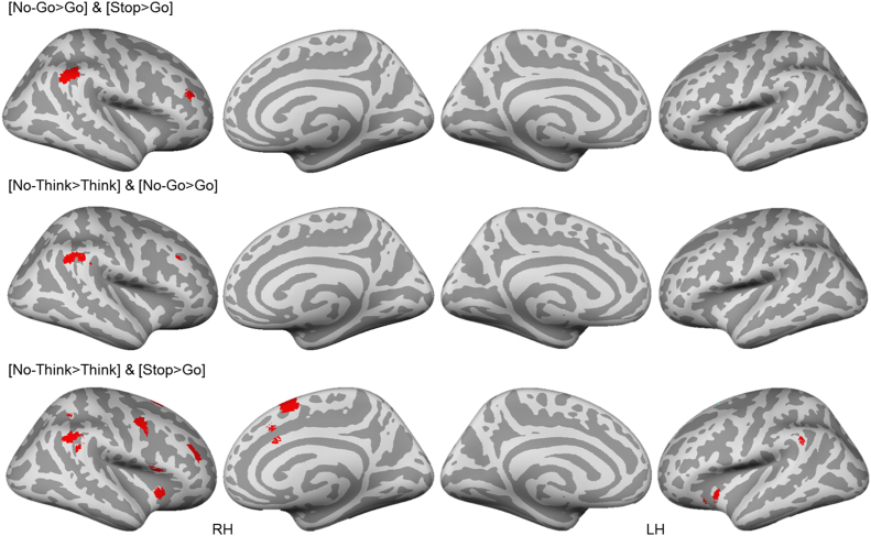
Fig. 3.2 Cross-task conjunction analysis. All clusters are thresholded using cluster-level inference (p<.05, uncorrected p<.001, threshold permutations=1000).
Fig. 3.2 Cross-task conjunction analysis. All clusters are thresholded using cluster-level inference (p<.05, uncorrected p<.001, threshold permutations=1000).CC BY · PubMed Central
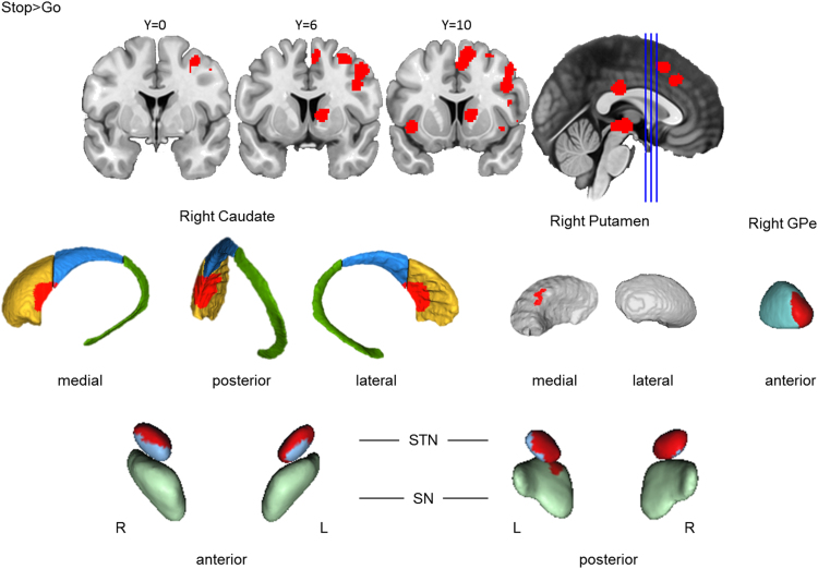
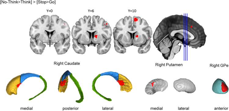
Fig. 3.3 Basal ganglia activation for action cancellation. Top row: Clusters are presented on coronal slices of a high-resolution MNI atlas. Reference lines for the coronal slices are presented in the sagittal plane. Middle row: Clusters are displayed on high-resolution parcellations of the caudate, putamen, and external globus pallidus (GPe). Bottom row: Clusters are displayed on high-resolution parcellaions of the subthalamic nucleus (STN) and substantia nigra (SN). All clusters are thresholded using cluster-level inference (p<.05, uncorrected p<.001, threshold permutations=1000).
Fig. 3.3 Basal ganglia activation for action cancellation. Top row: Clusters are presented on coronal slices of a high-resolution MNI atlas. Reference lines for the coronal slices are presented in the sagittal plane. Middle row: Clusters are displayed on high-resolution parcellations of the caudate, putamen, and external globus pallidus (GPe). Bottom row: Clusters are displayed on high-resolution parcellaions of the subthalamic nucleus (STN) and substantia nigra (SN). All clusters are thresholded using cluster-level inference (p<.05, uncorrected p<.001, threshold permutations=1000).CC BY · PubMed Central
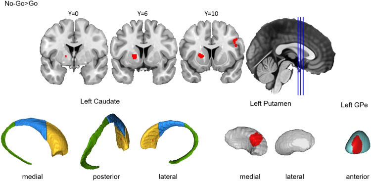
Fig. 3.4 Basal ganglia activation for action prevention. Top row: Clusters are presented on coronal slices of a high-resolution MNI atlas. Reference lines for the coronal slices are presented in the sagittal plane. Bottom row: Clusters are displayed on high-resolution parcellations of the caudate, putamen, and external globus pallidus (GPe). All clusters are thresholded using cluster-level inference (p<.05, uncorrected p<.001, threshold permutations=1000).
Fig. 3.4 Basal ganglia activation for action prevention. Top row: Clusters are presented on coronal slices of a high-resolution MNI atlas. Reference lines for the coronal slices are presented in the sagittal plane. Bottom row: Clusters are displayed on high-resolution parcellations of the caudate, putamen, and external globus pallidus (GPe). All clusters are thresholded using cluster-level inference (p<.05, uncorrected p<.001, threshold permutations=1000).CC BY · PubMed Central
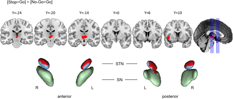
Fig. 3.5 Action cancellation reliably engaged STN and SN more than action prevention. Top row: Clusters are presented on coronal slices of a high-resolution MNI atlas. Reference lines for the coronal slices are presented in the sagittal plane. Bottom row: Clusters are displayed on high-resolution parcellaions of the subthalamic nucleus (STN) and substantia nigra (SN). The contrast analysis was computed using the thresholded ALE images from the individual analyses. All clusters are thresholded at uncorrected p<.001, with the p-value permutations of 10,000 iterations, and the minimum cluster volume of 200 mm3.
Fig. 3.5 Action cancellation reliably engaged STN and SN more than action prevention. Top row: Clusters are presented on coronal slices of a high-resolution MNI atlas. Reference lines for the coronal slices are presented in the sagittal plane. Bottom row: Clusters are displayed on high-resolution parcellaions of the subthalamic nucleus (STN) and substantia nigra (SN). The contrast analysis was computed using the thresholded ALE images from the individual analyses. All clusters are thresholded at uncorrected p<.001, with the p-value permutations of 10,000 iterations, and the minimum cluster volume of 200 mm3.CC BY · PubMed Central
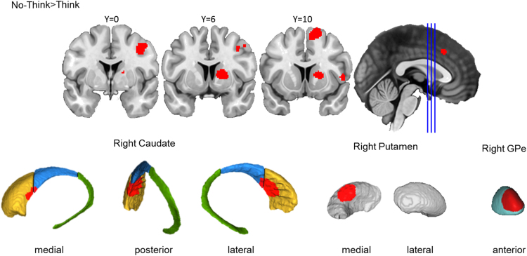
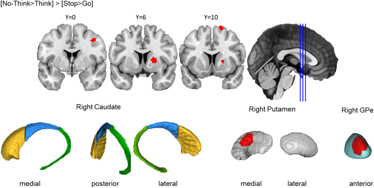
Fig. 3.6 Memory inhibition engaged the right basal ganglia. Top row: Clusters are presented on coronal slices of a high-resolution MNI atlas. Reference lines for the coronal slices are presented in the sagittal plane. Bottom row: Clusters are displayed on high-resolution parcellations of the caudate, putamen, and external globus pallidus (GPe). All clusters are thresholded using cluster-level inference (p<.05, uncorrected p<.001, threshold permutations=1000).
Fig. 3.6 Memory inhibition engaged the right basal ganglia. Top row: Clusters are presented on coronal slices of a high-resolution MNI atlas. Reference lines for the coronal slices are presented in the sagittal plane. Bottom row: Clusters are displayed on high-resolution parcellations of the caudate, putamen, and external globus pallidus (GPe). All clusters are thresholded using cluster-level inference (p<.05, uncorrected p<.001, threshold permutations=1000).CC BY · PubMed Central
Fig. 3.7 Spatial Co-localisation of memory inhibition and action cancellation in basal ganglia subregions. Top row: Clusters are presented on coronal slices of a high-resolution MNI atlas. Reference lines for the coronal slices are presented in the sagittal plane. Bottom row: Clusters are displayed on high-resolution parcellations of the caudate, putamen, and external globus pallidus (GPe). All clusters are thresholded using cluster-level inference (p<.05, uncorrected p<.001, threshold permutations=1000).
Fig. 3.7 Spatial Co-localisation of memory inhibition and action cancellation in basal ganglia subregions. Top row: Clusters are presented on coronal slices of a high-resolution MNI atlas. Reference lines for the coronal slices are presented in the sagittal plane. Bottom row: Clusters are displayed on high-resolution parcellations of the caudate, putamen, and external globus pallidus (GPe). All clusters are thresholded using cluster-level inference (p<.05, uncorrected p<.001, threshold permutations=1000).CC BY · PubMed Central
Fig. 3.8 Memory inhibition engaged putamen and GPe more reliably than action cancellation. Top row: Clusters are presented on coronal slices of a high-resolution MNI atlas. Reference lines for the coronal slices are presented in the sagittal plane. Bottom row: Clusters are displayed on high-resolution parcellations of the caudate, putamen, and external globus pallidus (GPe). All clusters are thresholded using cluster-level inference (p<.05, uncorrected p<.001, threshold permutations=1000).
Fig. 3.8 Memory inhibition engaged putamen and GPe more reliably than action cancellation. Top row: Clusters are presented on coronal slices of a high-resolution MNI atlas. Reference lines for the coronal slices are presented in the sagittal plane. Bottom row: Clusters are displayed on high-resolution parcellations of the caudate, putamen, and external globus pallidus (GPe). All clusters are thresholded using cluster-level inference (p<.05, uncorrected p<.001, threshold permutations=1000).CC BY · PubMed Central
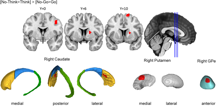
Fig. 3.9 Memory inhibition engaged caudate, putamen, and GPe more reliably than action prevention. Top row: Clusters are presented on coronal slices of a high-resolution MNI atlas. Reference lines for the coronal slices are presented in the sagittal plane. Bottom row: Clusters are displayed on high-resolution parcellations of the caudate, putamen, and external globus pallidus (GPe). All clusters are thresholded using cluster-level inference (p<.05, uncorrected p<.001, threshold permutations=1000).
Fig. 3.9 Memory inhibition engaged caudate, putamen, and GPe more reliably than action prevention. Top row: Clusters are presented on coronal slices of a high-resolution MNI atlas. Reference lines for the coronal slices are presented in the sagittal plane. Bottom row: Clusters are displayed on high-resolution parcellations of the caudate, putamen, and external globus pallidus (GPe). All clusters are thresholded using cluster-level inference (p<.05, uncorrected p<.001, threshold permutations=1000).CC BY · PubMed Central
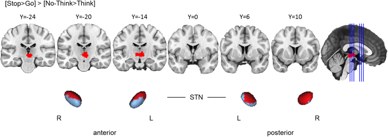
Fig. 3.10 Action cancellation engaged STN more reliably than memory inhibition. Top row: Clusters are presented on coronal slices of a high-resolution MNI atlas. Reference lines for the coronal slices are presented in the sagittal plane. Bottom row: Clusters are displayed on high-resolution parcellaions of the subthalamic nucleus (STN). The contrast analysis was computed using the thresholded ALE images from the individual analyses. All clusters are thresholded at uncorrected p<.001, with the p-value permutations of 10,000 iterations, and the minimum cluster volume of 200 mm3.
Fig. 3.10 Action cancellation engaged STN more reliably than memory inhibition. Top row: Clusters are presented on coronal slices of a high-resolution MNI atlas. Reference lines for the coronal slices are presented in the sagittal plane. Bottom row: Clusters are displayed on high-resolution parcellaions of the subthalamic nucleus (STN). The contrast analysis was computed using the thresholded ALE images from the individual analyses. All clusters are thresholded at uncorrected p<.001, with the p-value permutations of 10,000 iterations, and the minimum cluster volume of 200 mm3.CC BY · PubMed Central
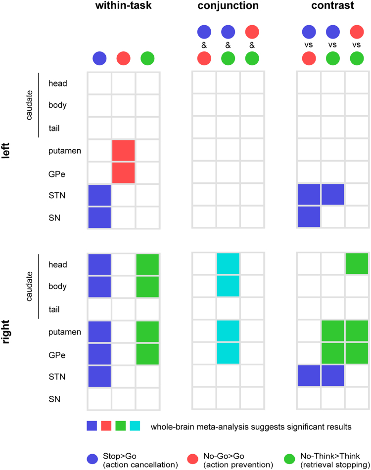
Fig. 3.11 Basal ganglia activations in the task-specific, conjunction, and contrast analyses. The left column shows basal ganglia activations from the task-specific meta-analyses, colour-coded by task contrasts (Blue=Stop>Go, Red=No-Go>Go, and Green=No-Think>Think). The middle column shows the conjunction analyses. Activations shared by two tasks are presented in the mixed colour based on the colours that we used to represent the individual tasks. The right column shows basal ganglia activations from the contrast analyses, with the colours denoting task-specific activity. For example, bilateral STN was activated more strongly in the Stop>Go contrast (blue) than the No-Go>Go and No-Think>Think contrasts. The top panel summarises activations in the left basal ganglia structures, while the bottom panel summaries those in the right.
Fig. 3.11 Basal ganglia activations in the task-specific, conjunction, and contrast analyses. The left column shows basal ganglia activations from the task-specific meta-analyses, colour-coded by task contrasts (Blue=Stop>Go, Red=No-Go>Go, and Green=No-Think>Think). The middle column shows the conjunction analyses. Activations shared by two tasks are presented in the mixed colour based on the colours that we used to represent the individual tasks. The right column shows basal ganglia activations from the contrast analyses, with the colours denoting task-specific activity. For example, bilateral STN was activated more strongly in the Stop>Go contrast (blue) than the No-Go>Go and No-Think>Think contrasts. The top panel summarises activations in the left basal ganglia structures, while the bottom panel summaries those in the right.CC BY · PubMed Central
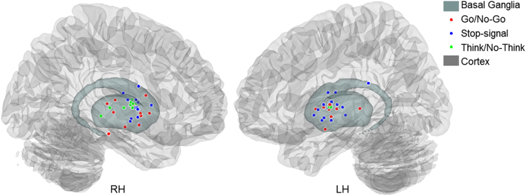
Fig. 3.12 Peak coordinates from the basal ganglia activations in the Go/No-Go, stop-signal, and Think/No-Think tasks.
Fig. 3.12 Peak coordinates from the basal ganglia activations in the Go/No-Go, stop-signal, and Think/No-Think tasks.CC BY · PubMed Central

Verification

Claims the paper makes

The claims and statistics of this paper haven’t been checked yet.

Run the check — free, no account needed. It reads the open-access text and checks the sources it cites.

A few free checks a day · papers already verified open instantly · results are public

Claims other papers make about this one

Each is checked against this paper’s own text: does it say what the citing paper says it does?

Self-check

Percentages, sums and reported differences re-computed from the paper’s own numbers.

The self-check needs the paper’s full text.

Citations

Cites

Show all 79 references

Research works printed but not linked: 2

  1. Levitt, 2002, An MRI study of caudate nucleus and lateral ventricle volume in first episode schizophrenia and affective psychosis, Biol. Psychiatry, 51, 59S
  2. Rodriguez, 2003, Path analysis of cortical-subcortical interactions in category learning with fMRI, Soc. Neurosci. Abstr., 400, 28

Cited by

Sorted by citations

See all 133

Metadata

Authors
Guo, Y., Schmitz, T. W., Mur, M., Ferreira, C. S., & Anderson, M. C.
Affiliations
  • MRC Cognition and Brain Sciences Unit (GB)
  • University of Birmingham (GB)
Corresponding author
Guo, Yuhua
Venue
Neuropsychologia
Publisher
Elsevier BV
Published
2018-01
Volume
108
Pages
117-134
ISSN
0028-3932 (ISSN-L)
OpenAlex
W2774718880
Cited by
133
References
79
Citations by year
2026: 9 · 2025: 24 · 2024: 17 · 2023: 12 · 2022: 15 · 2021: 16 · 2020: 24 · 2019: 8 · 2018: 7
FWCI
1.19
Type
journal article
Language
en
Open access
Locations
  • Neuropsychologia (publishedVersion, cc-by)
  • Europe PMC (PubMed Central) (publishedVersion, cc-by)
  • PubMed Central (publishedVersion, cc-by)
  • PubMed (publishedVersion)
  • Scholarship@Western (Western University) (publishedVersion, cc-by)
  • Scholarship@Western (Western University) (submittedVersion, cc-by)
  • University of Birmingham Research Portal (University of Birmingham) (submittedVersion, cc-by)
Funders
UK Medical Research Council (MC-A060-5PR00)
Topics
  • Life Sciences › Neuroscience › Cognitive Neuroscience › Neural and Behavioral Psychology Studies
  • Life Sciences › Neuroscience › Cellular and Molecular Neuroscience › Neurotransmitter Receptor Influence on Behavior
  • Health Sciences › Medicine › Clinical Neurology › Deep Brain Stimulation and Movement Disorders
Keywords
basal ganglia, action stopping, thought stopping, motor inhibition, go/no-go task, stop-signal task, Think/No-Think task, striatal subregions, prefrontal cortex, ventrolateral prefrontal cortex, anterior cingulate cortex, supplementary motor area, intrusive thoughts
MeSH terms
Basal Ganglia, Humans, Inhibition, Psychological, Magnetic Resonance Imaging, Memory, Motor Activity, Psychomotor Performance, Executive Function

About these checks

Registry, retraction, Cites and statistics results are computed, not judged, with one exception: a printed reference that no index lists is read once by a model before it counts as a citation error. Claims are extracted by a model and kept only when their quote appears verbatim in the text; each cited source is then checked, with a model’s help, for whether it says what the claim relies on. Those verdicts can be wrong — read the claim’s quote and the cited source before relying on one.

Statistics and arithmetic are recomputed, not judged.

Confidence bands are shown only for fields calibrated on our annotated test set. None is calibrated yet, so no band is shown.

What counts as a claim ›How we measure confidence ›How we check this ›

Are you an author, or do you see a problem with this page? Request a review