

The DOI matches its Crossref record; 51 of the 51 references it cites were found in a registry or an index.
VSV-G-Enveloped Vesicles for Traceless Delivery of CRISPR-Cas9
Open accessAbstract
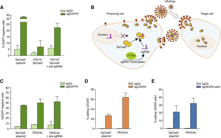
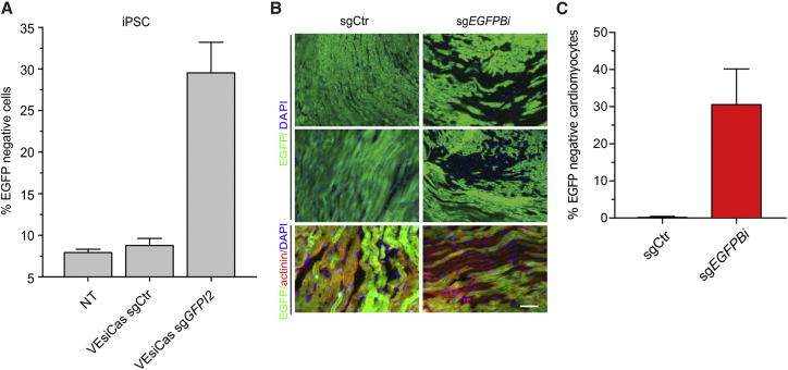
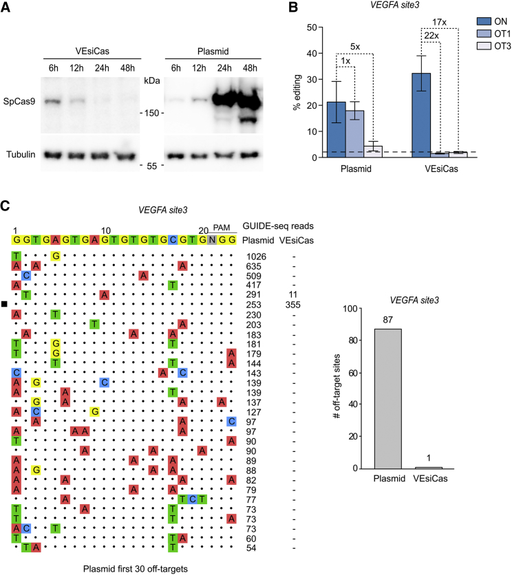
The method of delivery of CRISPR-Cas9 into target cells is a strong determinant of efficacy and specificity in genome editing. Even though high efficiency of Cas9 delivery is necessary for optimal editing, its long-term and high levels of expression correlate with increased off-target activity. We developed vesicles (VEsiCas) carrying CRISPR-SpCas9 ribonucleoprotein complexes (RNPs) that are efficiently delivered into target cells through the fusogenic glycoprotein of the vesicular stomatitis virus (VSV-G). A crucial step for VEsiCas production is the synthesis of the single guide RNA (sgRNA) mediated by the T7 RNA polymerase in the cytoplasm of producing cells as opposed to canonical U6-driven Pol III nuclear transcription. In VEsiCas, the absence of DNA encoding SpCas9 and sgRNA allows rapid clearance of the nuclease components in target cells, which correlates with reduced genome-wide off-target cleavages. Compared with SpCas9 RNPs electroporation, which is currently the method of choice to obtain transient SpCas9 activity, VEsiCas deliver the nuclease with higher efficiency and lower toxicity. We show that a wide variety of cells can be edited through VEsiCas, including a variety of transformed cells, induced pluripotent stem cells (iPSCs), and cardiomyocytes, in vivo. VEsiCas is a traceless CRISPR-Cas9 delivery tool for efficient and safe genome editing that represents a further advancement toward the therapeutic use of the CRISPR-Cas9 technology.
Overview
The claims and statistics of this paper haven’t been checked yet.
Run the check — free, no account needed. It reads the open-access text and checks the sources it cites.
Integrity signals
Registry
Cites
Statistics
Figures
Verification
Claims the paper makes
The claims and statistics of this paper haven’t been checked yet.
Run the check — free, no account needed. It reads the open-access text and checks the sources it cites.
Claims other papers make about this one
Each is checked against this paper’s own text: does it say what the citing paper says it does?
The check wasn’t confident either way, so it gives no verdict — never disputed.
In the citing paper
“Nevertheless, the HDR pathway is not highly active in human cells, thus strongly limiting the clinical efficacy of this application23,24.”
From Allele specific repair of splicing mutations in cystic fibrosis through AsCas12a genome editing (2019) · cites this paper as [23]
Experimental — accuracy not yet measured
Self-check
Percentages, sums and reported differences re-computed from the paper’s own numbers.
The self-check needs the paper’s full text.
Citations
Cites
Multiplex Genome Engineering Using CRISPR/Cas Systems
Cong, L., Ran, F. A., Cox, D., Lin, S., Barretto, R., Habib, N., Hsu, P. D., Wu, X., Jiang, W., Marraffini, L. A., et al. (2013) · Science · doi:10.1126/science.1231143 · Cited by 15,339
Double nicking by RNA-guided CRISPR Cas9 for enhanced genome editing specificity
Ran, F. A., Hsu, P. D., Lin, C.-Y., Gootenberg, J. S., Konermann, S., Trevino, A. E., Scott, D. A., Inoue, A., Matoba, S., Zhang, Y., et al. (2013) · Cell · doi:10.1016/j.cell.2013.08.021 · Cited by 3,299
High-Frequency off-Target Mutagenesis Induced by CRISPR-Cas Nucleases in Human Cells
Fu, Y., Foden, J. A., Khayter, C., Maeder, M. L., Reyon, D., Joung, J. K., & Sander, J. D. (2013) · Nature Biotechnology · doi:10.1038/nbt.2623 · Cited by 3,292
Easy quantitative assessment of genome editing by sequence trace decomposition
Brinkman, E. K., Chen, T., Amendola, M., & van Steensel, B. (2014) · Nucleic Acids Research · doi:10.1093/nar/gku936 · Cited by 2,390
GUIDE-seq enables genome-wide profiling of off-target cleavage by CRISPR-Cas nucleases
Tsai, S. Q., Zheng, Z., Nguyen, N. T., Liebers, M., Topkar, V. V., Thapar, V., Wyvekens, N., Khayter, C., Iafrate, A. J., Le, L. P., et al. (2015) · Nature Biotechnology · doi:10.1038/nbt.3117 · Cited by 2,268
RIG-I-mediated antiviral responses to single-stranded RNA bearing 5’-phosphates
Pichlmair, A., Schulz, O., Tan, C. P., Näslund, T. I., Liljeström, P., Weber, F., & Reis e Sousa, C. (2006) · Science · doi:10.1126/science.1132998 · Cited by 2,210
RNA-programmed genome editing in human cells
Jinek, M., East, A., Cheng, A., Lin, S., Ma, E., & Doudna, J. (2013) · eLife · doi:10.7554/elife.00471 · Cited by 2,098
Kim, S., Kim, D., Cho, S. W., Kim, J., & Kim, J.-S. (2014) · Genome Research · doi:10.1101/gr.171322.113 · Cited by 1,856
Zuris, J. A., Thompson, D. B., Shu, Y., Guilinger, J. P., Bessen, J. L., Hu, J. H., Maeder, M. L., Joung, J. K., Chen, Z.-Y., & Liu, D. R. (2015) · Nature Biotechnology · doi:10.1038/nbt.3081 · Cited by 1,460
CRISPR-Based Technologies for the Manipulation of Eukaryotic Genomes
Komor, A. C., Badran, A. H., & Liu, D. R. (2016) · Cell · doi:10.1016/j.cell.2016.10.044 · Cited by 1,037
Cited by
Sorted by citations
Base editing: advances and therapeutic opportunities
Porto, E. M., Komor, A. C., Slaymaker, I. M., & Yeo, G. W.-M. (2020) · Nature Reviews Drug Discovery · doi:10.1038/s41573-020-0084-6 · Cited by 499
Strategies in the delivery of Cas9 ribonucleoprotein for CRISPR/Cas9 genome editing
Zhang, S., Shen, J., Li, D., & Cheng, Y. (2021) · Theranostics · doi:10.7150/thno.47007 · Cited by 430
Gee, P., Lung, M. S. Y., Okuzaki, Y., Sasakawa, N., Iguchi, T., Makita, Y., Hozumi, H., Miura, Y., Yang, L. F., Iwasaki, M., et al. (2020) · Nature Communications · doi:10.1038/s41467-020-14957-y · Cited by 372
Recent Advances in CRISPR/Cas9 Delivery Strategies
Yip, B. (2020) · Biomolecules · doi:10.3390/biom10060839 · Cited by 292
Delivery of CRISPR-Cas tools for in vivo genome editing therapy: Trends and challenges
Taha, E. A., Lee, J., & Hotta, A. (2022) · Journal of Controlled Release · doi:10.1016/j.jconrel.2022.01.013 · Cited by 259
Metadata
- Authors
- Montagna, C., Petris, G., Casini, A., Maule, G., Franceschini, G. M., Zanella, I., Conti, L., Arnoldi, F., Burrone, O. R., Zentilin, L., Zacchigna, S., Giacca, M., & Cereseto, A.
- Affiliations
- University of Trento (IT)
- University of Trieste (IT)
- AREA Science Park (IT)
- Corresponding author
- Petris, Gianluca
- Cereseto, Anna
- ORCID
- Venue
- Molecular Therapy Nucleic Acids
- Publisher
- Elsevier BV
- Published
- 2018-09
- Volume
- 12
- Pages
- 453-462
- ISSN
- 2162-2531 (ISSN-L)
- PMC
- PMC6041463
- OpenAlex
- W2874925241
- Cited by
- 117
- References
- 51
- Citations by year
- 2026: 6 · 2025: 12 · 2024: 13 · 2023: 18 · 2022: 24 · 2021: 20 · 2020: 13 · 2019: 10 · 2018: 1
- FWCI
- 3.91
- Type
- journal article
- Language
- en
- Open access
- gold
- doi.org
- Licence
- Locations
- Molecular Therapy — Nucleic Acids (publishedVersion, cc-by-nc-nd)
- Europe PMC (PubMed Central) (publishedVersion, cc-by-nc-nd)
- PubMed Central (publishedVersion, cc-by-nc-nd)
- PubMed (publishedVersion)
- ArTS Archivio della ricerca di Trieste (University of Trieste https://www.units.it/) (submittedVersion, cc-by-nc-nd)
- Institutional Research Information System (Università degli Studi di Trento) (submittedVersion, cc-by-nc-nd)
- Lirias (submittedVersion, cc-by-nc-nd)
- DOAJ (DOAJ: Directory of Open Access Journals) (submittedVersion, cc-by-sa)
- Funders
- Università degli Studi di Trento
- Fondazione Cassa Di Risparmio Di Trento E Rovereto
- Topics
- Life Sciences › Biochemistry, Genetics and Molecular Biology › Molecular Biology › CRISPR and Genetic Engineering
- Life Sciences › Biochemistry, Genetics and Molecular Biology › Molecular Biology › RNA Interference and Gene Delivery
- Life Sciences › Biochemistry, Genetics and Molecular Biology › Genetics › Virus-based gene therapy research
- Keywords
- CRISPR/Cas9 delivery, vesicular stomatitis virus glycoprotein, ribonucleoprotein complexes, single guide RNA, T7 RNA polymerase, off-target effects, genome editing, induced pluripotent stem cells, cardiomyocytes
About these checks
Registry, retraction, Cites and statistics results are computed, not judged, with one exception: a printed reference that no index lists is read once by a model before it counts as a citation error. Claims are extracted by a model and kept only when their quote appears verbatim in the text; each cited source is then checked, with a model’s help, for whether it says what the claim relies on. Those verdicts can be wrong — read the claim’s quote and the cited source before relying on one.
Statistics and arithmetic are recomputed, not judged.
Confidence bands are shown only for fields calibrated on our annotated test set. None is calibrated yet, so no band is shown.
What counts as a claim ›How we measure confidence ›How we check this ›
Are you an author, or do you see a problem with this page? Request a review
