

The DOI matches its Crossref record; 57 of the 59 references it cites were found in a registry or an index.
Characterization of HIV-1 Nucleoside-Modified mRNA Vaccines in Rabbits and Rhesus Macaques
Open accessAbstract
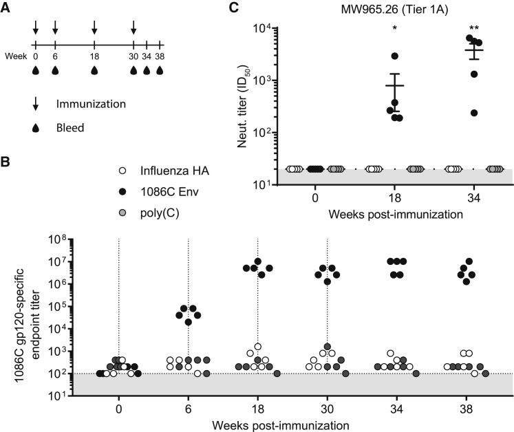
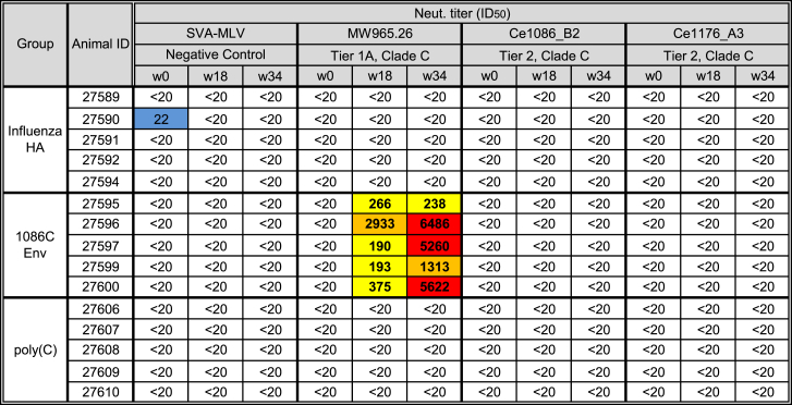
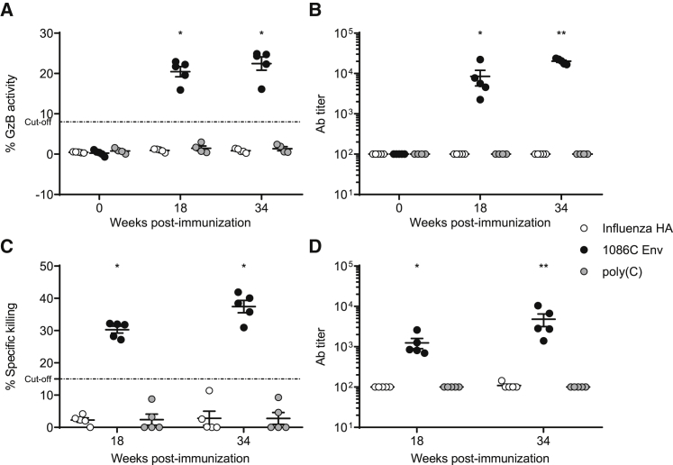
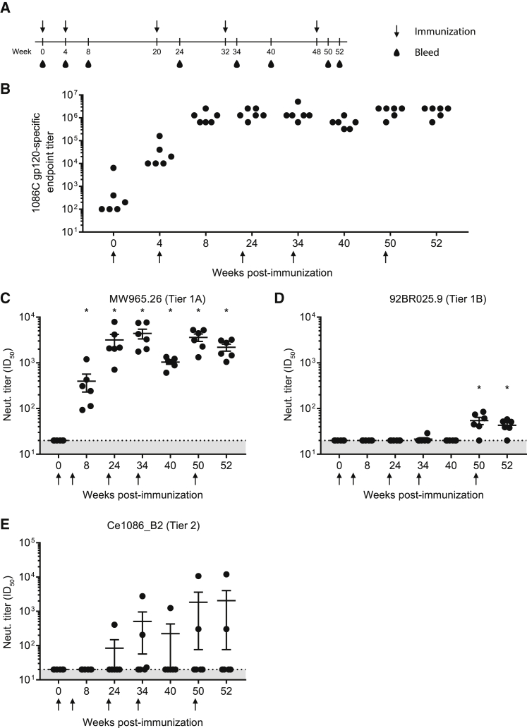
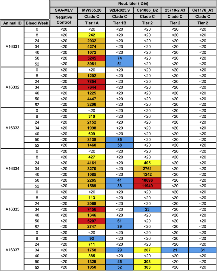
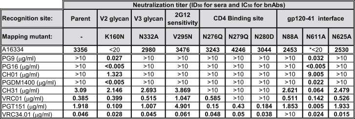
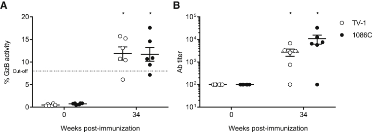
Despite the enormous effort in the development of effective vaccines against HIV-1, no vaccine candidate has elicited broadly neutralizing antibodies in humans. Thus, generation of more effective anti-HIV vaccines is critically needed. Here we characterize the immune responses induced by nucleoside-modified and purified mRNA-lipid nanoparticle (mRNA-LNP) vaccines encoding the clade C transmitted/founder HIV-1 envelope (Env) 1086C. Intradermal vaccination with nucleoside-modified 1086C Env mRNA-LNPs elicited high levels of gp120-specific antibodies in rabbits and rhesus macaques. Antibodies generated in rabbits neutralized a tier 1 virus, but no tier 2 neutralization activity could be measured. Importantly, three of six non-human primates developed antibodies that neutralized the autologous tier 2 strain. Despite stable anti-gp120 immunoglobulin G (IgG) levels, tier 2 neutralization titers started to drop 4 weeks after booster immunizations. Serum from both immunized rabbits and non-human primates demonstrated antibody-dependent cellular cytotoxicity activity. Collectively, these results are supportive of continued development of nucleoside-modified and purified mRNA-LNP vaccines for HIV. Optimization of Env immunogens and vaccination protocols are needed to increase antibody neutralization breadth and durability.
Overview
The claims and statistics of this paper haven’t been checked yet.
Run the check — free, no account needed. It reads the open-access text and checks the sources it cites.
Integrity signals
Registry
Cites
Statistics
Figures
Verification
Claims the paper makes
The claims and statistics of this paper haven’t been checked yet.
Run the check — free, no account needed. It reads the open-access text and checks the sources it cites.
Claims other papers make about this one
Each is checked against this paper’s own text: does it say what the citing paper says it does?
In the citing paper
“Several preclinical studies have delivered mRNA vaccines encoding HIV proteins using multiple delivery vehicles, including cationic nanoemulsions109,161, DOTAP/DOPE liposomes162, polymers84,163,164 and ionizable LNPs165,166, but they have had varied success.”
From mRNA vaccines for infectious diseases: principles, delivery and clinical translation (2021) · cites this paper as [166]
In the citing paper
“These studies suggest that novel antigens are necessary to effectively target HIV in addition to potent delivery vehicles.”
From mRNA vaccines for infectious diseases: principles, delivery and clinical translation (2021) · cites this paper as [166]
Self-check
Percentages, sums and reported differences re-computed from the paper’s own numbers.
The self-check needs the paper’s full text.
Citations
Cites
Antibody neutralization and escape by HIV-1
Wei, X., Decker, J. M., Wang, S., Hui, H., Kappes, J. C., Wu, X., Salazar-Gonzalez, J. F., Salazar, M. G., Kilby, J. M., Saag, M. S., et al. (2003) · Nature · doi:10.1038/nature01470 · Cited by 2,421
Keele, B. F., Giorgi, E. E., Salazar-Gonzalez, J. F., Decker, J. M., Pham, K. T., Salazar, M. G., Sun, C., Grayson, T. B., Wang, S., Li, H., et al. (2008) · Proceedings of the National Academy of Sciences · doi:10.1073/pnas.0802203105 · Cited by 1,913
Self-citationT follicular helper cell differentiation, function, and roles in disease
Crotty, S. (2014) · Immunity · doi:10.1016/j.immuni.2014.10.004 · Cited by 1,847
Broad neutralization coverage of HIV by multiple highly potent antibodies
Walker, L. M., Huber, M., Doores, K. J., Falkowska, E., Pejchal, R., Julien, J.-P., Wang, S.-K., Ramos, A., Chan-Hui, P.-Y., Moyle, M., et al. (2011) · Nature · doi:10.1038/nature10373 · Cited by 1,480
Structural Basis for Broad and Potent Neutralization of HIV-1 by Antibody VRC01
Zhou, T., Georgiev, I. S., Wu, X., Yang, Z., Dai, K., Finzi, A., Kwon, Y. D., Scheid, J. F., Shi, W., Xu, L., et al. (2010) · Science · doi:10.1126/science.1192819 · Cited by 1,123
Trkola, A., Purtscher, M., Muster, T., Ballaun, C., Buchacher, A., Sullivan, N. J., Srinivasan, K., Sodroski, J., Moore, J. P., & Katinger, H. W. D. (1996) · Journal of Virology · doi:10.1128/jvi.70.2.1100-1108.1996 · Cited by 1,115
Pardi, N., Tuyishime, S., Muramatsu, H., Kariko, K., Mui, B. L., Tam, Y. K., Madden, T. D., Hope, M. J., & Weissman, D. (2015) · Journal of Controlled Release · doi:10.1016/j.jconrel.2015.08.007 · Cited by 1,014
Self-citationCo-evolution of a broadly neutralizing HIV-1 antibody and founder virus
Liao, H., Lynch, R. M., Zhou, T., Gao, F., Alam, S. M., Boyd, S. D., Fire, A. Z., Roskin, K. M., Schramm, C. A., Zhang, Z., et al. (2013) · Nature · doi:10.1038/nature12053 · Cited by 986
Self-citationModified mRNA vaccines protect against zika virus infection
Richner, J. M., Himansu, S., Dowd, K. A., Butler, S. L., Salazar, V., Fox, J. M., Julander, J. G., Tang, W. W., Shresta, S., Pierson, T. C., et al. (2017) · Cell · doi:10.1016/j.cell.2017.02.017 · Cited by 909
Zika virus protection by a single low-dose nucleoside-modified mRNA vaccination
Pardi, N., Hogan, M. J., Pelc, R. S., Muramatsu, H., Andersen, H., DeMaso, C. R., Dowd, K. A., Sutherland, L. L., Scearce, R. M., Parks, R., et al. (2017) · Nature · doi:10.1038/nature21428 · Cited by 884
Self-citationCardiolipin Polyspecific Autoreactivity in Two Broadly Neutralizing HIV-1 Antibodies
Haynes, B. F., Fleming, J., Clair, E. W. S., Katinger, H., Stiegler, G. M., Kunert, R., Robinson, J. E., Scearce, R. M., Plonk, K., Staats, H. F., et al. (2005) · Science · doi:10.1126/science.1111781 · Cited by 746
Self-citationBahl, K., Senn, J. J., Yuzhakov, O., Bulychev, A., Brito, L. A., Hassett, K. J., Laska, M. E., Smith, M., Almarsson, Ö., Thompson, J., et al. (2017) · Molecular Therapy · doi:10.1016/j.ymthe.2017.03.035 · Cited by 642
Measuring HIV Neutralization in a Luciferase Reporter Gene Assay
Montefiori, D. C. (2009) · Methods in molecular biology · doi:10.1007/978-1-59745-170-3_26 · Cited by 585
Self-citationPardi, N., Hogan, M. J., Naradikian, M. S., Parkhouse, K., Cain, D. W., Jones, L., Moody, M. A., Verkerke, H. P., Myles, A., Willis, E., et al. (2018) · Journal of Experimental Medicine · doi:10.1084/jem.20171450 · Cited by 530
Self-citationThess, A., Grund, S., Mui, B. L., Hope, M. J., Baumhof, P., Fotin-Mleczek, M., & Schlake, T. (2015) · Molecular Therapy · doi:10.1038/mt.2015.103 · Cited by 513
Self-citationAntibodies in HIV-1 Vaccine Development and Therapy
Klein, F., Mouquet, H., Dosenovic, P., Scheid, J. F., Scharf, L., & Nussenzweig, M. C. (2013) · Science · doi:10.1126/science.1241144 · Cited by 452
Yates, N. L., Liao, H., Fong, Y., deCamp, A. C., Vandergrift, N., Williams, W. T., Alam, S. M., Ferrari, G., Yang, Z., Seaton, K. E., et al. (2014) · Science Translational Medicine · doi:10.1126/scitranslmed.3007730 · Cited by 449
Self-citationPolyreactivity increases the apparent affinity of anti-HIV antibodies by heteroligation
Mouquet, H., Scheid, J. F., Zöller, M., Krogsgaard, M., Ott, R. G., Shukair, S. A., Artyomov, M. N., Pietzsch, J., Connors, M., Pereyra, F., et al. (2010) · Nature · doi:10.1038/nature09385 · Cited by 432
Broad and potent HIV-1 neutralization by a human antibody that binds the gp41–gp120 interface
Huang, J., Kang, B. H., Pancera, M., Lee, J. H., Tong, T., Feng, Y., Imamichi, H., Georgiev, I. S., Chuang, G., Druz, A., et al. (2014) · Nature · doi:10.1038/nature13601 · Cited by 432
Self-citationWhittle, J. R. R., Zhang, R., Khurana, S., King, L. R., Manischewitz, J. F., Golding, H., Dormitzer, P. R., Haynes, B. F., Walter, E. B., Moody, M. A., et al. (2011) · Proceedings of the National Academy of Sciences · doi:10.1073/pnas.1111497108 · Cited by 430
Self-citationLiao, H., Bonsignori, M., Alam, S. M., McLellan, J. S., Tomaras, G. D., Moody, M. A., Kozink, D. M., Hwang, K.-K., Chen, X., Tsao, C.-Y., et al. (2013) · Immunity · doi:10.1016/j.immuni.2012.11.011 · Cited by 377
Self-citationFalkowska, E., Le, K. M., Ramos, A. G., Doores, K. J., Lee, J. H., Blattner, C., Ramirez, A., Derking, R., van Gils, M. J., Liang, C. J., et al. (2014) · Immunity · doi:10.1016/j.immuni.2014.04.009 · Cited by 359
CD4 T follicular helper cell dynamics during SIV infection
Petrovas, C., Yamamoto, T., Gerner, M. Y., Boswell, K. L., Wloka, K., Smith, E. C., Ambrozak, D. R., Utay, N. S., Timmer, K. J., Sun, X., et al. (2012) · Journal of Clinical Investigation · doi:10.1172/jci63039 · Cited by 328
HIV-1 Vaccines Based on Antibody Identification, B Cell Ontogeny, and Epitope Structure
Kwong, P. D., & Mascola, J. R. (2018) · Immunity · doi:10.1016/j.immuni.2018.04.029 · Cited by 308
Sullivan, N. R., Sun, Y., Sattentau, Q. J., Thali, M., Wu, D. T., Denisova, G. F., Gershoni, J. M., Robinson, J. E., Moore, J. P., & Sodroski, J. (1998) · Journal of Virology · doi:10.1128/jvi.72.6.4694-4703.1998 · Cited by 292
Pardi, N., Parkhouse, K., Kirkpatrick, E., McMahon, M., Zost, S. J., Mui, B. L., Tam, Y. K., Karikó, K., Barbosa, C. J., Madden, T. D., et al. (2018) · Nature Communications · doi:10.1038/s41467-018-05482-0 · Cited by 281
Self-citationLandais, E., Huang, X., Havenar‐Daughton, C., Murrell, B., Price, M. A., Wickramasinghe, L., Ramos, A. G., Bian, C. B., Simek, M. D., Allen, S. A., et al. (2016) · PLoS Pathogens · doi:10.1371/journal.ppat.1005369 · Cited by 265
Stable exposure of the coreceptor-binding site in a CD4-independent HIV-1 envelope protein
Hoffman, T. L., LaBranche, C. C., Zhang, W., Canziani, G., Robinson, J. E., CHAIKEN, I. M., Hoxie, J. A., & Doms, R. W. (1999) · Proceedings of the National Academy of Sciences · doi:10.1073/pnas.96.11.6359 · Cited by 263
Self-citationBehrens, A., Vasiljević, S., Pritchard, L. K., Harvey, D. J., Andev, R. S., Krumm, S. A., Struwe, W. B., Cupo, A., Kumar, A., Zitzmann, N., et al. (2016) · Cell Reports · doi:10.1016/j.celrep.2016.02.058 · Cited by 251
Mechanism of Neutralization by the Broadly Neutralizing HIV-1 Monoclonal Antibody VRC01
Li, Y., O'Dell, S., Walker, L. M., Wu, X., Guenaga, J., Feng, Y., Schmidt, S. D., McKee, K., Louder, M. K., Ledgerwood, J. E., et al. (2011) · Journal of Virology · doi:10.1128/jvi.00754-11 · Cited by 235
Self-citationMcCoy, L. E., van Gils, M. J., Ozorowski, G., Messmer, T., Briney, B. S., Voss, J. E., Kulp, D. W., Macauley, M. S., Sok, D., Pauthner, M. G., et al. (2016) · Cell Reports · doi:10.1016/j.celrep.2016.07.074 · Cited by 230
Native‐like Env trimers as a platform for HIV ‐1 vaccine design
Sanders, R. W., & Moore, J. P. (2017) · Immunological Reviews · doi:10.1111/imr.12481 · Cited by 227
In Vitro Transcription of Long RNA Containing Modified Nucleosides
Pardi, N., Muramatsu, H., Weissman, D., & Karikó, K. (2012) · Methods in molecular biology · doi:10.1007/978-1-62703-260-5_2 · Cited by 204
Self-citationHigh‐throughput quantitative analysis of HIV‐1 and SIV‐specific ADCC‐mediating antibody responses
Pollara, J., Hart, L., Brewer, F., Pickeral, J., Packard, B. Z., Hoxie, J. A., Komoriya, A., Ochsenbauer, C., Kappes, J. C., Roederer, M., et al. (2011) · Cytometry Part A · doi:10.1002/cyto.a.21084 · Cited by 202
Self-citationAndrabi, R., Voss, J. E., Liang, C. J., Briney, B. S., McCoy, L. E., Wu, C., Wong, C., Poignard, P., & Burton, D. R. (2015) · Immunity · doi:10.1016/j.immuni.2015.10.014 · Cited by 193
Sok, D., Doores, K. J., Briney, B. S., Le, K. M., Saye-Francisco, K. L., Ramos, A. G., Kulp, D. W., Julien, J., Menis, S., Wickramasinghe, L., et al. (2014) · Science Translational Medicine · doi:10.1126/scitranslmed.3008104 · Cited by 184
Sok, D., Laserson, U., Laserson, J., Liu, Y., Vigneault, F., Julien, J., Briney, B. S., Ramos, A. G., Saye-Francisco, K. L., Le, K. M., et al. (2013) · PLoS Pathogens · doi:10.1371/journal.ppat.1003754 · Cited by 183
HPLC purification of in vitro transcribed long RNA
Weissman, D., Pardi, N., Muramatsu, H., & Karikó, K. (2013) · Methods in molecular biology · doi:10.1007/978-1-62703-260-5_3 · Cited by 180
Self-citationHIV-1 Vaccine-Induced C1 and V2 Env-Specific Antibodies Synergize for Increased Antiviral Activities
Pollara, J., Bonsignori, M., Moody, M. A., Liu, P., Alam, S. M., Hwang, K., Gurley, T. C., Kozink, D. M., Armand, L. C., Marshall, D. J., et al. (2014) · Journal of Virology · doi:10.1128/jvi.00156-14 · Cited by 178
Self-citationMeyer, M., Huang, E., Yuzhakov, O., Ramanathan, P., Ciaramella, G., & Bukreyev, A. (2018) · The Journal of Infectious Diseases · doi:10.1093/infdis/jix592 · Cited by 178
Havenar‐Daughton, C., Lee, J. H., & Crotty, S. (2017) · Immunological Reviews · doi:10.1111/imr.12512 · Cited by 175
Polyreactivity and Autoreactivity among HIV-1 Antibodies
Liu, M., Yang, G., Wiehe, K. J., Nicely, N. I., Vandergrift, N., Rountree, W., Bonsignori, M., Alam, S. M., Gao, J., Haynes, B. F., et al. (2014) · Journal of Virology · doi:10.1128/jvi.02378-14 · Cited by 162
Self-citationNeutralizing antibodies to HIV-1 induced by immunization
McCoy, L. E., & Weiss, R. A. (2013) · The Journal of Experimental Medicine · doi:10.1084/jem.20121827 · Cited by 137
Lindgren, G., Ols, S., Liang, F., Thompson, E. A., Lin, A., Hellgren, F., Bahl, K., John, S., Yuzhakov, O., Hassett, K. J., et al. (2017) · Frontiers in Immunology · doi:10.3389/fimmu.2017.01539 · Cited by 132
Effect of the cytoplasmic domain on antigenic characteristics of HIV-1 envelope glycoprotein
Chen, J., Kovacs, J. M., Peng, H., Rits‐Volloch, S., Lü, J., Park, D., Zablowsky, E., Seaman, M. S., & Chen, B. (2015) · Science · doi:10.1126/science.aaa9804 · Cited by 125
Quality and quantity of T FH cells are critical for broad antibody development in SHIV AD8 infection
Yamamoto, T., Lynch, R. M., Gautam, R., Matus‐Nicodemos, R., Schmidt, S. D., Boswell, K. L., Darko, S. W., Wong, P., Sheng, Z., Petrovas, C., et al. (2015) · Science Translational Medicine · doi:10.1126/scitranslmed.aab3964 · Cited by 124
Klasse, P. J., Ketas, T. J., Cottrell, C. A., Ozorowski, G., Debnath, G., Camara, D., Francomano, E., Pugach, P., Ringe, R. P., LaBranche, C. C., et al. (2018) · PLoS Pathogens · doi:10.1371/journal.ppat.1006913 · Cited by 117
Self-citationAn autoreactive antibody from an SLE/HIV-1 individual broadly neutralizes HIV-1
Bonsignori, M., Wiehe, K. J., Grimm, S. K., Lynch, R. M., Yang, G., Kozink, D. M., Perrin, F., Cooper, A. J., Hwang, K., Chen, X., et al. (2014) · Journal of Clinical Investigation · doi:10.1172/jci73441 · Cited by 87
Self-citationChowdhury, A., Estrada, P. M. D. R., Tharp, G. K., Trible, R. P., Amara, R. R., Chahroudi, A., Reyes‐Terán, G., Bosinger, S. E., & Silvestri, G. (2015) · The Journal of Immunology · doi:10.4049/jimmunol.1402701 · Cited by 83
Pacheco, B., Alsahafi, N., Débbeche, O., Prévost, J., Ding, S., Chapleau, J.-P., Herschhorn, A., Madani, N., Princiotto, A. M., Melillo, B. N., et al. (2016) · Journal of Virology · doi:10.1128/jvi.02219-16 · Cited by 55
HIV-1 envelope glycoprotein immunogens to induce broadly neutralizing antibodies
Sliepen, K., & Sanders, R. W. (2015) · Expert Review of Vaccines · doi:10.1586/14760584.2016.1129905 · Cited by 50
Balla-Jhagjhoorsingh, S. S., Corti, D., Heyndríckx, L., Willems, E., Vereecken, K., Davis, D. A., & Vanham, G. L. (2013) · PLoS ONE · doi:10.1371/journal.pone.0068863 · Cited by 46
Hong, J. J., Amancha, P. K., Rogers, K. A., Courtney, C. L., Havenar‐Daughton, C., Crotty, S., Ansari, A. A., & Villinger, F. J. (2014) · The Journal of Immunology · doi:10.4049/jimmunol.1400749 · Cited by 44
Blackburn, M. J., Ma, Z.-M., Caccuri, F., McKinnon, K. M., Schifanella, L., Guan, Y., Gorini, G., Venzon, D. J., Fenizia, C., Binello, N., et al. (2015) · The Journal of Immunology · doi:10.4049/jimmunol.1402699 · Cited by 32
DNA Immunization for HIV Vaccine Development
Chen, Y., Wang, S., & Lu, S. (2014) · Vaccines · doi:10.3390/vaccines2010138 · Cited by 24
Behrens, A., Kumar, A., Medina-Ramírez, M., Cupo, A., Marshall, K., Portillo, V. M. C., Harvey, D. J., Ozorowski, G., Zitzmann, N., Wilson, I. A., et al. (2018) · Journal of Proteome Research · doi:10.1021/acs.jproteome.7b00639 · Cited by 22
Nguyen, H. T., Madani, N., Ding, H., Elder, E., Princiotto, A. M., Gu, C., Darby, P., Alin, J., Herschhorn, A., Kappes, J. C., et al. (2017) · Virology Journal · doi:10.1186/s12985-017-0704-x · Cited by 11
Cited by
Sorted by citations
COVID-19 vaccine BNT162b1 elicits human antibody and TH1 T cell responses
Sahin, U., Muik, A., Derhovanessian, E., Vogler, I., Kranz, L. M., Vormehr, M., Baum, A., Pascal, K., Quandt, J., Maurus, D., et al. (2020) · Nature · doi:10.1038/s41586-020-2814-7 · Cited by 1,977
Self-citationmRNA vaccines for infectious diseases: principles, delivery and clinical translation
Chaudhary, N., Weissman, D., & Whitehead, K. A. (2021) · Nature Reviews Drug Discovery · doi:10.1038/s41573-021-00283-5 · Cited by 1,555
Self-citationBNT162b2 vaccine induces neutralizing antibodies and poly-specific T cells in humans
Sahin, U., Muik, A., Vogler, I., Derhovanessian, E., Kranz, L. M., Vormehr, M., Quandt, J., Bidmon, N., Ulges, A., Baum, A., et al. (2021) · Nature · doi:10.1038/s41586-021-03653-6 · Cited by 919
Self-citationAlameh, M.-G., Tombácz, I., Bettini, E., Lederer, K., Ndeupen, S., Sittplangkoon, C., Wilmore, J. R., Gaudette, B. T., Soliman, O. Y., Pine, M., et al. (2021) · Immunity · doi:10.1016/j.immuni.2021.11.001 · Cited by 775
Self-citationBNT162b vaccines protect rhesus macaques from SARS-CoV-2
Vogel, A. B., Kanevsky, I., Che, Y., Swanson, K. A., Muik, A., Vormehr, M., Kranz, L. M., Walzer, K. C., Hein, S., Güler, A., et al. (2021) · Nature · doi:10.1038/s41586-021-03275-y · Cited by 694
Metadata
- Authors
- Pardi, N., LaBranche, C. C., Ferrari, G., Cain, D. W., Tombácz, I., Parks, R. J., Muramatsu, H., Mui, B. L., Tam, Y. K., Karikó, K., Polacino, P., Barbosa, C. J., Madden, T. D., Hope, M. J., Haynes, B. F., Montefiori, D. C., Hu, S.-L., & Weissman, D.
- Affiliations
- University of Pennsylvania (US)
- Duke Medical Center (US)
- Duke University (US)
- Acuitas Therapeutics (Canada) (CA)
- BioNTech (Germany) (DE)
- University of Washington (US)
- Corresponding author
- Pardi, Norbert
- Weissman, Drew
- ORCID
- Venue
- Molecular Therapy - Nucleic Acids
- Publisher
- Elsevier BV
- Published
- 2019-04
- Volume
- 15
- Pages
- 36-47
- ISSN
- 2162-2531 (ISSN-L)
- PMC
- PMC6454128
- OpenAlex
- W2924788224
- Cited by
- 103
- References
- 59
- Citations by year
- 2026: 4 · 2025: 11 · 2024: 13 · 2023: 16 · 2022: 17 · 2021: 26 · 2020: 14 · 2019: 2
- FWCI
- 8.26
- Type
- journal article
- Language
- en
- Open access
- gold
- doi.org
- Licence
- Locations
- Molecular Therapy — Nucleic Acids (publishedVersion, cc-by)
- Europe PMC (PubMed Central) (publishedVersion, cc-by)
- PubMed Central (publishedVersion, cc-by)
- PubMed (publishedVersion)
- DOAJ (DOAJ: Directory of Open Access Journals) (submittedVersion, cc-by-sa)
- Funders
- National Institute of Allergy and Infectious Diseases (UM1 AI100645, HHSN272201100016C)
- Bill and Melinda Gates Foundation
- National Institutes of Health (P51 OD010425)
- Topics
- Life Sciences › Immunology and Microbiology › Virology › HIV Research and Treatment
- Health Sciences › Medicine › Infectious Diseases › SARS-CoV-2 and COVID-19 Research
- Life Sciences › Immunology and Microbiology › Immunology › Immunotherapy and Immune Responses
- Keywords
- HIV-1 vaccine, nucleoside-modified mRNA, mRNA-LNP vaccine, HIV-1 envelope glycoprotein, neutralizing antibodies, antibody-dependent cellular cytotoxicity, intradermal vaccination, booster vaccination, antibody persistence, rabbits, rhesus monkeys
About these checks
Registry, retraction, Cites and statistics results are computed, not judged, with one exception: a printed reference that no index lists is read once by a model before it counts as a citation error. Claims are extracted by a model and kept only when their quote appears verbatim in the text; each cited source is then checked, with a model’s help, for whether it says what the claim relies on. Those verdicts can be wrong — read the claim’s quote and the cited source before relying on one.
Statistics and arithmetic are recomputed, not judged.
Confidence bands are shown only for fields calibrated on our annotated test set. None is calibrated yet, so no band is shown.
What counts as a claim ›How we measure confidence ›How we check this ›
Are you an author, or do you see a problem with this page? Request a review
