

The DOI matches its Crossref record; 75 of the 75 references it cites were found in a registry or an index.
Antibody structure prediction using interpretable deep learning
Open accessPublished version of a preprint
Abstract
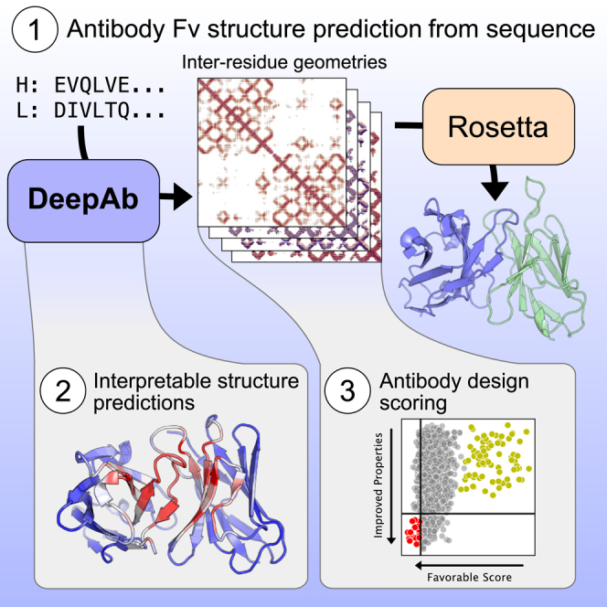
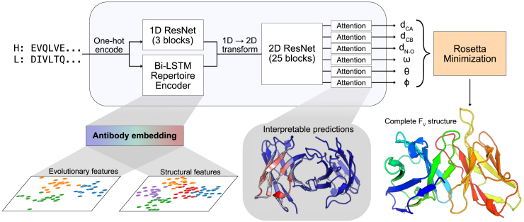
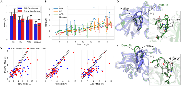
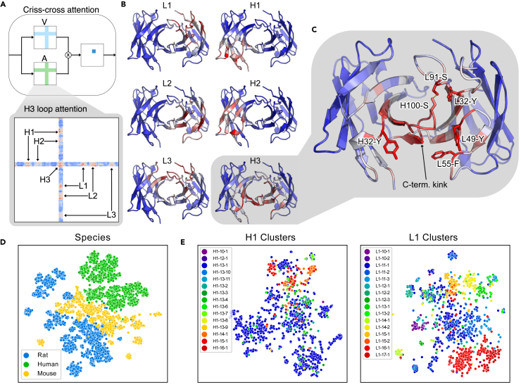
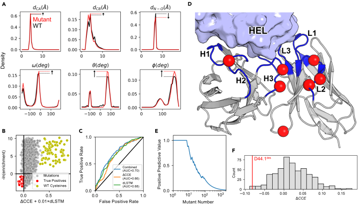
Abstract Therapeutic antibodies make up a rapidly growing segment of the biologics market. However, rational design of antibodies is hindered by reliance on experimental methods for determining antibody structures. In recent years, deep learning methods have driven significant advances in general protein structure prediction. Here, we present DeepAb, a deep learning method for predicting accurate antibody F V structures from sequence. We evaluate DeepAb on two benchmark sets – one balanced for structural diversity and the other composed of clinical-stage therapeutic antibodies – and find that our method consistently outperforms the leading alternatives. Previous deep learning methods have operated as “black boxes” and offered few insights into their predictions. By introducing a directly interpretable attention mechanism, we show that our network attends to physically important residue pairs. For example, in prediction of one CDR H3 residue conformation, the network attends to proximal aromatics and a key hydrogen bonding interaction that constrain the loop conformation. Finally, we present a novel mutant scoring metric derived from network confidence and show that for a particular antibody, all eight of the top-ranked mutations improve binding affinity. These results suggest that this model will be useful for a broad range of antibody prediction and design tasks. Significance Accurate structure models are critical for understanding the properties of potential therapeutic antibodies. Conventional methods for protein structure determination require significant investments of time and resources and may fail. Although greatly improved, methods for general protein structure prediction still cannot consistently provide the accuracy necessary to understand or design antibodies. We present a deep learning method for antibody structure prediction and demonstrate improvement over alternatives on diverse, therapeutically relevant benchmarks. In addition to its improved accuracy, our method reveals interpretable outputs about specific amino acids and residue interactions that should facilitate design of novel therapeutic antibodies.
Overview
The claims and statistics of this paper haven’t been checked yet.
Run the check — free, no account needed. It reads the open-access text and checks the sources it cites.
Integrity signals
Registry
Cites
Statistics
Figures
Verification
Claims the paper makes
The claims and statistics of this paper haven’t been checked yet.
Run the check — free, no account needed. It reads the open-access text and checks the sources it cites.
Self-check
Percentages, sums and reported differences re-computed from the paper’s own numbers.
The self-check needs the paper’s full text.
Citations
Cites
Adam: A Method for Stochastic Optimization
Kingma, D. P., & Ba, J. (2014) · arXiv · doi:10.48550/arxiv.1412.6980 · Cited by 98,946
Highly accurate protein structure prediction with AlphaFold
Jumper, J., Evans, R., Pritzel, A., Green, T., Figurnov, M., Ronneberger, O., Tunyasuvunakool, K., Bates, R., Žídek, A., Potapenko, A., et al. (2021) · Nature · doi:10.1038/s41586-021-03819-2 · Cited by 43,764
(2008) · Journal of Machine Learning Research · Cited by 33,051
Focal Loss for Dense Object Detection
Lin, T.-Y., Goyal, P., Girshick, R., He, K., & Dollar, P. (2017) · 2017 IEEE International Conference on Computer Vision (ICCV) · doi:10.1109/iccv.2017.324 · Cited by 30,581
Learning Phrase Representations using RNN Encoder–Decoder for Statistical Machine Translation
Cho, K., van Merrienboer, B., Gulcehre, C., Bahdanau, D., Bougares, F., Schwenk, H., & Bengio, Y. (2014) · Proceedings of the 2014 Conference on Empirical Methods in Natural Language Processing (EMNLP) · doi:10.3115/v1/d14-1179 · Cited by 25,545
Learning to Forget: Continual Prediction with LSTM
Gers, F. A., Schmidhuber, J., & Cummins, F. (2000) · Neural Computation · doi:10.1162/089976600300015015 · Cited by 6,122
Accurate prediction of protein structures and interactions using a 3-track network
Baek, M., DiMaio, F., Anishchenko, I., Dauparas, J., Ovchinnikov, S., Lee, G. R., Wang, J., Cong, Q., Kinch, L. N., Schaeffer, R. D., et al. (2021) · doi:10.1101/2021.06.14.448402 · Cited by 5,549
Lipton, Z. C. (2018) · Queue · doi:10.1145/3236386.3241340 · Cited by 5,427
Floyd, R. W. (1962) · Communications of the ACM · doi:10.1145/367766.368168 · Cited by 4,137
Improved protein structure prediction using potentials from deep learning
Senior, A. W., Evans, R., Jumper, J., Kirkpatrick, J., Sifre, L., Green, T., Qin, C., Žídek, A., Nelson, A. W. R., Bridgland, A., et al. (2020) · Nature · doi:10.1038/s41586-019-1923-7 · Cited by 3,378
Research works printed but not linked: 1
- Linder, 2020, Fast differentiable DNA and protein sequence optimization for molecular design, arXiv
Cited by
Sorted by citations
Fast, accurate antibody structure prediction from deep learning on massive set of natural antibodies
Ruffolo, J. A., Chu, L., Mahajan, S. P., & Gray, J. J. (2023) · Nature Communications · doi:10.1038/s41467-023-38063-x · Cited by 364
Self-citationImmuneBuilder: Deep-Learning models for predicting the structures of immune proteins
Abanades, B., Wong, W. K., Boyles, F., Georges, G. J., Bujotzek, A., & Deane, C. M. (2023) · Communications Biology · doi:10.1038/s42003-023-04927-7 · Cited by 323
Designing antibodies as therapeutics
Carter, P. J., & Rajpal, A. (2022) · Cell · doi:10.1016/j.cell.2022.05.029 · Cited by 282
Drug discovery and development: introduction to the general public and patient groups
Singh, N., Vayer, P., Tanwar, S., Poyet, J., Tsaioun, K., & Villoutreix, B. O. (2023) · Frontiers in Drug Discovery · doi:10.3389/fddsv.2023.1201419 · Cited by 235
Computational and artificial intelligence-based methods for antibody development
Kim, J., McFee, M., Fang, Q., Abdin, O., & Kim, P. M. (2023) · Trends in Pharmacological Sciences · doi:10.1016/j.tips.2022.12.005 · Cited by 178
Metadata
- Authors
- Ruffolo, J. A., Sulam, J., & Gray, J. J.
- Affiliations
- Johns Hopkins University (US)
- Corresponding author
- Gray, Jeffrey J.
- Venue
- Patterns
- Publisher
- Elsevier BV
- Published
- 2022-02
- Volume
- 3
- Issue
- 2
- Pages
- 100406
- ISSN
- 2666-3899 (ISSN-L)
- PMC
- PMC8848015
- OpenAlex
- W4226108318
- Cited by
- 216
- References
- 75
- Citations by year
- 2026: 26 · 2025: 42 · 2024: 50 · 2023: 58 · 2022: 37
- FWCI
- 11.62
- Type
- journal article
- Language
- en
- Open access
- gold
- doi.org
- Licence
- Locations
- Patterns (publishedVersion, cc-by-nc-nd)
- bioRxiv (Cold Spring Harbor Laboratory) (acceptedVersion, cc-by-nc)
- PubMed Central (publishedVersion, cc-by-nc-nd)
- PubMed (publishedVersion)
- Funders
- Maryland Advanced Research Computing Center
- National Institutes of Health (R01-GM078221, T32-GM008403)
- AstraZeneca plc
- Topics
- Life Sciences › Biochemistry, Genetics and Molecular Biology › Molecular Biology › Protein Structure and Dynamics
- Life Sciences › Immunology and Microbiology › Immunology › Monoclonal and Polyclonal Antibodies Research
- Life Sciences › Biochemistry, Genetics and Molecular Biology › Molecular Biology › vaccines and immunoinformatics approaches
- Keywords
- antibody structure prediction, therapeutic antibodies, explainable deep learning, attention mechanism, binding affinity, rational antibody design, hydrogen bonding
About these checks
Registry, retraction, Cites and statistics results are computed, not judged, with one exception: a printed reference that no index lists is read once by a model before it counts as a citation error. Claims are extracted by a model and kept only when their quote appears verbatim in the text; each cited source is then checked, with a model’s help, for whether it says what the claim relies on. Those verdicts can be wrong — read the claim’s quote and the cited source before relying on one.
Statistics and arithmetic are recomputed, not judged.
Confidence bands are shown only for fields calibrated on our annotated test set. None is calibrated yet, so no band is shown.
What counts as a claim ›How we measure confidence ›How we check this ›
Are you an author, or do you see a problem with this page? Request a review
