

The DOI matches its Crossref record; 163 of the 164 references it cites were found in a registry or an index.
Sex differences in inflammation, redox biology, mitochondria and autoimmunity
Open accessAbstract
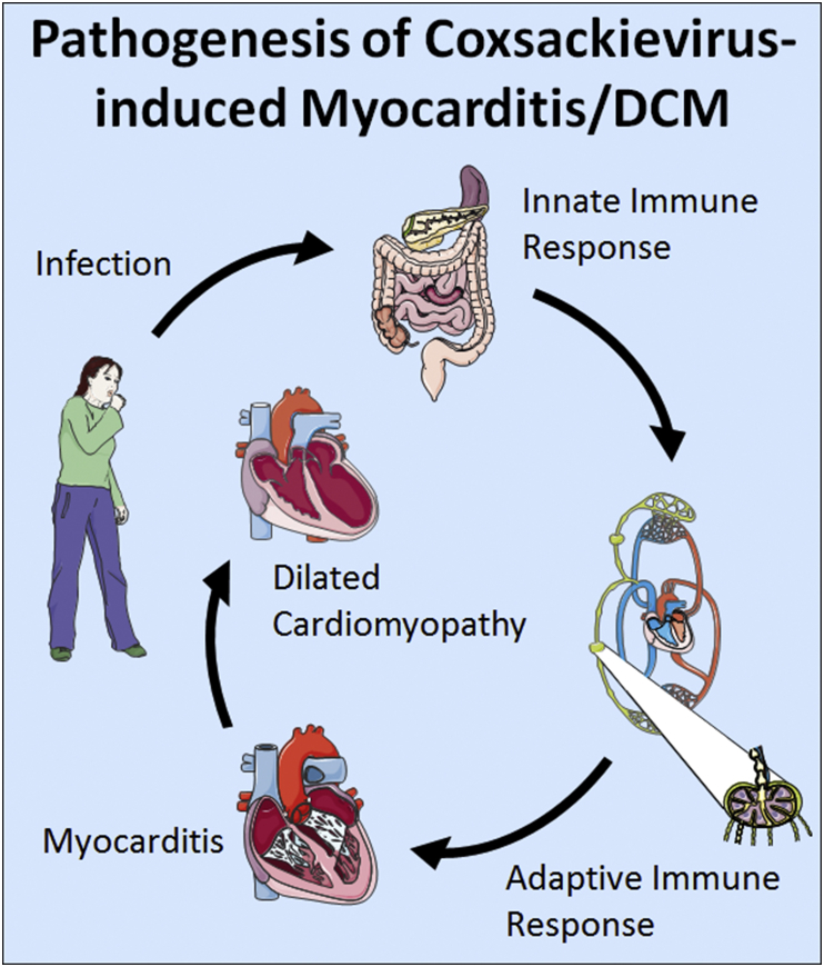
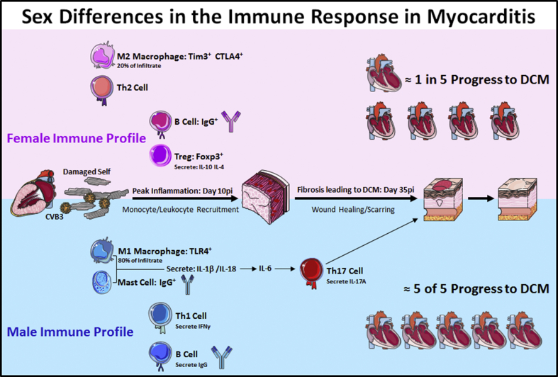
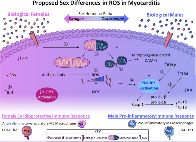
Autoimmune diseases are characterized by circulating antibodies and immune complexes directed against self-tissues that result in both systemic and organ-specific inflammation and pathology. Most autoimmune diseases occur more often in women than men. One exception is myocarditis, which is an inflammation of the myocardium that is typically caused by viral infections. Sex differences in the immune response and the role of the sex hormones estrogen and testosterone are well established based on animal models of autoimmune viral myocarditis as well as in mitochondrial function leading to reactive oxygen species production. RNA viruses like coxsackievirus B3, the primary cause of myocarditis in the US, activate the inflammasome through mitochondrial antiviral signaling protein located on the mitochondrial outer membrane. Toll-like receptor 4 and the inflammasome are the primary signaling pathways that increase inflammation during myocarditis, which is increased by testosterone. This review describes what is known about sex differences in inflammation, redox biology and mitochondrial function in the male-dominant autoimmune disease myocarditis and highlights gaps in the literature and future directions.
Overview
The claims and statistics of this paper haven’t been checked yet.
Run the check — free, no account needed. It reads the open-access text and checks the sources it cites.
Integrity signals
Registry
Cites
Statistics
Figures
Verification
Claims the paper makes
The claims and statistics of this paper haven’t been checked yet.
Run the check — free, no account needed. It reads the open-access text and checks the sources it cites.
Claims other papers make about this one
Each is checked against this paper’s own text: does it say what the citing paper says it does?
The source backs part of the claim, not all of it — or backs it for a different population, condition or comparison.
In the citing paper
“We have published previously that upregulation of complement and TLRs including TLR4 are key immune pathways that promote myocarditis in the autoimmune-CVB3 animal model”
From COVID-19, Myocarditis and Pericarditis (2023) · cites this paper as [63]
Experimental — accuracy not yet measured
Self-check
Percentages, sums and reported differences re-computed from the paper’s own numbers.
The self-check needs the paper’s full text.
Citations
Cites
Vos, T., Abajobir, A. A., Abate, K. H., Abbafati, C., Abbas, K. M., Abd-Allah, F., Abdulkader, R. S., Abdulle, A. M., Abebo, T. A., Abera, S. F., et al. (2017) · The Lancet · doi:10.1016/s0140-6736(17)32154-2 · Cited by 13,220
ROS function in redox signaling and oxidative stress
Schieber, M., & Chandel, N. S. (2014) · Current Biology · doi:10.1016/j.cub.2014.03.034 · Cited by 6,931
Sex differences in immune responses
Klein, S. L., & Flanagan, K. L. (2016) · Nature Reviews Immunology · doi:10.1038/nri.2016.90 · Cited by 6,370
Meister, A., & Anderson, M. E. (1983) · Annual Review of Biochemistry · doi:10.1146/annurev.bi.52.070183.003431 · Cited by 5,694
Neutrophil recruitment and function in health and inflammation
Kolaczkowska, E., & Kubes, P. (2013) · Nature Reviews Immunology · doi:10.1038/nri3399 · Cited by 5,472
The NLRP3 inflammasome: molecular activation and regulation to therapeutics
Swanson, K. V., Deng, M., & Ting, J. P.-Y. (2019) · Nature Reviews Immunology · doi:10.1038/s41577-019-0165-0 · Cited by 4,884
Macrophage biology in development, homeostasis and disease
Wynn, T. A., Chawla, A., & Pollard, J. W. (2013) · Nature · doi:10.1038/nature12034 · Cited by 4,684
Succinate is an inflammatory signal that induces IL-1β through HIF-1α
Tannahill, G. M., Curtis, A. M., Adamik, J., Palsson-McDermott, E. M., McGettrick, A. F., Goel, G., Frezza, C., Bernard, N. J., Kelly, B., Foley, N. H., et al. (2013) · Nature · doi:10.1038/nature11986 · Cited by 4,176
Glutathione Metabolism and Its Implications for Health
Wu, G., Lupton, J. R., Turner, N. D., Fang, Y.-Z., & Yang, S. (2004) · The Journal of Nutrition · doi:10.1093/jn/134.3.489 · Cited by 3,842
Nordberg, J., & Arnér, E. S. J. (2001) · Free Radical Biology and Medicine · doi:10.1016/s0891-5849(01)00724-9 · Cited by 2,720
Cited by
Sorted by citations
Zhu, H., Rhee, J., Cheng, P. P., Waliany, S., Chang, A., Witteles, R. M., Maecker, H. T., Davis, M. M., Nguyen, P. K. P., & Wu, S. M. (2020) · Current Cardiology Reports · doi:10.1007/s11886-020-01292-3 · Cited by 211
Cicco, S., Cicco, G., Racanelli, V., & Vacca, A. (2020) · Mediators of Inflammation · doi:10.1155/2020/7527953 · Cited by 195
COVID-19, Myocarditis and Pericarditis
Fairweather, D., Beetler, D. J., Di Florio, D. N., Musigk, N., Heidecker, B., & Cooper, L. T. (2023) · Circulation Research · doi:10.1161/circresaha.123.321878 · Cited by 194
Self-citationSeverely low testosterone in males with COVID‐19: A case‐control study
Salonia, A., Pontillo, M., Capogrosso, P., Gregori, S., Tassara, M. G., Boeri, L., Carenzi, C., Abbate, C., Cignoli, D., Ferrara, A. M., et al. (2021) · Andrology · doi:10.1111/andr.12993 · Cited by 146
Worse progression of COVID‐19 in men: Is testosterone a key factor?
Giagulli, V. A., Guastamacchia, E., Magrone, T., Jirillo, E., Lisco, G., Pergola, G. D., & Triggiani, V. (2020) · Andrology · doi:10.1111/andr.12836 · Cited by 142
Sex and gender differences in myocarditis and dilated cardiomyopathy: An update
Fairweather, D., Beetler, D. J., Musigk, N., Heidecker, B., Lyle, M. A., Cooper, L. T., & Bruno, K. A. (2023) · Frontiers in Cardiovascular Medicine · doi:10.3389/fcvm.2023.1129348 · Cited by 124
Self-citationChen, L., Huang, Q., Zhao, T., Sui, L., Wang, S., Xiao, Z., Nan, Y., & Ai, K. (2021) · Redox Biology · doi:10.1016/j.redox.2021.102046 · Cited by 99
Myopathic Cardiac Genotypes Increase Risk for Myocarditis
Kontorovich, A. R., Patel, N., Moscati, A., Richter, F., Peter, I., Purevjav, E., Selejan, S. R., Kindermann, I., Towbin, J. A., Bohm, M., et al. (2021) · JACC: Basic to Translational Science · doi:10.1016/j.jacbts.2021.06.001 · Cited by 95
Sex and gender differences in the outcome of patients with COVID‐19
Falahi, S., & Kenarkoohi, A. (2020) · Journal of Medical Virology · doi:10.1002/jmv.26243 · Cited by 84
Testosterone in males with COVID‐19: A 7‐month cohort study
Salonia, A., Pontillo, M., Capogrosso, P., Gregori, S., Carenzi, C., Ferrara, A. M., Rowe, I., Boeri, L., Larcher, A., Ramirez, G. A., et al. (2021) · Andrology · doi:10.1111/andr.13097 · Cited by 84
The Interplay of Sex Steroids, the Immune Response, and the Intestinal Microbiota
de Pace, F., & Watnick, P. I. (2020) · Trends in Microbiology · doi:10.1016/j.tim.2020.11.001 · Cited by 79
Myocarditis associated with COVID-19 vaccination
Buoninfante, A., Andeweg, A. C., Genov, G., & Cavaleri, M. (2024) · npj Vaccines · doi:10.1038/s41541-024-00893-1 · Cited by 81
Immunomodulatory role of estrogen in ischemic stroke: neuroinflammation and effect of sex
Zhong, X., Sun, Y., Lu, Y., & Xu, L. (2023) · Frontiers in Immunology · doi:10.3389/fimmu.2023.1164258 · Cited by 63
How Does Vitamin D Affect Immune Cells Crosstalk in Autoimmune Diseases?
Gallo, D., Baci, D., Kuštrimović, N. Z., Lanzo, N., Patera, B., Tanda, M. L., Piantanida, E., & Mortara, L. (2023) · International Journal of Molecular Sciences · doi:10.3390/ijms24054689 · Cited by 65
Metaflammation in glucolipid metabolic disorders: Pathogenesis and treatment
Xiong, P., Zhang, F., Liu, F., Zhao, J., Huang, X., Luo, D., & Guo, J. (2023) · Biomedicine & Pharmacotherapy · doi:10.1016/j.biopha.2023.114545 · Cited by 59
Belonwu, S., Li, Y., Bunis, D. G., Rao, A. A., Solsberg, C. W., Tang, A., Fragiadakis, G. K., Dubal, D. B., Oskotsky, T., & Sirota, M. (2021) · Molecular Neurobiology · doi:10.1007/s12035-021-02591-8 · Cited by 55
Lang, H., Noble, K. V., Barth, J. L., Rumschlag, J. A., Jenkins, T. R., Storm, S., Eckert, M. A., Dubno, J. R., & Schulte, B. A. (2023) · Journal of Neuroscience · doi:10.1523/jneurosci.2234-22.2023 · Cited by 58
Tanabe, S., O’Brien, J. M., Tollefsen, K. E., Kim, Y. J., Chauhan, V., Yauk, C. L., Huliganga, E., Rudel, R. A., Kay, J. E., Helm, J. S., et al. (2022) · Frontiers in Toxicology · doi:10.3389/ftox.2022.887135 · Cited by 57
17β‐estradiol reduces SARS‐CoV‐2 infection in vitro
Lemes, R. M. R., Costa, A. J., Bartolomeo, C. S., Bassani, T. B., Nishino, M. S., Pereira, G. J. S., Smaili, S. S., Maciel, R. M. B., Braconi, C. T., da Cruz, E. F., et al. (2021) · Physiological Reports · doi:10.14814/phy2.14707 · Cited by 51
Sex-Specific Differences of the Inflammatory State in Experimental Autoimmune Myocarditis
Barcena, M. L., Jeuthe, S., Niehues, M. H., Pozdniakova, S., Haritonow, N., Kühl, A. A., Messroghli, D. R., & Regitz-Zagrosek, V. (2021) · Frontiers in Immunology · doi:10.3389/fimmu.2021.686384 · Cited by 47
Sex Differences, Genetic and Environmental Influences on Dilated Cardiomyopathy
Jain, A., Norton, N., Bruno, K. A., Cooper, L. T., Atwal, P. S., & Fairweather, D. (2021) · Journal of Clinical Medicine · doi:10.3390/jcm10112289 · Cited by 48
Self-citationLiao, T.-L., Lee, Y.-C., Tzeng, C.-R., Wang, Y.-P., Chang, H.-Y., Lin, Y.-F., & Kao, S.-H. (2019) · Free Radical Biology and Medicine · doi:10.1016/j.freeradbiomed.2019.01.022 · Cited by 44
Lynch, S. E., Boyett, J. E., Smith, M. R., & Giordano‐Mooga, S. (2021) · Frontiers in Cell and Developmental Biology · doi:10.3389/fcell.2020.610516 · Cited by 46
COVID-19 mRNA Vaccines: The Molecular Basis of Some Adverse Events
Giannotta, G., Murrone, A., & Giannotta, N. (2023) · Vaccines · doi:10.3390/vaccines11040747 · Cited by 45
Astre, G., Atlan, T., Goshtchevsky, U., Oron-Gottesman, A., Smirnov, M., Shapira, K., Velan, A., Deelen, J., Levy, T., Levanon, E. Y., et al. (2023) · Developmental Cell · doi:10.1016/j.devcel.2023.05.015 · Cited by 44
Zhou, W., Zhao, Z., Yu, Z., Hou, Y., Keerthiga, R., & Fu, A. (2022) · Mitochondrion · doi:10.1016/j.mito.2022.04.004 · Cited by 40
Zhang, Z., & Guo, J. (2024) · Antioxidants · doi:10.3390/antiox14010038 · Cited by 40
Loss of CTRP4 alters adiposity and food intake behaviors in obese mice
Sarver, D. C., Stewart, A. N., Rodriguez, S., Little, H. C., Aja, S. M., & Wong, G. W. (2020) · American Journal of Physiology-Endocrinology and Metabolism · doi:10.1152/ajpendo.00448.2020 · Cited by 38
Testosterone in males with COVID‐19: a 12‐month cohort study
Salonia, A., Pontillo, M., Capogrosso, P., Pozzi, E., Ferrara, A. M., Cotellessa, A., Belladelli, F., Corsini, C., Gregori, S., Rowe, I., et al. (2022) · Andrology · doi:10.1111/andr.13322 · Cited by 35
Differences in Risk Factors for Coronary Atherosclerosis According to Sex
Kim, H. (2024) · Journal of Lipid and Atherosclerosis · doi:10.12997/jla.2024.13.2.97 · Cited by 40
Luo, X., Chang, X., Zhou, H., Lin, H., & Fan, H. (2021) · Veterinary Microbiology · doi:10.1016/j.vetmic.2021.109057 · Cited by 36
Salciccia, S., Giudice, F. D., Eisenberg, M. L., Mastroianni, C. M., BERARDINIS, E. D., Ricciuti, G. P., Viscuso, P., Zingaropoli, M. A., Pasculli, P., Ciardi, M. R., et al. (2021) · Therapeutic Advances in Endocrinology and Metabolism · doi:10.1177/20420188211010105 · Cited by 38
Gao, A., Peng, B., Gao, Y., Yang, Z., Li, Z., Guo, T., Qiu, H., & Gao, R. (2025) · Diabetology & Metabolic Syndrome · doi:10.1186/s13098-025-01687-8 · Cited by 34
Zhang, H., Zhangsun, Z., Xue, C.-X., Li, X.-Y., Ren, J. T., Jiang, Y., Liu, T., Yao, H.-R., Zhang, J., Gou, T.-T., et al. (2022) · Acta Pharmacologica Sinica · doi:10.1038/s41401-022-00991-7 · Cited by 38
Li, C., Cai, B., Lu, D., & Zeng, H. (2023) · Mitochondrion · doi:10.1016/j.mito.2023.01.009 · Cited by 33
Sex Differences Define the Vulnerability to Atherosclerosis
Poznyak, A. V., Sukhorukov, V. N., Guo, S., Postnov, A. Y., & Orekhov, A. N. (2023) · Clinical Medicine Insights Cardiology · doi:10.1177/11795468231189044 · Cited by 32
Extracellular vesicles as carriers for mitochondria: Biological functions and clinical applications
Liao, Z., Tong, B., Ke, W., Cao, Y., Wu, X., & Lei, M. (2024) · Mitochondrion · doi:10.1016/j.mito.2024.101935 · Cited by 35
Cortellini, A., Ricciuti, B., Borghaei, H., Naqash, A. R., D’Alessio, A., Fulgenzi, C. A. M., Addeo, A., Banna, G. L., & Pinato, D. J. (2022) · Cancer · doi:10.1002/cncr.34348 · Cited by 31
You, M., Li, S., Yan, S., Yao, D., Wang, T., & Wang, Y. (2023) · Environment International · doi:10.1016/j.envint.2023.108228 · Cited by 27
The role of extracellular DNA in COVID-19: Clues from inflamm-aging
Storci, G., Bonifazi, F., Garagnani, P., Olivieri, F., & Bonafè, M. (2020) · Ageing Research Reviews · doi:10.1016/j.arr.2020.101234 · Cited by 30
Ribeiro, P. S. S., Willemen, H. L. D. M., Versteeg, S., Gil, C. M., & Eijkelkamp, N. (2023) · Immunotherapy Advances · doi:10.1093/immadv/ltad022 · Cited by 29
Gut microbiome-immune interactions and their role in rheumatoid arthritis development
Nurgaziyev, M., Issilbayeva, A., Bersimbaev, R. I., Ilderbayev, O. Z., Vinogradova, E. B., Jarmukhanov, Z., Nurgozhina, A., Sergazy, S., Kozhabergen, N., Akhmetova, Z., et al. (2024) · PeerJ · doi:10.7717/peerj.17477 · Cited by 26
The Metabolomic Underpinnings of Symptom Burden in Patients With Multiple Chronic Conditions
Corwin, E. J., Brewster, G. S., Dunbar, S. B., Wells, J., Hertzberg, V., Holstad, M. M., Song, M., & Jones, D. P. (2020) · Biological Research For Nursing · doi:10.1177/1099800420958196 · Cited by 25
Zangeneh, F. Z., & Shoushtari, M. S. (2021) · Journal of Family & Reproductive Health · doi:10.18502/jfrh.v15i3.7132 · Cited by 23
Stewart, A. N., Glaser, E. P., Mott, C. A., Bailey, W. M., Sullivan, P. G., Patel, S. P., & Gensel, J. C. (2022) · Journal of Neurotrauma · doi:10.1089/neu.2022.0010 · Cited by 22
Alterations of the intestinal microbiome and metabolome in women with rheumatoid arthritis
Yun, H., Wang, X., Wei, C., Liu, Q., Li, X., Li, N., Zhang, G., Cui, D., & Liu, R. (2023) · Clinical and Experimental Medicine · doi:10.1007/s10238-023-01161-7 · Cited by 22
Tan, S. Y., Lei, X., Little, H. C., Rodriguez, S., Sarver, D. C., Cao, X., & Wong, G. W. (2020) · American Journal of Physiology-Endocrinology and Metabolism · doi:10.1152/ajpendo.00533.2019 · Cited by 22
Cancer and Tumour Suppressor p53 Encounters at the Juncture of Sex Disparity
Haupt, S., & Haupt, Y. (2021) · Frontiers in Genetics · doi:10.3389/fgene.2021.632719 · Cited by 22
Huang, Y., Li, H., Liang, R., Chen, J., & Tang, Q. (2024) · Biogerontology · doi:10.1007/s10522-024-10121-x · Cited by 23
Aging, sex and NLRP3 inflammasome in cardiac ischaemic disease
Alloatti, G., Penna, C. R. R., Comità, S., Tullio, F., Aragno, M., Biasi, F., & Pagliaro, P. (2022) · Vascular Pharmacology · doi:10.1016/j.vph.2022.107001 · Cited by 21
Lou, K., Liu, S., Zhang, F., Sun, W., Su, X., Bi, W., Yin, Q., Yang, P., Qiu, Y., Zhang, Z., et al. (2023) · Journal of Translational Medicine · doi:10.1186/s12967-023-04511-x · Cited by 25
Łukawska-Tatarczuk, M. M., Franek, E., Czupryniak, L., Joniec‐Maciejak, I., Pawlak, A., Wojnar, E., Zieliński, J., Mirowska-Guzel, D. M., & Mrozikiewicz‐Rakowska, B. (2021) · Biomolecules · doi:10.3390/biom11081110 · Cited by 22
Duan, X., Nie, Y., Xie, X., Zhang, Q., Zhu, C., Zhu, H., Chen, R., Xu, J. J., Zhang, J., Yang, C., et al. (2024) · Frontiers in Microbiology · doi:10.3389/fmicb.2024.1421608 · Cited by 23
Li, Y. E., Wang, S., Reiter, R. J., & Ren, J. T. (2022) · International Journal of Infectious Diseases · doi:10.1016/j.ijid.2022.08.026 · Cited by 23
COVID-19-Induced Myocarditis: Pathophysiological Roles of ACE2 and Toll-like Receptors
Pannucci, P., Jefferson, S. R., Hampshire, J., Cooper, S. L., Hill, S. J., & Woolard, J. (2023) · International Journal of Molecular Sciences · doi:10.3390/ijms24065374 · Cited by 21
Sexual Dimorphism in Cardiometabolic Diseases: The Role of AMPK
Kvandová, M., Púzserová, A., & Bališ, P. (2023) · International Journal of Molecular Sciences · doi:10.3390/ijms241511986 · Cited by 20
Cumulative social disadvantage and its impact on long COVID: insights from a U.S. national survey
Xiang, J., Zheng, H., Cai, Y., Chen, S., Wang, Y., & Chen, R. (2025) · BMC Medicine · doi:10.1186/s12916-025-04039-5 · Cited by 18
Zhou, Y., Zheng, K. I., Ma, H., Li, G., Pan, X.-Y., Zhu, P., Targher, G., Byrne, C. D., Wang, X., Chen, Y., et al. (2020) · Nutrition Metabolism and Cardiovascular Diseases · doi:10.1016/j.numecd.2020.10.004 · Cited by 17
Lan, X., Wang, C., Li, W., Chao, Z., Lao, G., Wu, K., Li, G., Ning, Y., & Zhou, Y. (2022) · Journal of Affective Disorders · doi:10.1016/j.jad.2022.06.073 · Cited by 17
Kim, H., & Kim, M. (2023) · Korean Circulation Journal · doi:10.4070/kcj.2023.0205 · Cited by 18
Cardiomyopathy Associated with Anti-SARS-CoV-2 Vaccination: What Do We Know?
Parra-Lucares, A., Toro, L., Weitz-Muñoz, S., & Ramos, C. (2021) · Viruses · doi:10.3390/v13122493 · Cited by 15
Sex and age differences in sST2 in cardiovascular disease
Beetler, D. J., Bruno, K. A., Florio, D. N. D., Douglass, E. J., Shrestha, S., Tschöpe, C., Cunningham, M. W., Krejčí, J., Vašků, J. B., Pankuweit, S., et al. (2023) · Frontiers in Cardiovascular Medicine · doi:10.3389/fcvm.2022.1073814 · Cited by 15
Self-citationTargeting the Toll-like receptor pathway as a therapeutic strategy for neonatal infection
Dias, M. L., O’Connor, K. M., Dempsey, E. M., O’Halloran, K. D., & McDonald, F. B. (2021) · American Journal of Physiology-Regulatory, Integrative and Comparative Physiology · doi:10.1152/ajpregu.00307.2020 · Cited by 15
Sex differences in neuroimmunoendocrine communication. Involvement on longevity
Suárez, L. M., Cerro, E. D., Félix, J., González-Sánchez, M., Ceprián, N., Guerra‐Pérez, N., Novelle, M. G., de Toda, I. M., & la Fuente, M. D. (2023) · Mechanisms of Ageing and Development · doi:10.1016/j.mad.2023.111798 · Cited by 17
Sex differences in immune responses to viruses, bacteria and vaccines
Chaulagain, S., Liu, J., McCombs, J. E., & Klein, S. L. (2026) · Nature Immunology · doi:10.1038/s41590-026-02470-1 · Cited by 16
Wang, S., Guo, J., Liu, X., Tian, W., Zhang, Y., Wang, Y., Liu, Y., Mingyan, E., & Fang, S. (2023) · Diabetology & Metabolic Syndrome · doi:10.1186/s13098-023-01090-1 · Cited by 14
The autoimmune tautology revisited
Anaya, J., & Beltrán, S. (2023) · Journal of Translational Autoimmunity · doi:10.1016/j.jtauto.2023.100204 · Cited by 14
Wang, X., Ma, R., Shi, R., Qin, H., Chen, W., Yu, Z., Ding, Y., Peng, C., & Shi, Y. (2023) · Journal of Translational Medicine · doi:10.1186/s12967-023-04726-y · Cited by 14
Editorial: Sex Differences in Inflammatory Diseases
Trabace, L., Roviezzo, F., & Rossi, A. (2022) · Frontiers in Pharmacology · doi:10.3389/fphar.2022.962869 · Cited by 14
Long-term e-cigarette aerosol exposure causes pulmonary emphysema in adult female and male mice
Rodríguez-Herrera, A. J., de Souza, A. B. F., Castro, T. D. F., Júnior, P. A. M., Marcano-Gómez, E. C., Menezes, T. P., Castro, M. L. D. C., Talvani, A., Costa, D. C., Cangussú, S. D., et al. (2023) · Regulatory Toxicology and Pharmacology · doi:10.1016/j.yrtph.2023.105412 · Cited by 11
Wang, H., Chen, X., Lin, F., Zheng, J., Chen, K., Wang, X., Lin, X., Chen, X., Ye, Q., Wang, Y.-Q., et al. (2023) · Journal of Affective Disorders · doi:10.1016/j.jad.2023.12.050 · Cited by 12
Exercise After Acute Myocarditis
Bryde, R. E., Cooper, L. T., Fairweather, D., Florio, D. N. D., & Martinez, M. W. (2022) · Cardiology Clinics · doi:10.1016/j.ccl.2022.08.009 · Cited by 13
Self-citationSex Hormone–Dependent Lipid Mediator Formation in Male and Female Mice During Peritonitis
Troisi, F., Pace, S., Jordan, P. M., Meyer, K. P. L., Bilancia, R., Ialenti, A., Borrelli, F., Rossi, A., Sautebin, L., Serhan, C. N., et al. (2022) · Frontiers in Pharmacology · doi:10.3389/fphar.2021.818544 · Cited by 12
Zhou, Y., Li, Z., Li, J., Wang, X., Qi, K., Zhang, S., & Zhou, C. (2023) · Journal of the American Medical Directors Association · doi:10.1016/j.jamda.2023.03.022 · Cited by 11
Inflammatory Cytokine Levels in Patients with Autosomal Dominant Polycystic Kidney Disease
Arjune, S., Lettenmeier, K., Todorova, P., Späth, M. R., Majjouti, M., Mahabir, E., Grundmann, F., & Müller, R. (2024) · Kidney360 · doi:10.34067/kid.0000000000000525 · Cited by 11
Zhao, S., Wang, J., Xue, M., Wu, B., Sheng, L., Wen, Y., Wang, G., Song, J., Du, D., & Xu, J. (2026) · Bone Research · doi:10.1038/s41413-025-00502-4 · Cited by 11
Lan, X., Wu, F., Wang, C., Wu, K., Fang, Z., Lao, G., Zhang, B., Ning, Y., & Zhou, Y. (2022) · Journal of Affective Disorders · doi:10.1016/j.jad.2022.11.016 · Cited by 9
Sex- specific differences in suspected myocarditis presentations and outcomes
Schütze, J., Greisser, N., Joss, P., Gebhard, C., Bernhard, B., Greulich, S., Stark, A. W., Safarkhanlo, Y., Pavlicek, M., Hundertmark, M., et al. (2024) · International Journal of Cardiology · doi:10.1016/j.ijcard.2024.132593 · Cited by 10
The Epidemiology of COVID-19 Vaccine-Induced Myocarditis
Costa, C., & Moniati, F. (2024) · Advances in Medicine · doi:10.1155/2024/4470326 · Cited by 10
Impact of endocrine disruptors on peripheral blood mononuclear cells in vitro: role of gender
Maddalon, A., Cari, L., Iulini, M., Alhosseini, M. N., Galbiati, V., Marinovich, M., Nocentini, G., & Corsini, E. (2023) · Archives of Toxicology · doi:10.1007/s00204-023-03592-3 · Cited by 10
Sex differences in mitochondrial gene expression during viral myocarditis
Florio, D. N. D., Weigel, G. J., Gorelov, D. J., McCabe, E. J., Beetler, D. J., Shapiro, K. A., Bruno, K. A., Chekuri, I., Jain, A., Whelan, E. R., et al. (2024) · Biology of Sex Differences · doi:10.1186/s13293-024-00678-0 · Cited by 10
Self-citationFang, X., Yang, J., Liu, Y., Lin, Y., Li, Y., Yin, X., Dou, X., & Miao, C. (2025) · Frontiers in Immunology · doi:10.3389/fimmu.2025.1569271 · Cited by 9
Tong, Y., Teng, Y., Zhang, Y., Huang, C., Liao, W., Wan, B., Zong, S., & Zeng, G. (2025) · Innovation in Aging · doi:10.1093/geroni/igaf095 · Cited by 9
Sex Dimorphism of Allergen-Induced Secreted Proteins in Murine and Human Lungs
Hemshekhar, M., Mostafa, D. H. D., Spicer, V., Piyadasa, H., Maestre‐Batlle, D., Bølling, A. K., Halayko, A. J., Carlsten, C., & Mookherjee, N. (2022) · Frontiers in Immunology · doi:10.3389/fimmu.2022.923986 · Cited by 9
Maes, M. E., Colombo, G., Uiterkamp, F. E. S., Sternberg, F., Venturino, A., Pohl, E. E., & Siegert, S. (2023) · iScience · doi:10.1016/j.isci.2023.107780 · Cited by 9
Sex-Biased Control of Inflammation and Metabolism by a Mitochondrial Nod-Like Receptor
Snäkä, T., Bekkar, A., Desponds, C., Prével, F., Claudinot, S., Isorce, N., Teixeira, F., Grasset, C., Xénarios, I., López‐Mejía, I. C., et al. (2022) · Frontiers in Immunology · doi:10.3389/fimmu.2022.882867 · Cited by 9
Effect of Sex Differences in Silicotic Mice
Jin, F., Li, Y., Wang, X., Yang, X., Li, T., Xu, H., Wei, Z., & Liu, H. (2022) · International Journal of Molecular Sciences · doi:10.3390/ijms232214203 · Cited by 9
Noah, M. L. N., Mprah, R., Wowui, P. I., Adekunle, A. O., Adu‐Amankwaah, J., Tan, R., Gong, Z., Li, T., Fu, L., Machuki, J. O., et al. (2024) · Cell Communication and Signaling · doi:10.1186/s12964-024-01535-8 · Cited by 8
Zhou, L., Luo, J., Wei, Y., Han, L., Sun, A., Zhang, M., Hu, D., Yang, H., Tang, R., Xie, P., et al. (2025) · Journal of Ethnopharmacology · doi:10.1016/j.jep.2025.120272 · Cited by 8
Araújo, M. R., Mendes‐Frias, A., & Silvestre, R. (2026) · Biology of Sex Differences · doi:10.1186/s13293-026-00857-1 · Cited by 8
Immunodiagnosis — the promise of personalized immunotherapy
Wang, R., Xiong, K., Wang, Z., Wu, D., Hu, B., Ruan, J., Sun, C., Ma, D., Li, L., & Liao, S. (2023) · Frontiers in Immunology · doi:10.3389/fimmu.2023.1216901 · Cited by 9
Bourguiba, R., Deshayes, S., Amaryan, G., Koné‐Paut, I., Bélot, A., Sarkisyan, T., Guedri, R., Mejbri, M., Melki, I., Meinzer, U., et al. (2024) · Rheumatology International · doi:10.1007/s00296-024-05706-1 · Cited by 7
Roy, S., Banerjee, D., Banerjee, A., & Choudhury, L. (2025) · Biologia Futura · doi:10.1007/s42977-025-00291-1 · Cited by 11
Bynum, H., & Edwards, K. S. (2026) · International Journal of Molecular Sciences · doi:10.3390/ijms27114966 · Cited by 8
Sexual dimorphisms in redox biology
Asmis, R., & Giordano‐Mooga, S. (2020) · Redox Biology · doi:10.1016/j.redox.2020.101533 · Cited by 7
Epididymal embryonic development harbors TLR4/NFKB signaling pathway as a morphogenetic player
Ferreira, L. G. A., Nishino, F. A., Fernandes, S. G., Ribeiro, C. M., Hinton, B. T., & Avellar, M. C. W. (2021) · Journal of Reproductive Immunology · doi:10.1016/j.jri.2021.103456 · Cited by 7
Disease comorbidities associated with chemical intolerance
Palmer, R., Walker, T., Perales, R. B., Rincón, R., Jaén, C. R., & Miller, C. (2021) · Environmental Disease · doi:10.4103/ed.ed_18_21 · Cited by 7
Ramiro, L., Faura, J., Simats, A., García‐Rodríguez, P., Ma, F., Martín, L., Canals, F., Rosell, A., & Montaner, J. (2023) · BMC Neuroscience · doi:10.1186/s12868-023-00775-7 · Cited by 7
Ricci, J. F., Farahani, N. A. S., Davis, C., Ritter, K. G., Parrow, L. M., Tomerlin, P. I., Darakjian, A. A., Gegoutchadze, K., Gupta, D., & Bruno, K. A. (2025) · Frontiers in Immunology · doi:10.3389/fimmu.2025.1601307 · Cited by 8
Ciani, M., Rigillo, G., Bertarini, B., Benatti, C., Alboni, S., & Tascedda, F. (2025) · Frontiers in Immunology · doi:10.3389/fimmu.2025.1711656 · Cited by 6
Metadata
- Authors
- Di Florio, D. N., Sin, J., Coronado, M. J., Atwal, P. S., & Fairweather, D.
- Affiliations
- Cedars-Sinai Medical Center (US)
- Whitman College (US)
- Mayo Clinic (US)
- Corresponding author
- Fairweather, DeLisa
- Venue
- Redox Biology
- Publisher
- Elsevier BV
- Published
- 2020-04
- Volume
- 31
- Pages
- 101482
- ISSN
- 2213-2317 (ISSN-L)
- PMC
- PMC7212489
- OpenAlex
- W3010344840
- Cited by
- 185
- References
- 164
- Citations by year
- 2026: 30 · 2025: 24 · 2024: 26 · 2023: 44 · 2022: 29 · 2021: 19 · 2020: 12
- FWCI
- 4.41
- Type
- journal article
- Language
- en
- Open access
- gold
- doi.org
- Licence
- Locations
- Redox Biology (publishedVersion, cc-by-nc-nd)
- PubMed Central (publishedVersion, cc-by-nc-nd)
- PubMed (publishedVersion)
- DOAJ (DOAJ: Directory of Open Access Journals) (submittedVersion, cc-by-sa)
- Funders
- Mayo Clinic
- National Institutes of Health (R21 AI145356, R21 AI152318, TL1 TR002380)
- Topics
- Health Sciences › Medicine › Infectious Diseases › Viral Infections and Immunology Research
- Life Sciences › Biochemistry, Genetics and Molecular Biology › Molecular Biology › Inflammasome and immune disorders
- Life Sciences › Biochemistry, Genetics and Molecular Biology › Molecular Biology › Mitochondrial Function and Pathology
- Keywords
- sex differences, autoimmune myocarditis, viral myocarditis, coxsackievirus B3, inflammasome, TLR4, reactive oxygen species, mitochondrial function, estrogen, testosterone, inflammation, autoimmunity
- MeSH terms
- Animals, Autoimmune Diseases, Biology, Disease Models, Animal, Female, Humans, Inflammation, Male, Mitochondria, Myocardium, Oxidation-Reduction, Sex Characteristics, Autoimmunity
About these checks
Registry, retraction, Cites and statistics results are computed, not judged, with one exception: a printed reference that no index lists is read once by a model before it counts as a citation error. Claims are extracted by a model and kept only when their quote appears verbatim in the text; each cited source is then checked, with a model’s help, for whether it says what the claim relies on. Those verdicts can be wrong — read the claim’s quote and the cited source before relying on one.
Statistics and arithmetic are recomputed, not judged.
Confidence bands are shown only for fields calibrated on our annotated test set. None is calibrated yet, so no band is shown.
What counts as a claim ›How we measure confidence ›How we check this ›
Are you an author, or do you see a problem with this page? Request a review
