

The DOI matches its Crossref record; 50 of the 50 references it cites were found in a registry or an index.
Nonadhesive Alginate Hydrogels Support Growth of Pluripotent Stem Cell-Derived Intestinal Organoids
Open accessPublished version of a preprint
Abstract
Human intestinal organoids (HIOs) represent a powerful system to study human development and are promising candidates for clinical translation as drug-screening tools or engineered tissue. Experimental control and clinical use of HIOs is limited by growth in expensive and poorly defined tumor-cell-derived extracellular matrices, prompting investigation of synthetic ECM-mimetics for HIO culture. Since HIOs possess an inner epithelium and outer mesenchyme, we hypothesized that adhesive cues provided by the matrix may be dispensable for HIO culture. Here, we demonstrate that alginate, a minimally supportive hydrogel with no inherent cell instructive properties, supports HIO growth in vitro and leads to HIO epithelial differentiation that is virtually indistinguishable from Matrigel-grown HIOs. In addition, alginate-grown HIOs mature to a similar degree as Matrigel-grown HIOs when transplanted in vivo, both resembling human fetal intestine. This work demonstrates that purely mechanical support from a simple-to-use and inexpensive hydrogel is sufficient to promote HIO survival and development.
Overview
The claims and statistics of this paper haven’t been checked yet.
Run the check — free, no account needed. It reads the open-access text and checks the sources it cites.
Integrity signals
Registry
Cites
Statistics
Figures
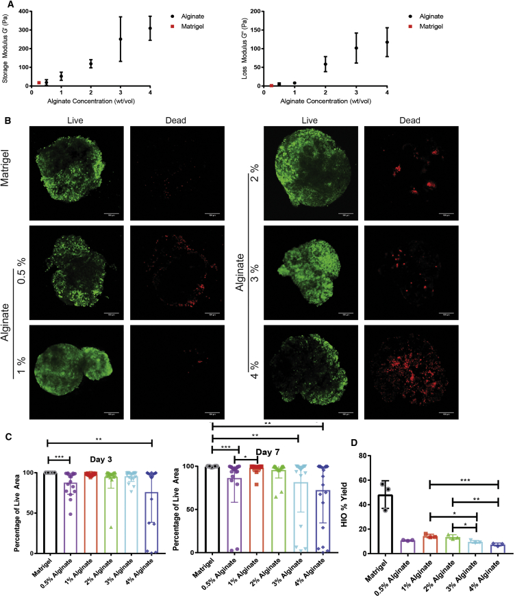
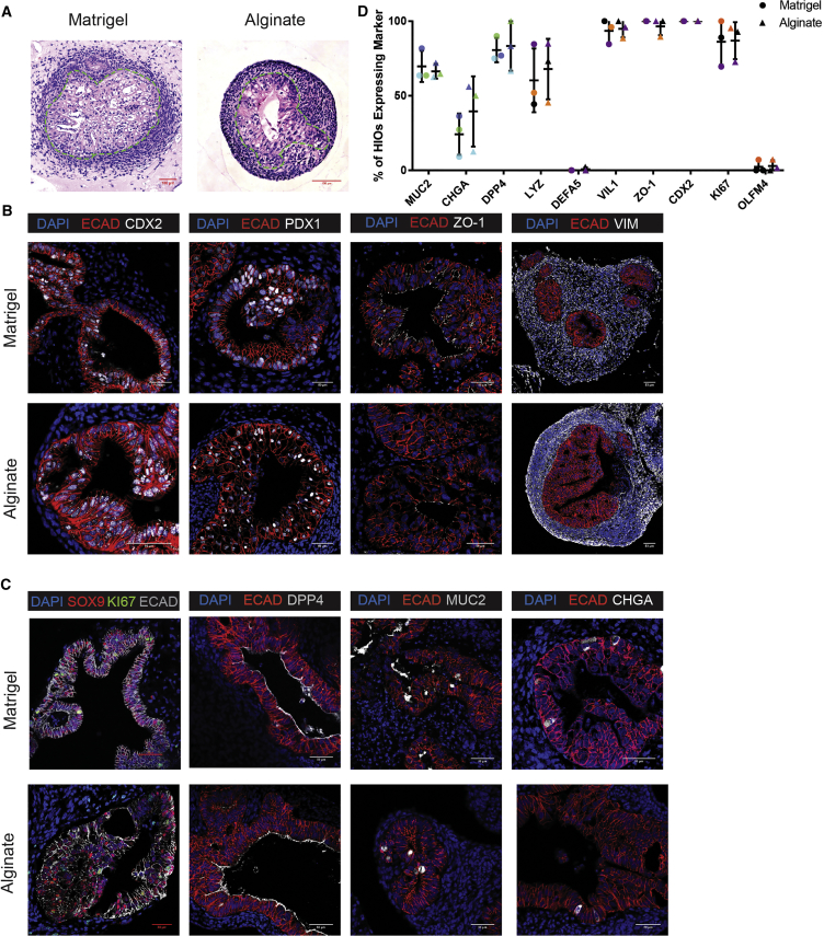
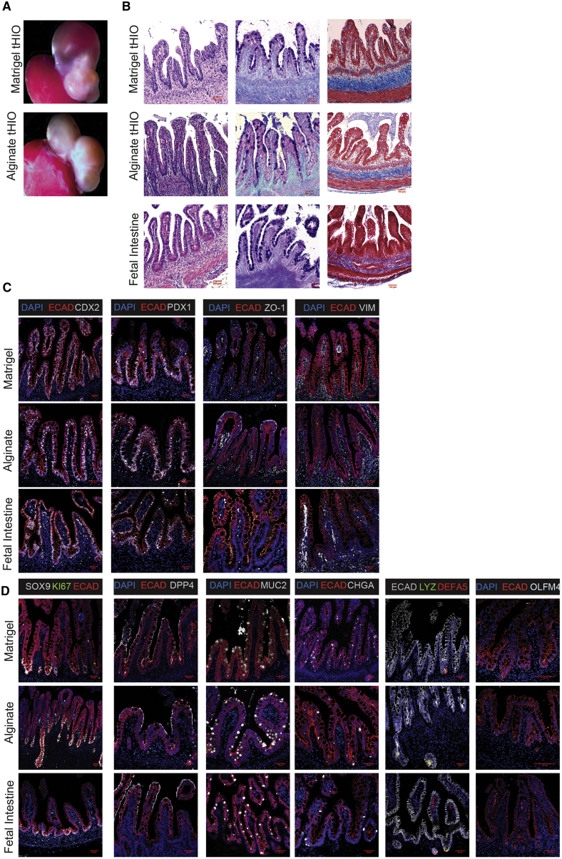
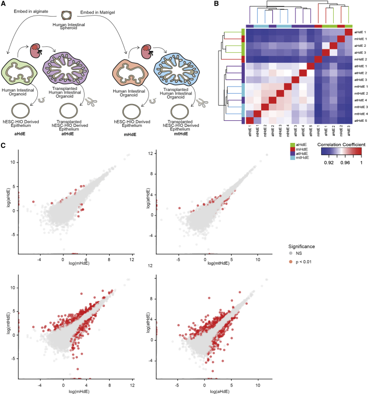
Verification
Claims the paper makes
The claims and statistics of this paper haven’t been checked yet.
Run the check — free, no account needed. It reads the open-access text and checks the sources it cites.
Claims other papers make about this one
Each is checked against this paper’s own text: does it say what the citing paper says it does?
The cited source says what the claim attributes to it.
In the citing paper
“Mechanical properties, e.g., ECM stiffness, of the cell and tissue microenvironment are known to play an important role in cell survival [53,54], proliferation[55], migration[56], and tissue differentiation[57,58].”
In the citing paper
“Organoid types grown in alginate include human lung99,100, human brain101, murine intestinal102, human intestinal102,103, human pancreatic104, and human and murine vascular105.”
From Towards organoid culture without Matrigel (2021) · cites this paper as [103]
In the citing paper
“In addition to organoids grown in naturally-derived proteins, several laboratories have grown a wide range of organoids in polysaccharides such as alginate or alginate-chitosan mixtures.”
From Towards organoid culture without Matrigel (2021) · cites this paper as [103]
In the citing paper
“Capeling and colleagues grew HIOs on an alginate substrate and found that differentiation of human pluripotent cells into HIOs could be supported without the alginate providing chemical cues to the cells.”
From Towards organoid culture without Matrigel (2021) · cites this paper as [103]
In the citing paper
“There have been a number of studies showing that organoids can be grown on unmodified surfaces such as alginate, but this requires more research103,105,110,175,183.”
From Towards organoid culture without Matrigel (2021) · cites this paper as [103]
Self-check
Percentages, sums and reported differences re-computed from the paper’s own numbers.
The self-check needs the paper’s full text.
Citations
Cites
Moderated estimation of fold change and dispersion for RNA-seq data with DESeq2
Love, M. I., Huber, W., & Anders, S. (2014) · Genome Biology · doi:10.1186/s13059-014-0550-8 · Cited by 95,106
Near-optimal probabilistic RNA-seq quantification
Bray, N. L., Pimentel, H., Melsted, P., & Pachter, L. (2016) · Nature Biotechnology · doi:10.1038/nbt.3519 · Cited by 9,723
Alginate: Properties and biomedical applications
Lee, K. Y., & Mooney, D. J. (2011) · Progress in Polymer Science · doi:10.1016/j.progpolymsci.2011.06.003 · Cited by 8,477
Cerebral organoids model human brain development and microcephaly
Lancaster, M. A., Renner, M., Martin, C.-A., Wenzel, D., Bicknell, L. S., Hurles, M. E., Homfray, T., Penninger, J. M., Jackson, A. P., & Knoblich, J. A. (2013) · Nature · doi:10.1038/nature12517 · Cited by 5,372
Alginate hydrogels as synthetic extracellular matrix materials
Rowley, J. A., Madlambayan, G., & Mooney, D. J. (1999) · Biomaterials · doi:10.1016/s0142-9612(98)00107-0 · Cited by 2,342
Vascularized and functional human liver from an iPSC-derived organ bud transplant
Takebe, T., Sekine, K., Enomura, M., Koike, H., Kimura, M., Ogaeri, T., Zhang, R.-R., Ueno, Y., Zheng, Y.-W., Koike, N., et al. (2013) · Nature · doi:10.1038/nature12271 · Cited by 2,170
Self-organizing optic-cup morphogenesis in three-dimensional culture
Eiraku, M., Takata, N., Ishibashi, H., Kawada, M., Sakakura, E., Okuda, S., Sekiguchi, K., Adachi, T., & Sasai, Y. (2011) · Nature · doi:10.1038/nature09941 · Cited by 2,001
Directed differentiation of human pluripotent stem cells into intestinal tissue in vitro
Spence, J. R., Mayhew, C. N., Rankin, S. A., Kuhar, M. F., Vallance, J. E., Tolle, K. A., Hoskins, E. E., Kalinichenko, V. V., Wells, S. I., Zorn, A. M., et al. (2010) · Nature · doi:10.1038/nature09691 · Cited by 1,911
Self-citationAlginate Hydrogels as Biomaterials
Augst, A., Kong, H. J., & Mooney, D. J. (2006) · Macromolecular Bioscience · doi:10.1002/mabi.200600069 · Cited by 1,905
Designer matrices for intestinal stem cell and organoid culture
Gjorevski, N., Sachs, N., Manfrin, A., Giger, S., Bragina, M. E., Ordóñez-Morán, P., Clevers, H., & Lutolf, M. P. (2016) · Nature · doi:10.1038/nature20168 · Cited by 1,426
Cited by
Sorted by citations
Alginate: From Food Industry to Biomedical Applications and Management of Metabolic Disorders
Gheorghita, R. E., Lobiuc, A., Dimian, M., & Covașă, M. (2020) · Polymers · doi:10.3390/polym12102417 · Cited by 492
Next-generation cancer organoids
LeSavage, B. L., Suhar, R. A., Broguière, N., Lütolf, M. P., & Heilshorn, S. C. (2021) · Nature Materials · doi:10.1038/s41563-021-01057-5 · Cited by 479
Engineered materials for organoid systems
Kratochvil, M. J., Seymour, A. J., Li, T. L., Pașca, S. P., Kuo, C. J., & Heilshorn, S. C. (2019) · Nature Reviews Materials · doi:10.1038/s41578-019-0129-9 · Cited by 430
Towards organoid culture without Matrigel
Kozlowski, M. T., Crook, C. J., & Ku, H. T. (2021) · Communications Biology · doi:10.1038/s42003-021-02910-8 · Cited by 409
Engineering Stem Cell Self-organization to Build Better Organoids
Brassard, J. A., & Lütolf, M. P. (2019) · Cell stem cell · doi:10.1016/j.stem.2019.05.005 · Cited by 392
Metadata
- Authors
- Capeling, M. M., Czerwinski, M., Huang, S., Tsai, Y.-H., Wu, A., Nagy, M. S., Juliar, B., Sundaram, N., Song, Y., Han, W. M., Takayama, S., Alsberg, E., Garcia, A. J., Helmrath, M., Putnam, A. J., & Spence, J. R.
- Affiliations
- University of Michigan (US)
- Cincinnati Children's Hospital Medical Center (US)
- Georgia Institute of Technology (US)
- Case Western Reserve University (US)
- Corresponding author
- Spence, Jason R.
- ORCID
- Venue
- Stem Cell Reports
- Publisher
- Elsevier BV
- Published
- 2019-02
- Volume
- 12
- Issue
- 2
- Pages
- 381-394
- ISSN
- 2213-6711 (ISSN-L)
- PMC
- PMC6373433
- OpenAlex
- W2949559557
- Cited by
- 226
- References
- 50
- Citations by year
- 2026: 11 · 2025: 39 · 2024: 19 · 2023: 30 · 2022: 35 · 2021: 45 · 2020: 33 · 2019: 14
- FWCI
- 10.98
- Type
- journal article
- Language
- en
- Open access
- gold
- doi.org
- Licence
- Locations
- Stem Cell Reports (publishedVersion, cc-by-nc-nd)
- bioRxiv (Cold Spring Harbor Laboratory) (acceptedVersion, cc-by-nc-nd)
- Europe PMC (PubMed Central) (publishedVersion, cc-by-nc-nd)
- PubMed Central (publishedVersion, cc-by-nc-nd)
- PubMed (publishedVersion)
- DOAJ (DOAJ: Directory of Open Access Journals) (submittedVersion, cc-by-sa)
- Funders
- Intestinal Stem Cell Consortium (U01DK103141)
- National Institute of Diabetes and Digestive and Kidney Diseases
- National Institute of Allergy and Infectious Diseases
- Novel Alternative Model Systems for Enteric Diseases (U19AI116482)
- Cellular and Biotechnology Training Program (T32GM008353)
- Training Program in Organogenesis Fellowship (T32HD007505)
- NICHD (R24HD000836)
- Topics
- Physical Sciences › Materials Science › Biomaterials › Hydrogels: synthesis, properties, applications
- Health Sciences › Medicine › Surgery › Tissue Engineering and Regenerative Medicine
- Physical Sciences › Engineering › Biomedical Engineering › 3D Printing in Biomedical Research
- Keywords
- human intestinal organoids, alginate hydrogel, extracellular matrix mimics, pluripotent stem cells, organoid transplantation, mechanical support, organoid maturation
- MeSH terms
- Alginates, Animals, Cell Differentiation, Cell Line, Collagen, Drug Combinations, Epithelium, Extracellular Matrix, Humans, Intestines, Laminin, Organoids, Proteoglycans, Mice, SCID, Mice, Inbred NOD, Hydrogels, Tissue Engineering, Pluripotent Stem Cells, Mice
About these checks
Registry, retraction, Cites and statistics results are computed, not judged, with one exception: a printed reference that no index lists is read once by a model before it counts as a citation error. Claims are extracted by a model and kept only when their quote appears verbatim in the text; each cited source is then checked, with a model’s help, for whether it says what the claim relies on. Those verdicts can be wrong — read the claim’s quote and the cited source before relying on one.
Statistics and arithmetic are recomputed, not judged.
Confidence bands are shown only for fields calibrated on our annotated test set. None is calibrated yet, so no band is shown.
What counts as a claim ›How we measure confidence ›How we check this ›
Are you an author, or do you see a problem with this page? Request a review
