

The DOI matches its Crossref record; 17 of the 17 references it cites were found in a registry or an index.
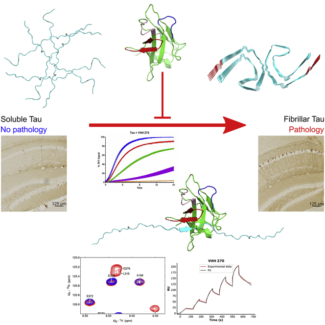
Inhibition of Tau seeding by targeting Tau nucleation core within neurons with a single domain antibody fragment
Published version of a preprint
Overview
The claims and statistics of this paper haven’t been checked yet.
Run the check — free, no account needed. It reads the open-access text and checks the sources it cites.
Integrity signals
Registry
Cites
Statistics
Figures
Verification
Claims the paper makes
The claims and statistics of this paper haven’t been checked yet.
Run the check — free, no account needed. It reads the open-access text and checks the sources it cites.
Claims other papers make about this one
Each is checked against this paper’s own text: does it say what the citing paper says it does?
In the citing paper
“Our previous studies have shown that these VHHs interact with short tau epitopes.[19][20][21]”
From A Proline-Rich-Domain-Binding Single Domain Antibody Selectively Inhibits RNA-Induced Phase Separation of Tau (2026) · cites this paper as [19]
In the citing paper
“VHH Z70 binds to the PHF6 sequence involved in tau nucleation and inhibits tau aggregation in the presence of heparin, as well as in cell and mouse seeding models.19”
From A Proline-Rich-Domain-Binding Single Domain Antibody Selectively Inhibits RNA-Induced Phase Separation of Tau (2026) · cites this paper as [19]
Self-check
Percentages, sums and reported differences re-computed from the paper’s own numbers.
The self-check needs the paper’s full text.
Citations
Cites
Naturally occurring antibodies devoid of light chains
Hamers-Casterman, C., Atarhouch, T., Muyldermans, S., Robinson, G., Hammers, C., Songa, E. B., Bendahman, N., & Hammers, R. (1993) · Nature · doi:10.1038/363446a0 · Cited by 3,241
Braak, H., Alafuzoff, I., Arzberger, T., Kretzschmar, H., & Del Tredici, K. (2006) · Acta Neuropathologica · doi:10.1007/s00401-006-0127-z · Cited by 3,172
Cryo-EM structures of tau filaments from Alzheimer’s disease
Fitzpatrick, A. W. P., Falcon, B., He, S., Murzin, A. G., Murshudov, G., Garringer, H. J., Crowther, R. A., Ghetti, B., Goedert, M., & Scheres, S. H. W. (2017) · Nature · doi:10.1038/nature23002 · Cited by 2,061
Nelson, P. T., Alafuzoff, I., Bigio, E. H., Bouras, C., Braak, H., Cairns, N. J., Castellani, R. J., Crain, B. J., Davies, P., Tredici, K. D., et al. (2012) · Journal of Neuropathology & Experimental Neurology · doi:10.1097/nen.0b013e31825018f7 · Cited by 2,050 · cited 1×
“In AD, the severity of cognitive decline is better correlated with the evolution of NFTs than amyloid deposits 3–5 .”
Transmission and spreading of tauopathy in transgenic mouse brain
Clavaguera, F., Bolmont, T., Crowther, R. A., Abramowski, D., Frank, S., Probst, A., Fraser, G., Stalder, A. K., Beibel, M., Staufenbiel, M., et al. (2009) · Nature Cell Biology · doi:10.1038/ncb1901 · Cited by 1,803
Propagation of Tau Misfolding from the Outside to the Inside of a Cell
Frost, B., Jacks, R. L., & Diamond, M. I. (2009) · Journal of Biological Chemistry · doi:10.1074/jbc.m808759200 · Cited by 1,252
Tau-targeting therapies for Alzheimer disease
Congdon, E. E., & Sigurdsson, E. M. (2018) · Nature Reviews Neurology · doi:10.1038/s41582-018-0013-z · Cited by 1,113 · cited 2×
“…decreasing Tau accumulation and in some case ensuring recovery of cognitive or motor functions 18–25 , have motivated several pharmaceutical companies to launch clinical trials of active and passive immunization, the latter with various anti-Tau monoclonal antibodies 26,27 (Immunoglobulin G, IgG).”
Structure-based classification of tauopathies
Shi, Y., Zhang, W., Yang, Y., Murzin, A. G., Falcon, B., Kotecha, A., van Beers, M., Tarutani, A., Kametani, F., Garringer, H. J., et al. (2021) · Nature · doi:10.1038/s41586-021-03911-7 · Cited by 878
Structural Polymorphism of 441-Residue Tau at Single Residue Resolution
Mukrasch, M. D., Bibow, S., Korukottu, J., Jeganathan, S., Biernat, J., Griesinger, C., Mandelkow, E., & Zweckstetter, M. (2009) · PLoS Biology · doi:10.1371/journal.pbio.1000034 · Cited by 652
Trans-cellular Propagation of Tau Aggregation by Fibrillar Species
Kfoury, N., Holmes, B. B., Jiang, H., Holtzman, D. M., & Diamond, M. I. (2012) · Journal of Biological Chemistry · doi:10.1074/jbc.m112.346072 · Cited by 552
Alzheimer's disease drug development pipeline: 2017
Cummings, J., Lee, G., Mortsdorf, T., Ritter, A., & Zhong, K. (2017) · Alzheimer's & Dementia: Translational Research & Clinical Interventions · doi:10.1016/j.trci.2017.05.002 · Cited by 549 · cited 1×
“Intervention strategies based on the amyloid cascade hypothesis had, up to date, limited success despite their being the primary target of clinical assays 2 .”
Asuni, A. A., Boutajangout, A., Quartermain, D., & Sigurdsson, E. M. (2007) · The Journal of Neuroscience · doi:10.1523/jneurosci.2361-07.2007 · Cited by 506
Schwarz, A. J., Yu, P., Miller, B. B., Shcherbinin, S., Dickson, J., Navitsky, M., Joshi, A. D., Devous, M. D., & Mintun, M. S. (2016) · Brain · doi:10.1093/brain/aww023 · Cited by 425
Zommer, N., Sergeant, N., Schraen-Maschke, S., Blum, D., & Buee, L. (2012) · Current Alzheimer Research · doi:10.2174/156720512800492503 · Cited by 193 · cited 2×
“…transgenic (Tg) mouse models of tauopathies, in decreasing Tau accumulation and in some case ensuring recovery of cognitive or motor functions 18–25 , have motivated several pharmaceutical companies to launch clinical trials of active and passive immunization, the latter with various anti-Tau…”
Bi, M., Ittner, A., Ke, Y. D., Götz, J., & Ittner, L. M. (2011) · PLoS ONE · doi:10.1371/journal.pone.0026860 · Cited by 153
Roberts, M., Sevastou, I., Imaizumi, Y., Mistry, K., Talma, S., Dey, M., Gartlon, J., Ochiai, H., Zhou, Z., Akasofu, S., et al. (2020) · Acta Neuropathologica Communications · doi:10.1186/s40478-020-0884-2 · Cited by 135
Calpain-mediated tau fragmentation is altered in Alzheimer’s disease progression
Chen, H.-H., Liu, P., Auger, P., Lee, S.-H., Adolfsson, O., Rey-Bellet, L., Lafrance-Vanasse, J., Friedman, B. A., Pihlgren, M., Muhs, A., et al. (2018) · Scientific Reports · doi:10.1038/s41598-018-35130-y · Cited by 51
Research works printed but not linked: 68
- Messer, 2020, Optimizing intracellular antibodies (intrabodies/nanobodies) to treat neurodegenerative disorders, Neurobiol. Dis., 134, 104619, 10.1016/j.nbd.2019.104619 · DOI
- Goedert, 2006, A century of Alzheimer's disease, Science, 314, 777, 10.1126/science.1132814 · DOI
- Brion, 1986, Alzheimer's disease and tau proteins, Lancet, 2, 1098, 10.1016/S0140-6736(86)90495-2 · DOI
- Grundke-Iqbal, 1986, Microtubule-associated protein tau. A component of Alzheimer paired helical filaments, J. Biol. Chem., 261, 6084, 10.1016/S0021-9258(17)38495-8 · DOI
- Mudher, 2017, What is the evidence that tau pathology spreads through prion-like propagation?, Acta Neuropathol. Commun., 5, 99, 10.1186/s40478-017-0488-7 · DOI · Paper page
- Wilcock, 1982, Plaques, tangles and dementia. A quantitative study, J. Neurol. Sci., 56, 343, 10.1016/0022-510X(82)90155-1 · DOI · Paper page
- Boutajangout, 2010, Immunotherapy targeting pathological tau prevents cognitive decline in a new tangle mouse model, J. Neurosci., 30, 16559, 10.1523/JNEUROSCI.4363-10.2010 · DOI · Paper page
- Dai, 2017, Tau passive immunization inhibits not only tau but also Aβ pathology, Alzheimers Res. Ther., 9, 1, 10.1186/s13195-016-0227-5 · DOI
- Chai, 2011, Passive immunization with anti-Tau antibodies in two transgenic models: reduction of Tau pathology and delay of disease progression, J. Biol. Chem., 286, 34457, 10.1074/jbc.M111.229633 · DOI
- Yanamandra, 2013, Anti-tau antibodies that block tau aggregate seeding in vitro markedly decrease pathology and improve cognition in vivo, Neuron, 80, 402, 10.1016/j.neuron.2013.07.046 · DOI · Paper page
- Albert, 2019, Prevention of tau seeding and propagation by immunotherapy with a central tau epitope antibody, Brain, 142, 1736, 10.1093/brain/awz100 · DOI
- Jadhav, 2019, A walk through tau therapeutic strategies, Acta Neuropathol. Commun., 7, 22, 10.1186/s40478-019-0664-z · DOI
- Dujardin, 2014, Neuron-to-neuron wild-type Tau protein transfer through a trans-synaptic mechanism: relevance to sporadic tauopathies, Acta Neuropathol. Commun., 2, 14, 10.1186/2051-5960-2-14 · DOI
- Colin, 2020, From the prion-like propagation hypothesis to therapeutic strategies of anti-tau immunotherapy, Acta Neuropathol., 139, 3, 10.1007/s00401-019-02087-9 · DOI · Paper page
- Clavaguera, 2014, Intercellular transfer of tau aggregates and spreading of tau pathology: implications for therapeutic strategies, Neuropharmacology, 76, 9, 10.1016/j.neuropharm.2013.08.037 · DOI
- Dujardin, 2014, Ectosomes: a new mechanism for non-exosomal secretion of tau protein, PLoS One, 9, e100760, 10.1371/journal.pone.0100760 · DOI
- Leroux, 2021, Extracellular vesicles: major actors of heterogeneity in tau spreading among human tauopathies, Mol. Ther. J. Am. Soc. Gene Ther.
- Tardivel, 2016, Tunneling nanotube (TNT)-mediated neuron-to neuron transfer of pathological Tau protein assemblies, Acta Neuropathol. Commun., 4, 117, 10.1186/s40478-016-0386-4 · DOI · Paper page
- Pain, 2015, Camelid single-domain antibody fragments: uses and prospects to investigate protein misfolding and aggregation, and to treat diseases associated with these phenomena, Biochimie, 111, 82, 10.1016/j.biochi.2015.01.012 · DOI
- Herce, 2017, Cell-permeable nanobodies for targeted immunolabelling and antigen manipulation in living cells, Nat. Chem., 9, 762, 10.1038/nchem.2811 · DOI
- Li, 2016, Cell-penetrating anti-GFAP VHH and corresponding fluorescent fusion protein VHH-GFP spontaneously cross the blood-brain barrier and specifically recognize astrocytes: application to brain imaging, FASEB J., 26, 3969, 10.1096/fj.11-201384 · DOI
- Gormal, 2020, Modular transient nanoclustering of activated β2-adrenergic receptors revealed by single-molecule tracking of conformation-specific nanobodies, Proc. Natl. Acad. Sci. U S A, 117, 30476, 10.1073/pnas.2007443117 · DOI
- Li, 2016, Camelid single-domain antibodies: a versatile tool for in vivo imaging of extracellular and intracellular brain targets, J. Control Release, 243, 1, 10.1016/j.jconrel.2016.09.019 · DOI
- Vitale, 2018, Anti-tau conformational scFv MC1 antibody efficiently reduces pathological tau species in adult JNPL3 mice, Acta Neuropathol. Commun., 6, 82, 10.1186/s40478-018-0585-2 · DOI
- Goodwin, 2020, Anti-tau scFvs targeted to the cytoplasm or secretory pathway variably modify pathology and neurodegenerative phenotypes, Mol. Ther. J. Am. Soc. Gene Ther.
- von Bergen, 2000, Assembly of tau protein into Alzheimer paired helical filaments depends on a local sequence motif ((306)VQIVYK(311)) forming beta structure, Proc. Natl. Acad. Sci. U S A, 97, 5129, 10.1073/pnas.97.10.5129 · DOI · Paper page
- Moutel, 2016, NaLi-H1: a universal synthetic library of humanized nanobodies providing highly functional antibodies and intrabodies, eLife, 5, e16228, 10.7554/eLife.16228 · DOI
- Dupré, 2019, Single domain antibody fragments as new tools for the detection of neuronal tau protein in cells and in mice studies, ACS Chem. Neurosci., 10, 3997, 10.1021/acschemneuro.9b00217 · DOI
- Dingus, 2021, A general approach for stabilizing nanobodies for intracellular expression, biorxiv
- Vielemeyer, 2010, Characterization of single chain antibody targets through yeast two hybrid, BMC Biotechnol., 10, 59, 10.1186/1472-6750-10-59 · DOI
- Zhang, 2001, Regulation of Smad degradation and activity by Smurf2, an E3 ubiquitin ligase, Proc. Natl. Acad. Sci. U S A, 98, 974, 10.1073/pnas.98.3.974 · DOI
- Holmes, 2014, Proteopathic tau seeding predicts tauopathy in vivo, Proc. Natl. Acad. Sci. U S A, 111, E4376, 10.1073/pnas.1411649111 · DOI · Paper page
- Vandermeeren, 2018, Anti-tau monoclonal antibodies derived from soluble and filamentous tau show diverse functional properties in vitro and in vivo, J. Alzheimers Dis., 65, 265, 10.3233/JAD-180404 · DOI
- Malia, 2016, Epitope mapping and structural basis for the recognition of phosphorylated tau by the anti-tau antibody AT8, Proteins, 84, 427, 10.1002/prot.24988 · DOI
- Gallardo, 2019, Targeting tauopathy with engineered tau-degrading intrabodies, Mol. Neurodegener., 14, 38, 10.1186/s13024-019-0340-6 · DOI
- Nobuhara, 2017, Tau antibody-targeting pathological species block neuronal uptake and interneuron propagation of tau in vitro, Am. J. Pathol., 187, 1399, 10.1016/j.ajpath.2017.01.022 · DOI · Paper page
- Spencer, 2018, Selective targeting of 3 repeat Tau with brain penetrating single chain antibodies for the treatment of neurodegenerative disorders, Acta Neuropathol., 136, 69, 10.1007/s00401-018-1869-0 · DOI
- Dam, 2021, Safety and efficacy of anti-tau monoclonal antibody gosuranemab in progressive supranuclear palsy: a phase 2, randomized, placebo-controlled trial, Nat. Med., 27, 1451, 10.1038/s41591-021-01455-x · DOI
- Höglinger, 2021, Safety and efficacy of tilavonemab in progressive supranuclear palsy: a phase 2, randomised, placebo-controlled trial, Lancet Neurol., 20, 182, 10.1016/S1474-4422(20)30489-0 · DOI
- Jabbari, 2021, Tau-targeting antibody therapies: too late, wrong epitope or wrong target?, Nat. Med., 27, 1341, 10.1038/s41591-021-01465-9 · DOI
- Courade, 2018, Epitope determines efficacy of therapeutic anti-Tau antibodies in a functional assay with human Alzheimer Tau, Acta Neuropathol., 136, 729, 10.1007/s00401-018-1911-2 · DOI
- Arakhamia, 2020, Posttranslational modifications mediate the structural diversity of tauopathy strains, Cell, 180, 633, 10.1016/j.cell.2020.01.027 · DOI
- Falcon, 2018, Structures of filaments from Pick's disease reveal a novel tau protein fold, Nature, 561, 137, 10.1038/s41586-018-0454-y · DOI
- Falcon, 2019, Novel tau filament fold in chronic traumatic encephalopathy encloses hydrophobic molecules, Nature, 568, 420, 10.1038/s41586-019-1026-5 · DOI
- Sawaya, 2007, Atomic structures of amyloid cross-beta spines reveal varied steric zippers, Nature, 447, 453, 10.1038/nature05695 · DOI
- Seidler, 2018, Structure-based inhibitors of tau aggregation, Nat. Chem., 10, 170, 10.1038/nchem.2889 · DOI
- Mirbaha, 2018, Inert and seed-competent tau monomers suggest structural origins of aggregation, eLife, 7, e36584, 10.7554/eLife.36584 · DOI
- Moreira, 2021, Dynamic interactions and Ca2+-binding modulate the holdase-type chaperone activity of S100B preventing tau aggregation and seeding, Nat. Commun., 12, 6292, 10.1038/s41467-021-26584-2 · DOI · Paper page
- Mok, 2018, Mapping interactions with the chaperone network reveals factors that protect against tau aggregation, Nat. Struct. Mol. Biol., 25, 384, 10.1038/s41594-018-0057-1 · DOI
- Carlomagno, 2021, The AD tau core spontaneously self-assembles and recruits full-length tau to filaments, Cell Rep., 34, 108843, 10.1016/j.celrep.2021.108843 · DOI
- Zhang, 2019, Heparin-induced tau filaments are polymorphic and differ from those in Alzheimer's and Pick's diseases, eLife, 8, e43584, 10.7554/eLife.43584 · DOI · Paper page
- Dutt, 2020, Real-world evidence of caplacizumab use in the management of acute TTP, Blood, 137, 1731, 10.1182/blood.2020007599 · DOI
- Matz, 2012, Phage display and selections on purified antigens, Methods Mol. Biol., 907, 213, 10.1007/978-1-61779-974-7_11 · DOI
- Danis, 2016, Nuclear magnetic resonance spectroscopy for the identification of multiple phosphorylations of intrinsically disordered proteins, J. Vis. Exp., 10.3791/55001 · DOI
- Luna-Vargas, 2011, Enabling high-throughput ligation-independent cloning and protein expression for the family of ubiquitin specific proteases, J. Struct. Biol., 175, 113, 10.1016/j.jsb.2011.03.017 · DOI
- Smet, 2004, Accepting its random coil nature allows a partial NMR assignment of the neuronal Tau protein, Chembiochem., 5, 1639, 10.1002/cbic.200400145 · DOI
- Lippens, 2004, Proline-directed random-coil chemical shift values as a tool for the NMR assignment of the tau phosphorylation sites, Chembiochem., 5, 73, 10.1002/cbic.200300763 · DOI
- Cadwell, 1992, Randomization of genes by PCR mutagenesis, PCR Methods Appl., 2, 28, 10.1101/gr.2.1.28 · DOI
- Vojtek, 1995, Ras-Raf interaction: two-hybrid analysis, Methods Enzymol., 255, 331, 10.1016/S0076-6879(95)55036-4 · DOI
- Fromont-Racine, 1997, Toward a functional analysis of the yeast genome through exhaustive two-hybrid screens, Nat. Genet., 16, 277, 10.1038/ng0797-277 · DOI
- Karlsson, 2006, Analyzing a kinetic titration series using affinity biosensors, Anal. Biochem., 349, 136, 10.1016/j.ab.2005.09.034 · DOI · Paper page
- Vagin, 2010, Molecular replacement with MOLREP, Acta Crystallogr. D Biol. Crystallogr., 66, 22, 10.1107/S0907444909042589 · DOI
- Murshudov, 2011, REFMAC5 for the refinement of macromolecular crystal structures, Acta Crystallogr. D Biol. Crystallogr., 67, 355, 10.1107/S0907444911001314 · DOI
- Emsley, 2010, Features and development of coot, Acta Crystallogr. D Biol. Crystallogr., 66, 486, 10.1107/S0907444910007493 · DOI · Paper page
- Krissinel, 2007, Inference of macromolecular assemblies from crystalline state, J. Mol. Biol., 372, 774, 10.1016/j.jmb.2007.05.022 · DOI
- Banning, 2010, A flow cytometry-based FRET assay to identify and analyse protein-protein interactions in living cells, PLoS One, 5, e9344, 10.1371/journal.pone.0009344 · DOI
- Schindowski, 2006, Alzheimer's disease-like tau neuropathology leads to memory deficits and loss of functional synapses in a novel mutated tau transgenic mouse without any motor deficits, Am. J. Pathol., 169, 599, 10.2353/ajpath.2006.060002 · DOI
- Leroy, 2007, Early axonopathy preceding neurofibrillary tangles in mutant tau transgenic mice, Am. J. Pathol., 171, 976, 10.2353/ajpath.2007.070345 · DOI · Paper page
Cited by
Tau-targeting therapies for Alzheimer disease: Current status and future directions
Congdon, E. E., Ji, C., Tetlow, A. M., Jiang, Y., & Sigurdsson, E. M. (2023) · Nature Reviews Neurology · doi:10.1038/s41582-023-00883-2 · Cited by 371
Spontaneous pathology in PS19 tauopathy mice progresses via brain networks
Ramirez, D. M. O., Whitesell, J. D., Bhagwat, N., Thomas, T. L., Ajay, A. D., Nawaby, A., Delatour, B., Bay, S., LaFaye, P., Harris, J. A., et al. (2025) · Neurobiology of Disease · doi:10.1016/j.nbd.2025.107072 · Cited by 5
Thiou, S., Martin, L., Manousaki, E., Nguyen, M., Mortelecque, J., Heidsieck, L., Cantrelle, F.-X., Blum, D., Buée-Scherrer, V., Buée, L., et al. (2026) · ACS Chemical Neuroscience · doi:10.1021/acschemneuro.6c00110 · Cited by 1
Self-citation
Metadata
- Authors
- Danis, C., Dupré, E., Zejneli, O., Caillierez, R., Arrial, A., Bégard, S., Mortelecque, J., Eddarkaoui, S., Loyens, A., Cantrelle, F.-X., Hanoulle, X., Rain, J.-C., Colin, M., Buée, L., & Landrieu, I.
- Venue
- Molecular Therapy
- Publisher
- Elsevier BV
- Published
- 2022-04
- Volume
- 30
- Issue
- 4
- Pages
- 1484-1499
- ISSN
- 1525-0016
- PMC
- PMC9077319
- Cited by
- 76
- References
- 17
- Language
- en
- Licence
- Funders
- ANR
About these checks
Registry, retraction, Cites and statistics results are computed, not judged, with one exception: a printed reference that no index lists is read once by a model before it counts as a citation error. Claims are extracted by a model and kept only when their quote appears verbatim in the text; each cited source is then checked, with a model’s help, for whether it says what the claim relies on. Those verdicts can be wrong — read the claim’s quote and the cited source before relying on one.
Statistics and arithmetic are recomputed, not judged.
Confidence bands are shown only for fields calibrated on our annotated test set. None is calibrated yet, so no band is shown.
What counts as a claim ›How we measure confidence ›How we check this ›
Are you an author, or do you see a problem with this page? Request a review
Data sources
Bibliographic data: Semantic Scholar, OpenAlex, Crossref.
Contains information from the Semantic Scholar Academic Graph Datasets, made available under the ODC Attribution License (ODC-BY 1.0).
Semantic Scholar’s data is described in: Kinney, R., et al. (2023). The Semantic Scholar Open Data Platform. arXiv:2301.10140.
