Cited by 12Protein Tyrosine PhosphatasesProtein Structure and DynamicsProtein Kinase Regulation and GTPase Signaling

The DOI matches its Crossref record; 2 of the 2 references it cites were found in a registry or an index.

Sequence – dynamics – function relationships in protein tyrosine phosphatases

Open access

Published version of a preprint

Abstract

Protein tyrosine phosphatases (PTPs) are crucial regulators of cellular signaling. Their activity is regulated by the motion of a conserved loop, the WPD-loop, from a catalytically inactive open to a catalytically active closed conformation. WPD-loop motion optimally positions a catalytically critical residue into the active site, and is directly linked to the turnover number of these enzymes. Crystal structures of chimeric PTPs constructed by grafting parts of the WPD-loop sequence of PTP1B onto the scaffold of YopH showed WPD-loops in a wide-open conformation never previously observed in either parent enzyme. This wide-open conformation has, however, been observed upon binding of small molecule inhibitors to other PTPs, suggesting the potential of targeting it for drug discovery efforts. Here, we have performed simulations of both enzymes and show that there are negligible energetic differences in the chemical step of catalysis, but significant differences in the dynamical properties of the WPD-loop. Detailed interaction network analysis provides insight into the molecular basis for this population shift to a wide-open conformation. Taken together, our study provides insight into the links between loop dynamics and chemistry in these YopH variants specifically, and how WPD-loop dynamic can be engineered through modification of the internal protein interaction network.

Overview

The claims and statistics of this paper haven’t been checked yet.

Run the check — free, no account needed. It reads the open-access text and checks the sources it cites.

A few free checks a day · papers already verified open instantly · results are public

Integrity signals

Registry

Crossref · 10.1017/qrd.2024.3

Cites

2 of 2 references verified0 citation errors · 0 not checkable

Statistics

Figures

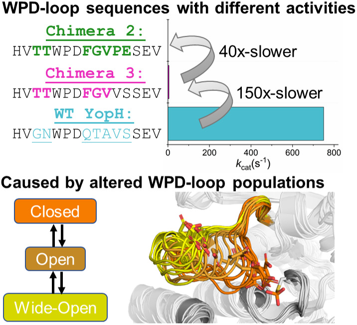
Graphical abstract
Graphical abstract CC BY · PubMed Central
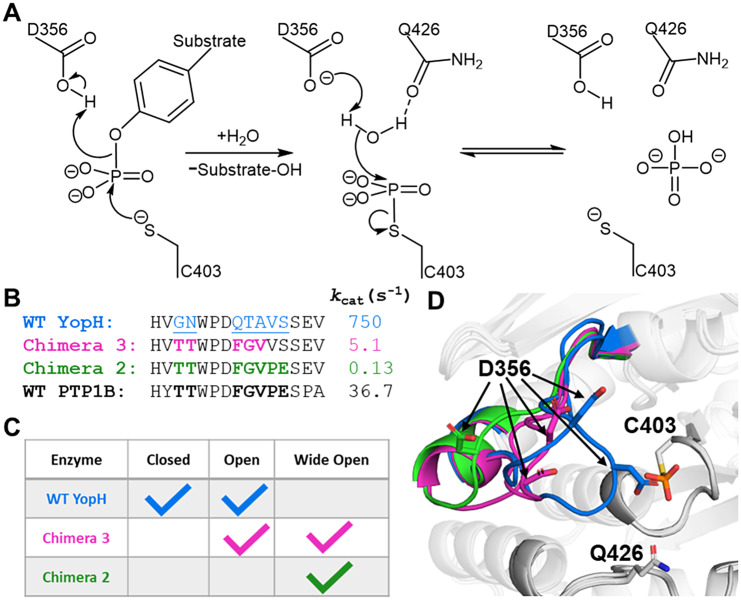
Figure 1. (a) Mechanism of PTP catalysis, with the residue numbering of wild-type (WT) YopH. (b) WPD-loop sequence alignments and corresponding turnover numbers of wild-type YopH, and YopH Chimeras 3 and 2, and wild-type PTP1B (from which the substitutions in the chimeras were obtained). Mutated chimera residues are shown in bold. Turnover numbers were measured at pH 5.5 and 25 °C using the substrate pNPP (Moise et al., 2018. (c) Summary of the available crystal structures for each PTP in the different WPD-loop conformational states available. A tick indicates the availability of a crystal structure for a given PTP in a given conformational state. (d) A visualization of the diversity of WPD-loop conformational states captured by X-ray crystallography for wild-type YopH and Chimeras 2 and 3, and associated conformational diversity of the D356 side chain. Color coding of the structures WPD-loop matches that in panels (b) and (c). C403 is the nucleophilic cysteine, which is located on the P-loop, while Q426 is located on the Q-loop.
Figure 1. (a) Mechanism of PTP catalysis, with the residue numbering of wild-type (WT) YopH. (b) WPD-loop sequence alignments and corresponding turnover numbers of wild-type YopH, and YopH Chimeras 3 and 2, and wild-type PTP1B (from which the substitutions in the chimeras were obtained). Mutated chimera residues are shown in bold. Turnover numbers were measured at pH 5.5 and 25 °C using the substrate pNPP (Moise et al., 2018. (c) Summary of the available crystal structures for each PTP in the different WPD-loop conformational states available. A tick indicates the availability of a crystal structure for a given PTP in a given conformational state. (d) A visualization of the diversity of WPD-loop conformational states captured by X-ray crystallography for wild-type YopH and Chimeras 2 and 3, and associated conformational diversity of the D356 side chain. Color coding of the structures WPD-loop matches that in panels (b) and (c). C403 is the nucleophilic cysteine, which is located on the P-loop, while Q426 is located on the Q-loop.CC BY · PubMed Central
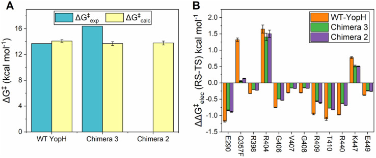
Figure 2. (a) Comparison of the calculated (ΔG‡calc) and experimental (ΔG‡exp) activation free energies calculated for the hydrolysis step of the reaction catalyzed by wild-type (WT) YopH and Chimeras 3 and 2. Simulation data are presented in kcal mol−1 as the average values and standard error of the mean obtained from 30 EVB simulation replicas. Experimental data are obtained from (Zhang et al., 1992; Stuckey et al., 1994; Whittier et al., 2013; Moise et al., 2018) The raw data for this figure are presented in Supplementary Table S1. Note that no experimental data are presented in panel A for Chimera 2, as this Chimera is catalytically inactive (Moise et al., 2018). (b) Electrostatic contributions of selected amino acids (∆∆G‡elec, kcal mol−1) to the calculated activation free energies for the hydrolysis steps catalyzed by WT-YopH and Chimeras 2 and 3. All electrostatic contributions were scaled assuming an internal dielectric constant of 4 (Li et al., 2013). Data were obtained from the calculated EVB trajectories using the linear response approximation (LRA)(Lee et al., 1992; Muegge et al., 1997) and are represented as average and standard error of the mean over 30 individual trajectories per system. The amino acids that directly participate in the reaction (the catalytic cysteine and aspartic acid) are not shown. Chimeras 2 and 3 carry a Q357F substitution. The corresponding raw data for this plot are presented in Supplementary Table S3.
Figure 2. (a) Comparison of the calculated (ΔG‡calc) and experimental (ΔG‡exp) activation free energies calculated for the hydrolysis step of the reaction catalyzed by wild-type (WT) YopH and Chimeras 3 and 2. Simulation data are presented in kcal mol−1 as the average values and standard error of the mean obtained from 30 EVB simulation replicas. Experimental data are obtained from (Zhang et al., 1992; Stuckey et al., 1994; Whittier et al., 2013; Moise et al., 2018) The raw data for this figure are presented in Supplementary Table S1. Note that no experimental data are presented in panel A for Chimera 2, as this Chimera is catalytically inactive (Moise et al., 2018). (b) Electrostatic contributions of selected amino acids (∆∆G‡elec, kcal mol−1) to the calculated activation free energies for the hydrolysis steps catalyzed by WT-YopH and Chimeras 2 and 3. All electrostatic contributions were scaled assuming an internal dielectric constant of 4 (Li et al., 2013). Data were obtained from the calculated EVB trajectories using the linear response approximation (LRA)(Lee et al., 1992; Muegge et al., 1997) and are represented as average and standard error of the mean over 30 individual trajectories per system. The amino acids that directly participate in the reaction (the catalytic cysteine and aspartic acid) are not shown. Chimeras 2 and 3 carry a Q357F substitution. The corresponding raw data for this plot are presented in Supplementary Table S3.CC BY · PubMed Central
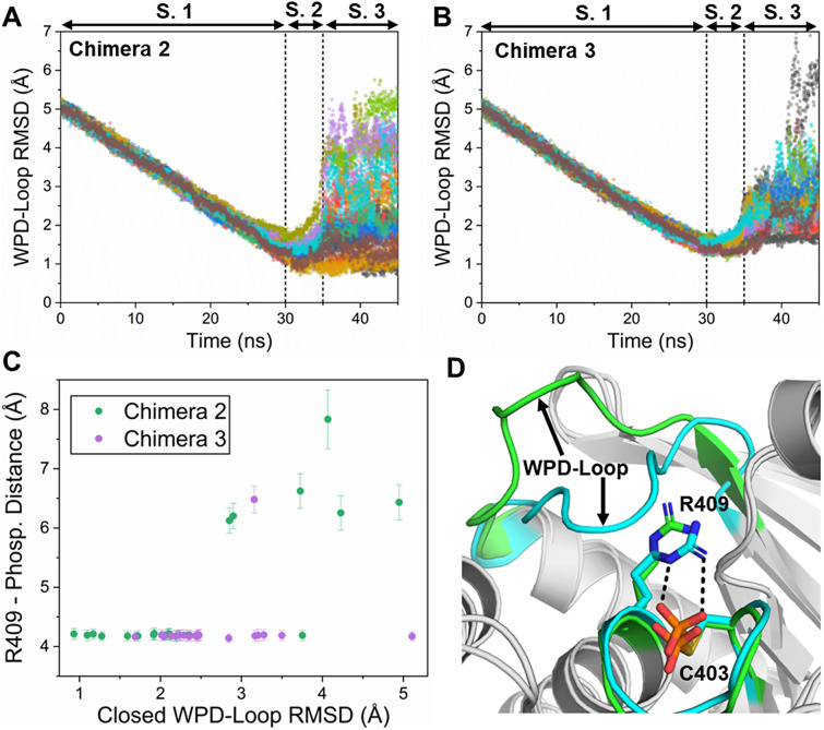
Figure 3. (a,b) The RMSD to the closed WPD-loop conformation over the course of our targeted MD (tMD) simulations of YopH Chimera 2 (a) and Chimera 3 (b). The reference state used is the backbone atoms of the closed WPD-loop structure of YopH. (c) Center of mass distance between the P-loop R409 side chain and the phosphorylated cysteine against the RMSD to the closed WPD-loop for the last 5 ns of each tMD simulation for both Chimeras 2 and 3. Errors are the standard deviation obtained from each frame. (d) Representative structures of the productive and non-productive R409 side-chain conformations, with the productive (for WPD-loop closure) structure colored in cyan and the non-productive structure (the R409 would sterically clash with the WPD-loop if it was closed) colored in green.
Figure 3. (a,b) The RMSD to the closed WPD-loop conformation over the course of our targeted MD (tMD) simulations of YopH Chimera 2 (a) and Chimera 3 (b). The reference state used is the backbone atoms of the closed WPD-loop structure of YopH. (c) Center of mass distance between the P-loop R409 side chain and the phosphorylated cysteine against the RMSD to the closed WPD-loop for the last 5 ns of each tMD simulation for both Chimeras 2 and 3. Errors are the standard deviation obtained from each frame. (d) Representative structures of the productive and non-productive R409 side-chain conformations, with the productive (for WPD-loop closure) structure colored in cyan and the non-productive structure (the R409 would sterically clash with the WPD-loop if it was closed) colored in green.CC BY · PubMed Central
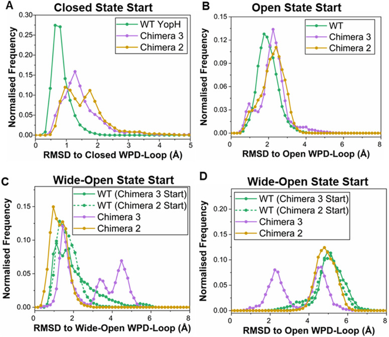
Figure 4. Evaluating the stability of the closed, open, and wide-open WPD-loop conformational states in simulations of wild-type (WT) YopH and Chimeras 2 and 3. (a) Histograms of the WPD-loop Cα-atom RMSD to the closed WPD-loop conformational state, with simulations starting from the closed conformational state. The closed conformation Chimera structures were generated by introducing the relevant substitutions into the wild-type YopH WPD-loop in silico, as described in the Methods section. (b) Histograms of the WPD-loop Cα-atom RMSD to the open WPD-loop conformational state, with simulations starting from the open conformational state. (c) Histogram of the WPD-loop Cα-atom RMSD to the wide-open conformational state for simulations initiated from the wide-open conformational state of the WPD-loop. (d) Histogram of the WPD-loop Cα-atom RMSD to the open conformational state for simulations initiated from the wide-open conformational state of the WPD-loop. For panels (c) and (d) which refer to the wide-open simulations, the two wild-type YopH wide-open loop conformations were constructed by introducing the relevant substitutions into both the Chimera 3 and Chimera 2 WPD-loop residues in silico, see the Methods section. These are indicated on the graph legend as ‘WT (Chimera 3 Start)’ if produced using the Chimera 3 crystal structure or ‘WT (Chimera 2 Start)’ if produced using the Chimera 2 crystal structure. In all cases, each histogram was constructed using 15 × 500 ns long MD simulation replicas, sampling data every 100 ps, with a bin width of 0.16 Å.
Figure 4. Evaluating the stability of the closed, open, and wide-open WPD-loop conformational states in simulations of wild-type (WT) YopH and Chimeras 2 and 3. (a) Histograms of the WPD-loop Cα-atom RMSD to the closed WPD-loop conformational state, with simulations starting from the closed conformational state. The closed conformation Chimera structures were generated by introducing the relevant substitutions into the wild-type YopH WPD-loop in silico, as described in the Methods section. (b) Histograms of the WPD-loop Cα-atom RMSD to the open WPD-loop conformational state, with simulations starting from the open conformational state. (c) Histogram of the WPD-loop Cα-atom RMSD to the wide-open conformational state for simulations initiated from the wide-open conformational state of the WPD-loop. (d) Histogram of the WPD-loop Cα-atom RMSD to the open conformational state for simulations initiated from the wide-open conformational state of the WPD-loop. For panels (c) and (d) which refer to the wide-open simulations, the two wild-type YopH wide-open loop conformations were constructed by introducing the relevant substitutions into both the Chimera 3 and Chimera 2 WPD-loop residues in silico, see the Methods section. These are indicated on the graph legend as ‘WT (Chimera 3 Start)’ if produced using the Chimera 3 crystal structure or ‘WT (Chimera 2 Start)’ if produced using the Chimera 2 crystal structure. In all cases, each histogram was constructed using 15 × 500 ns long MD simulation replicas, sampling data every 100 ps, with a bin width of 0.16 Å.CC BY · PubMed Central
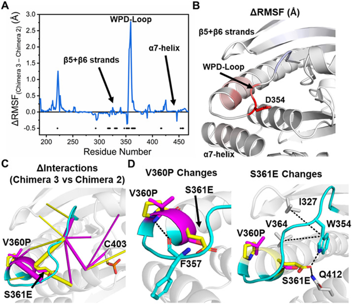
Figure 5. (a) Difference in the calculated root-mean-squared fluctuations (ΔRMSF) of Chimeras 3 and 2 when sampling the wide-open WPD-loop conformational state. A two-sided t-test was performed to validate the significance of the calculated ΔRMSFs, and those residues identified as significant have a black dot placed at the bottom of the graph. To account for the usage of multiple t-tests, the Benjamini–Hochberg correction (Benjamini and Hochberg, 1995) was applied, using a false discovery rate of 5%, see the Methods section for further details. (b) Projection of the calculated ΔRMSFs identified as significantly different onto the structure of YopH, with residues colored from blue (more rigid in the Chimera 3), to white (equally rigid or no significant difference) to red (more rigid in Chimera 2). RMSF profiles of both enzymes are provided in Supplementary Figure S12. (c) Differences in the non-covalent interaction network between Chimera 3 and Chimera 2 when sampling the wide-open WPD-loop conformational state as determined by KIF (Crean et al., 2023). Interactions which are on average stronger in the Chimera 3 are colored magenta, while those that favor Chimera 2 are colored yellow. These data are shown graphically in Supplementary Figure S12. (d) Illustration of the major changes in interactions at the two substitution sites (V360P and S361E), which were depicted in panel (c).
Figure 5. (a) Difference in the calculated root-mean-squared fluctuations (ΔRMSF) of Chimeras 3 and 2 when sampling the wide-open WPD-loop conformational state. A two-sided t-test was performed to validate the significance of the calculated ΔRMSFs, and those residues identified as significant have a black dot placed at the bottom of the graph. To account for the usage of multiple t-tests, the Benjamini–Hochberg correction (Benjamini and Hochberg, 1995) was applied, using a false discovery rate of 5%, see the Methods section for further details. (b) Projection of the calculated ΔRMSFs identified as significantly different onto the structure of YopH, with residues colored from blue (more rigid in the Chimera 3), to white (equally rigid or no significant difference) to red (more rigid in Chimera 2). RMSF profiles of both enzymes are provided in Supplementary Figure S12. (c) Differences in the non-covalent interaction network between Chimera 3 and Chimera 2 when sampling the wide-open WPD-loop conformational state as determined by KIF (Crean et al., 2023). Interactions which are on average stronger in the Chimera 3 are colored magenta, while those that favor Chimera 2 are colored yellow. These data are shown graphically in Supplementary Figure S12. (d) Illustration of the major changes in interactions at the two substitution sites (V360P and S361E), which were depicted in panel (c).CC BY · PubMed Central

Verification

Claims the paper makes

The claims and statistics of this paper haven’t been checked yet.

Run the check — free, no account needed. It reads the open-access text and checks the sources it cites.

A few free checks a day · papers already verified open instantly · results are public

Self-check

Percentages, sums and reported differences re-computed from the paper’s own numbers.

The self-check needs the paper’s full text.

Citations

Cites

Research works printed but not linked: 77

  1. 10.1021/ct400341p · DOI
  2. 10.1093/protein/5.3.215 · DOI
  3. 10.1016/j.cell.2008.11.038 · DOI
  4. 10.1046/j.1432-1327.1999.00090.x · DOI
  5. 10.1038/s41467-021-23450-z · DOI
  6. 10.1021/acs.biochem.8b00663 · DOI
  7. 10.1038/nchembio.2175 · DOI · Paper page
  8. 10.1126/science.1241735 · DOI
  9. 10.1002/pro.4064 · DOI
  10. 10.1371/journal.pone.0052283 · DOI
  11. 10.1111/febs.14954 · DOI
  12. 10.1021/acscentsci.9b00909 · DOI
  13. 10.1126/science.1118953 · DOI
  14. 10.1021/bi900830g · DOI
  15. 10.1038/370571a0 · DOI
  16. 10.1515/hsz-2018-0348 · DOI
  17. 10.1016/S0021-9258(18)35903-9 · DOI
  18. 10.1093/protein/10.12.1363 · DOI
  19. 10.4155/fmc.10.241 · DOI
  20. 10.1021/bi300694t · DOI
  21. 10.1093/protein/gzab009 · DOI
  22. 10.1021/jacs.9b04470 · DOI
  23. 10.1021/jacsau.1c00054 · DOI
  24. 10.1038/s41598-017-05931-8 · DOI
  25. 10.1016/S0014-5793(03)00603-3 · DOI
  26. 10.1146/annurev.pharmtox.42.083001.144616 · DOI
  27. 10.1016/j.softx.2017.12.001 · DOI
  28. 10.1016/S0092-8674(00)81688-8 · DOI · Paper page
  29. 10.7554/eLife.36307 · DOI
  30. Case, 2018, AMBER 2018
  31. Zhang, 1994, Protein tyrosine phosphatases: Mechanism of catalysis and substrate specificity, Advances in Enzymology and Related Areas of Molecular Biology, 68, 1
  32. 10.1021/jacs.8b09378 · DOI
  33. 10.1016/S0968-0004(03)00135-X · DOI · Paper page
  34. 10.1021/acscatal.8b00624 · DOI
  35. 10.1002/pro.3379 · DOI
  36. 10.1016/j.str.2018.01.015 · DOI
  37. 10.1016/0263-7855(94)80072-3 · DOI
  38. 10.1021/jm2003766 · DOI
  39. 10.1016/S0021-9258(18)68703-4 · DOI
  40. 10.1038/nrd.2018.201 · DOI
  41. 10.1016/j.abb.2012.06.002 · DOI
  42. 10.1073/pnas.93.6.2493 · DOI
  43. 10.1021/ja00540a008 · DOI
  44. 10.1016/j.crstbi.2020.12.002 · DOI
  45. 10.1080/10409239891204161 · DOI
  46. 10.1038/nsmb803 · DOI
  47. 10.1021/jacs.2c04624 · DOI
  48. 10.1021/cs500325p · DOI
  49. 10.1021/jacs.0c04924 · DOI
  50. 10.1016/j.bbapap.2015.03.010 · DOI
  51. 10.1039/D2SC04135A · DOI
  52. 10.1038/nchem.2474 · DOI
  53. 10.1021/bi00043a003 · DOI
  54. 10.1093/nar/28.1.235 · DOI · Paper page
  55. 10.1038/nrc1837 · DOI
  56. 10.1126/science.1123883 · DOI
  57. 10.1016/j.cpc.2013.09.018 · DOI
  58. 10.1021/jacs.0c11806 · DOI
  59. 10.1038/nrd895 · DOI
  60. 10.1073/pnas.0233793100 · DOI
  61. 10.1063/1.445869 · DOI
  62. 10.1016/j.tibs.2021.08.008 · DOI
  63. 10.1074/jbc.M210194200 · DOI
  64. 10.1080/00268976.2013.824126 · DOI
  65. 10.1002/anie.202110793 · DOI
  66. 10.1126/science.1546324 · DOI
  67. 10.1038/s41557-021-00763-6 · DOI
  68. 10.1111/j.2517-6161.1995.tb02031.x · DOI · Paper page
  69. 10.1021/ct400065j · DOI
  70. 10.1021/acs.jctc.5b00255 · DOI
  71. 10.2174/1568026033452302 · DOI
  72. 10.1016/j.drudis.2007.03.011 · DOI
  73. 10.1063/5.0140882 · DOI · Paper page
  74. 10.1021/ja9621760 · DOI
  75. 10.1093/nar/gkac249 · DOI
  76. 10.1016/j.tem.2014.10.004 · DOI
  77. 10.1016/0092-8674(95)90405-0 · DOI

Metadata

Authors
Crean, R. M., Corbella, M., Calixto, A. R., Hengge, A. C., & Kamerlin, S. C. L.
Affiliations
  • Uppsala University (SE)
  • Utah State University (US)
Corresponding author
  • Hengge, Alvan C.
  • Kamerlin, Shina C. L.
Venue
QRB Discovery
Published
2024
Volume
5
Pages
e4-e4
ISSN
2633-2892 (ISSN-L)
OpenAlex
W4391173948
Cited by
12
References
2
Citations by year
2026: 2 · 2025: 3 · 2024: 1
FWCI
0.81
Type
journal article
Language
en
Open access
Locations
  • QRB Discovery (publishedVersion, cc-by)
  • ChemRxiv (acceptedVersion, cc-by)
  • PubMed Central (publishedVersion, cc-by)
  • PubMed (publishedVersion)
  • DOAJ (DOAJ: Directory of Open Access Journals) (submittedVersion, cc-by-sa)
Funders
  • Knut och Alice Wallenbergs Stiftelse (2018.0140, 2019.0431)
  • Vetenskapsrådet (2019-03499)
  • Horizon 2020 Framework Programme (890562)
Topics
  • Life Sciences › Biochemistry, Genetics and Molecular Biology › Molecular Biology › Protein Tyrosine Phosphatases
  • Life Sciences › Biochemistry, Genetics and Molecular Biology › Molecular Biology › Protein Structure and Dynamics
  • Life Sciences › Biochemistry, Genetics and Molecular Biology › Molecular Biology › Protein Kinase Regulation and GTPase Signaling
Keywords
protein tyrosine phosphatase, WPD loop, loop dynamics, conformational change, PTP1B, YopH, chimeric enzymes, molecular dynamics simulation, catalytic mechanism, enzyme turnover number, small molecule inhibitors

About these checks

Registry, retraction, Cites and statistics results are computed, not judged, with one exception: a printed reference that no index lists is read once by a model before it counts as a citation error. Claims are extracted by a model and kept only when their quote appears verbatim in the text; each cited source is then checked, with a model’s help, for whether it says what the claim relies on. Those verdicts can be wrong — read the claim’s quote and the cited source before relying on one.

Statistics and arithmetic are recomputed, not judged.

Confidence bands are shown only for fields calibrated on our annotated test set. None is calibrated yet, so no band is shown.

What counts as a claim ›How we measure confidence ›How we check this ›

Are you an author, or do you see a problem with this page? Request a review