

The DOI matches its Crossref record; 12 of the 12 references it cites were found in a registry or an index.
The Potential Contribution of Hexavalent Chromium to the Carcinogenicity of Chrysotile Asbestos
Overview
The claims and statistics of this paper haven’t been checked yet.
Run the check — free, no account needed. It reads the open-access text and checks the sources it cites.
Integrity signals
Registry
Cites
Statistics
Figures
Verification
Claims the paper makes
The claims and statistics of this paper haven’t been checked yet.
Run the check — free, no account needed. It reads the open-access text and checks the sources it cites.
Claims other papers make about this one
Each is checked against this paper’s own text: does it say what the citing paper says it does?
In the citing paper
“Recently, tetrahedrally coordinated Fe (Fe tet ) in the fibers' Si sheets was identified as the principal Fe species to catalyze this process.”
From Redox Cycling of Tetrahedral Iron Drives the Fenton Reactivity of Chrysotile Asbestos (2024) · cites this paper as [39]
In the citing paper
“On dissolving chrysotile surfaces at circumneutral pH, Fe 3+ tet was demonstrated to be the most efficient Fe species (on a molar basis) at catalyzing the decomposition of H 2 O 2 and the only Fe species capable of catalyzing the generation of HO • from H 2 O 2 .18,38,39”
From Redox Cycling of Tetrahedral Iron Drives the Fenton Reactivity of Chrysotile Asbestos (2024) · cites this paper as [39]
In the citing paper
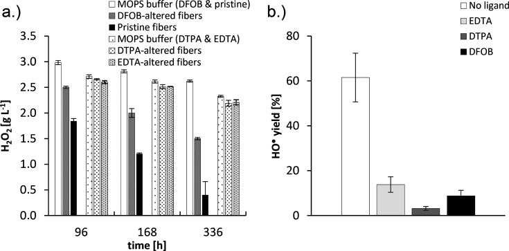
“DFOB-altered fibers had a HO • yield of 9% relative to pristine fibers, which is in accordance with published values for the same fiber type.18,38,39”
From Redox Cycling of Tetrahedral Iron Drives the Fenton Reactivity of Chrysotile Asbestos (2024) · cites this paper as [39]
In the citing paper
“Consequently, redox cycling of the low-abundant Fe tet surface sites drives the fibers' high catalytic activity in generating HO • .18,38,39,[48][49][50][51]”
From Redox Cycling of Tetrahedral Iron Drives the Fenton Reactivity of Chrysotile Asbestos (2024) · cites this paper as [39]
In the citing paper
“DFOB has previously been shown to effectively dissolve Fe 3+ tet from Shijiazhuang chrysotile 18,38,39 and to generate vacant sites in the exposed Si sheet which promote Si dissolution.18”
From Redox Cycling of Tetrahedral Iron Drives the Fenton Reactivity of Chrysotile Asbestos (2024) · cites this paper as [39]
In the citing paper
“which, based on this and earlier studies, can be discerned as the ultimate Fenton-reactive metal species on chrysotile fiber surfaces.18,38,39”
From Redox Cycling of Tetrahedral Iron Drives the Fenton Reactivity of Chrysotile Asbestos (2024) · cites this paper as [39]
In the citing paper
“Increased dissolution kinetics of Fe tet from chrysotile, e.g.facilitated by chelating ligands, renders fiber surfaces depleted of Fe tet sites and consequently diminishes the fibers' Fenton reactivity.18,38,39”
From Redox Cycling of Tetrahedral Iron Drives the Fenton Reactivity of Chrysotile Asbestos (2024) · cites this paper as [39]
In the citing paper
“The ability of Fe tet to fully redox cycle drives its high potency in catalyzing Haber-Weiss cycles.18,38,39”
From Redox Cycling of Tetrahedral Iron Drives the Fenton Reactivity of Chrysotile Asbestos (2024) · cites this paper as [39]
In the citing paper
“In conclusion, this study together with previous studies 18,38,39 supports the notion that Fe tet in the exposed Si lattice of pathogenic silicate minerals such as chrysotile asbestos is the principal Fe species catalyzing particle-mediated HO • generation at the physiological pH of 7.4, through cycling between its ferric and ferrous redox states.”
From Redox Cycling of Tetrahedral Iron Drives the Fenton Reactivity of Chrysotile Asbestos (2024) · cites this paper as [39]
Self-check
Percentages, sums and reported differences re-computed from the paper’s own numbers.
The self-check needs the paper’s full text.
Citations
Cites
De Laat, J., & Gallard, H. (1999) · Environmental Science & Technology · doi:10.1021/es981171v · Cited by 749
�ber die Katalyse des Hydroperoxydes
Haber, F., & Weiss, J. (1932) · Die Naturwissenschaften · doi:10.1007/bf01504715 · Cited by 513
The molecular basis of asbestos induced lung injury
Kamp, D. W., & Weitzman, S. A. (1999) · Thorax · doi:10.1136/thx.54.7.638 · Cited by 345
Pathology of Asbestos-Associated Diseases
(2014) · doi:10.1007/978-3-642-41193-9 · Cited by 199
Free Radical Generation at the Solid/Liquid Interface in Iron Containing Minerals
Fubini, B., Mollo, L., & Giamello, E. (1995) · Free Radical Research · doi:10.3109/10715769509065280 · Cited by 177
Dissolution kinetics of chrysotile at pH 7 to 10
Bales, R. C., & Morgan, J. J. (1985) · Geochimica et Cosmochimica Acta · doi:10.1016/0016-7037(85)90228-5 · Cited by 152
Gazzano, E., Turci, F., Foresti, E., Putzu, M. G., Aldieri, E., Silvagno, F., Lesci, I. G., Tomatis, M., Riganti, C., Romano, C., et al. (2007) · Chemical Research in Toxicology · doi:10.1021/tx600354f · Cited by 88
Self-citationChrysotile dissolution rates: Implications for carbon sequestration
Thom, J. G. M., Dipple, G. M., Power, I. M., & Harrison, A. L. (2013) · Applied Geochemistry · doi:10.1016/j.apgeochem.2013.04.016 · Cited by 86
Landrigan, P. J. (1998) · New England Journal of Medicine · doi:10.1056/nejm199805283382209 · Cited by 70
A Biomimetic Approach to the Chemical Inactivation of Chrysotile Fibres by Lichen Metabolites
Turci, F., Favero‐Longo, S. E., Tomatis, M., Martra, G., Castelli, D., Piervittori, R., & Fubini, B. (2007) · Chemistry – A European Journal · doi:10.1002/chem.200600991 · Cited by 49
Self-citation
Research works printed but not linked: 75
- 10.1177/1420326X03037003 · DOI
- Frank, A.The History of the Extraction and Uses of Asbestos. 1-7. 2005. · DOI
- WHO-IARC, 2012, IARC Monogr. Eval. Carcinog. Risks Hum., 100, 219
- 10.1136/bmj.b3209 · DOI
- WHO, 2014, International programme on chemical safety: Chrysotile asbestos
- 10.2486/indhealth.39.57 · DOI
- 10.1080/10937404.2011.556046 · DOI · Paper page
- 10.1289/ehp.11272 · DOI
- 10.1111/j.1440-1843.2011.01975.x · DOI
- Virta R. L., 2006, Worldwide asbestos supply and consumption trends from1900 through 2003, 1298
- 10.1021/es00142a013 · DOI · Paper page
- 10.2747/0020-6814.46.6.479 · DOI · Paper page
- 10.1180/claymin.1987.022.1.03 · DOI · Paper page
- 10.1016/0013-9351(71)90016-8 · DOI
- 10.1016/0891-5849(92)90117-Y · DOI · Paper page
- 10.1021/cr00033a005 · DOI · Paper page
- 10.1016/j.redox.2016.12.035 · DOI · Paper page
- 10.1016/0378-4274(95)03531-1 · DOI · Paper page
- 10.1021/tx600364d · DOI
- 10.1002/(SICI)1097-0274(199712)32:6<592::AID-AJIM3>3.0.CO;2-S · DOI
- Schreier H., 1989, Asbestos in the natural environment
- 10.1080/0002889718506461 · DOI · Paper page
- 10.1016/j.envpol.2016.06.007 · DOI
- 10.1016/j.envres.2019.01.011 · DOI · Paper page
- Morgan A., 1973, Ann. Occup. Hyg., 16, 231
- 10.1016/0013-9351(85)90091-X · DOI
- 10.1080/00039896.1967.10664930 · DOI
- 10.1038/215441b0 · DOI
- 10.1021/acs.est.6b03484 · DOI
- 10.2747/0020-6814.46.2.97 · DOI
- 10.4319/lo.1990.35.3.0730 · DOI
- 10.1021/es010597y · DOI
- 10.1191/096032701682693062 · DOI
- 10.1016/j.cbi.2010.04.018 · DOI
- 10.1016/0165-1110(90)90007-X · DOI
- 10.1016/0277-5387(96)00141-6 · DOI
- 10.1021/acs.analchem.6b03810 · DOI
- 10.1007/BF02783843 · DOI
- 10.1080/02772248709357205 · DOI
- 10.2147/OTT.S139262 · DOI
- 10.3109/10408449309105012 · DOI
- Bal W., 2011, Met Ions Life Sci, 8, 319
- 10.1021/tx049774+ · DOI
- 10.1021/tx010096q · DOI
- 10.2174/0929867053764635 · DOI · Paper page
- WHO-IARC, 2012, IARC Monogr. Eval. Carcinog. Risks Hum., 100, 147
- WHO-IARC, 2012, IARC Monogr. Eval. Carcinog. Risks Hum., 100, 169
- 10.15430/JCP.2015.20.4.232 · DOI
- 10.1016/0378-4274(94)90048-5 · DOI
- 10.1515/reveh.2011.012 · DOI
- 10.1080/00028896709342664 · DOI
- 10.1080/00028896809343057 · DOI
- 10.1111/j.1749-6632.1965.tb41125.x · DOI
- 10.1021/ja01485a009 · DOI
- 10.1016/j.jhazmat.2021.128068 · DOI
- 10.1002/chem.200500046 · DOI
- 10.1007/s12633-014-9260-9 · DOI
- 10.1021/ic50011a024 · DOI
- 10.1080/00958979109409459 · DOI
- 10.1021/ac9902594 · DOI
- 10.1006/abbi.1994.1009 · DOI
- 10.1093/carcin/9.9.1643 · DOI
- 10.1039/C3EM00717K · DOI
- 10.1016/j.ab.2005.10.005 · DOI
- 10.1080/15287390802529864 · DOI
- 10.1164/rccm.201404-0658OC · DOI
- 10.1158/1078-0432.CCR-19-3573 · DOI
- 10.1113/jphysiol.1993.sp019919 · DOI
- 10.1016/j.bbamcr.2016.04.002 · DOI · Paper page
- Parkhurst, D. L., Appelo, C. A. J. (1999.) User’s Guide to PHREEQC (version 2). A Computer Program for Speciation, Batch Reaction, One-Dimensional Transport, and Inverse Geochemical Calculations ; U.S. Geological Survey Water-Resources Investigations Report pp. 99–4259.
- 10.3184/095422909X12548400846521 · DOI
- Maurizio P., 2008, Geochim. Cosmochim. Acta, 72, 5692, 10.1016/j.gca.2008.09.009 · DOI
- 10.1021/acs.est.6b05408 · DOI
- 10.1371/journal.pone.0027730 · DOI
- 10.3109/08958378.2013.807321 · DOI
Cited by
Sorted by citations
Redox Cycling of Tetrahedral Iron Drives the Fenton Reactivity of Chrysotile Asbestos
Walter, M., Schenkeveld, W. D. C., Geroldinger, G., Gille, L., & Kraemer, S. M. (2024) · ACS Earth and Space Chemistry · doi:10.1021/acsearthspacechem.3c00189 · Cited by 7
Self-citation
Metadata
- Authors
- Walter, M., Schenkeveld, W. D. C., Tomatis, M., Schelch, K., Peter-Vörösmarty, B., Geroldinger, G., Gille, L., Bruzzoniti, M. C., Turci, F., Kraemer, S. M., & Grusch, M.
- Venue
- Chemical Research in Toxicology
- Publisher
- American Chemical Society (ACS)
- Published
- 2022-11-21
- Volume
- 35
- Issue
- 12
- Pages
- 2335-2347
- ISSN
- 0893-228X
- 1520-5010
- PMC
- PMC9768810
- Cited by
- 10
- References
- 12
- Language
- en
- Funders
- Austrian Science Fund (T 1062-B33)
- Istituto Nazionale per l'Assicurazione Contro Gli Infortuni sul Lavoro (Bric2019 ID 57.1)
- Universit?t Wien
- Berndorf Private Foundation
- Stadt Wien
About these checks
Registry, retraction, Cites and statistics results are computed, not judged, with one exception: a printed reference that no index lists is read once by a model before it counts as a citation error. Claims are extracted by a model and kept only when their quote appears verbatim in the text; each cited source is then checked, with a model’s help, for whether it says what the claim relies on. Those verdicts can be wrong — read the claim’s quote and the cited source before relying on one.
Statistics and arithmetic are recomputed, not judged.
Confidence bands are shown only for fields calibrated on our annotated test set. None is calibrated yet, so no band is shown.
What counts as a claim ›How we measure confidence ›How we check this ›
Are you an author, or do you see a problem with this page? Request a review
How to cite
Cite CoChat for the checks on this page, and the paper for its own findings.
- This page
- CoChat. (2026). Citation check: The Potential Contribution of Hexavalent Chromium to the Carcinogenicity of Chrysotile Asbestos. https://app.cochat.ai/papers/10.1021/acs.chemrestox.2c00314
- The paper
- Walter, M., Schenkeveld, W. D. C., Tomatis, M., Schelch, K., Peter-Vörösmarty, B., Geroldinger, G., Gille, L., Bruzzoniti, M. C., Turci, F., Kraemer, S. M., & Grusch, M. (2022). The Potential Contribution of Hexavalent Chromium to the Carcinogenicity of Chrysotile Asbestos. Chemical Research in Toxicology. https://doi.org/10.1021/acs.chemrestox.2c00314
