

The DOI matches its Crossref record; 5 of the 5 references it cites were found in a registry or an index.
WIPI-dependent autophagy during neutrophil differentiation of NB4 acute promyelocytic leukemia cells
Overview
The claims and statistics of this paper haven’t been checked yet.
Run the check — free, no account needed. It reads the open-access text and checks the sources it cites.
Integrity signals
Registry
Cites
Statistics
Figures
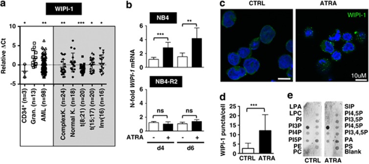
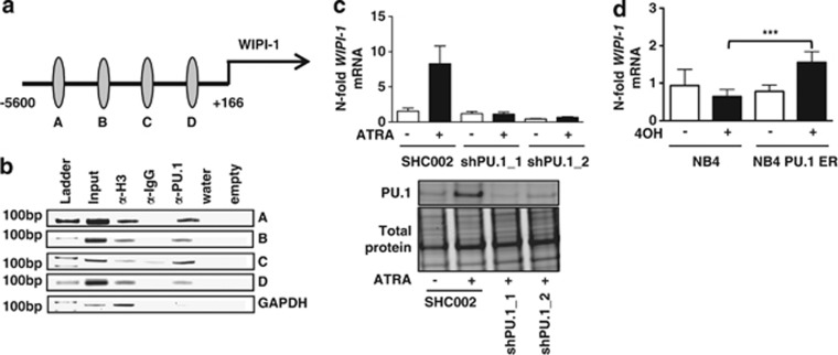
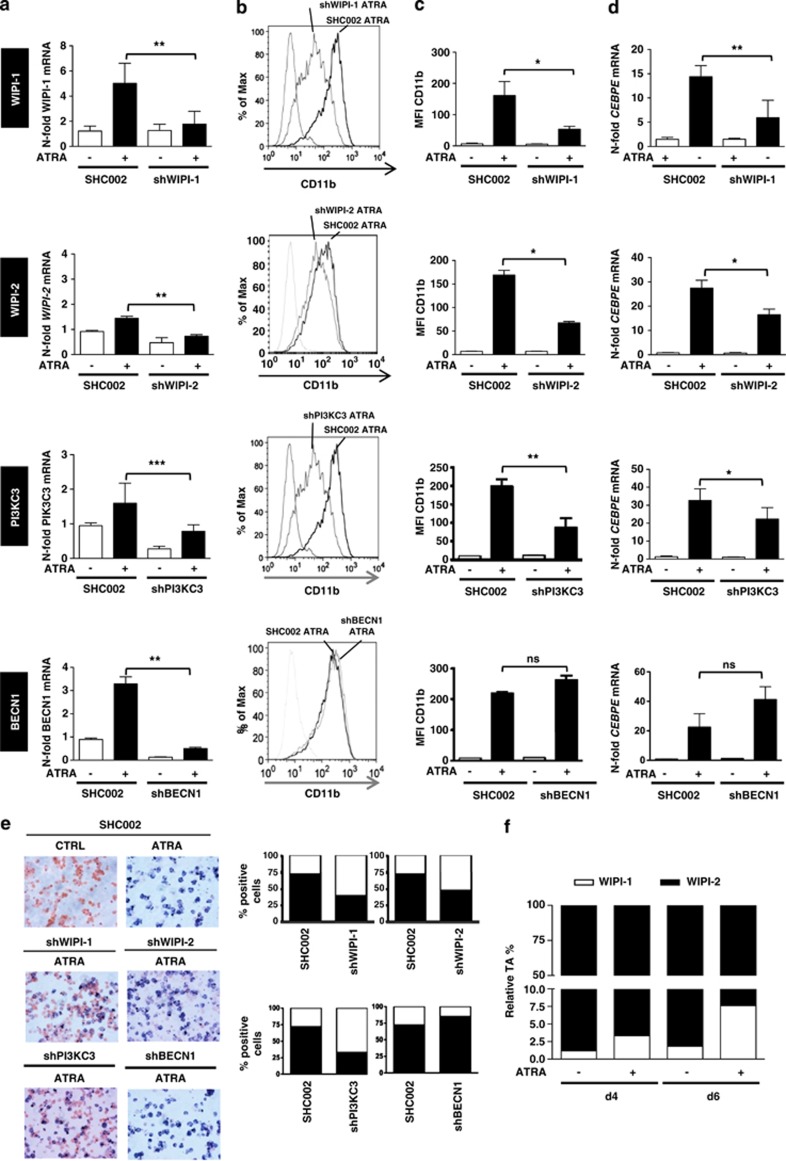
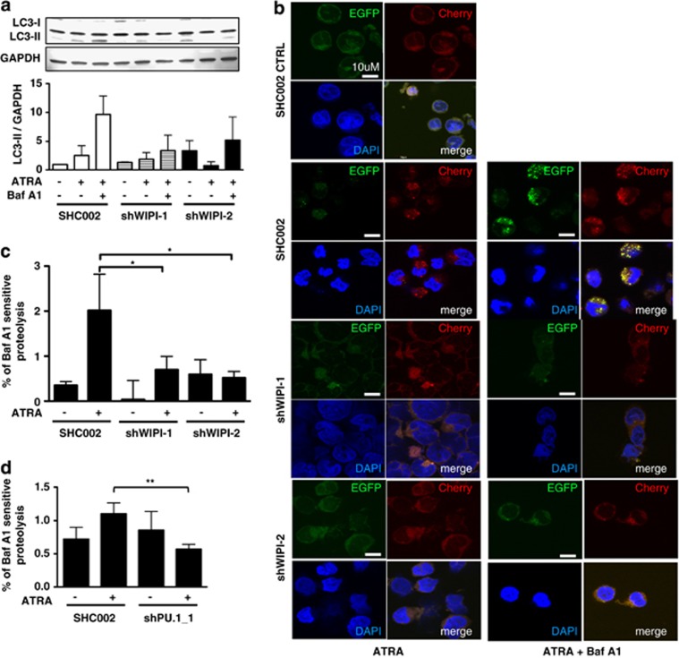
Verification
Claims the paper makes
The claims and statistics of this paper haven’t been checked yet.
Run the check — free, no account needed. It reads the open-access text and checks the sources it cites.
Claims other papers make about this one
Each is checked against this paper’s own text: does it say what the citing paper says it does?
In the citing paper
“Moreover, NFE2L2/NRF2 is a regulator of Lamp2a transcription, and therefore, it controls CMA [982].”
From Guidelines for the use and interpretation of assays for monitoring autophagy (4th edition) 1 (2021) · cites this paper as [982]
Self-check
Percentages, sums and reported differences re-computed from the paper’s own numbers.
The self-check needs the paper’s full text.
Citations
Cites
Guidelines for the use and interpretation of assays for monitoring autophagy
Klionsky, D. J., Abdalla, F. C., Abeliovich, H., Abraham, R. T., Acevedo-Arozena, A., Adeli, K., Agholme, L., Agnello, M., Agostinis, P., Aguirre-Ghiso, J. A., et al. (2012) · Autophagy · doi:10.4161/auto.19496 · Cited by 3,996 · cited 2×
“Moreover, we employed a biochemical standard method to determine the turnover of long-lived proteins by autophagy and measured the proteolysis of radiolabeled proteins in Beclin 1-knockdown cells.(23) The proteolysis in Beclin 1knockdown cells under basal conditions was not significantly decreased upon bafilomycin A1…”
Self-citationRekhtman, N., Radparvar, F., Evans, T., & Skoultchi, A. I. (1999) · Genes & Development · doi:10.1101/gad.13.11.1398 · Cited by 454
PU.1 inhibits GATA-1 function and erythroid differentiation by blocking GATA-1 DNA binding
Zhang, P., Zhang, X., Iwama, A., Yu, C., Smith, K. A., Mueller, B. U., Narravula, S., Torbett, B. E., Orkin, S. H., & Tenen, D. G. (2000) · Blood · doi:10.1182/blood.v96.8.2641 · Cited by 368
Regulation of autophagy by phosphatidylinositol 3‐phosphate
Burman, C., & Ktistakis, N. T. (2010) · FEBS Letters · doi:10.1016/j.febslet.2010.01.011 · Cited by 234
Human WIPI‐1 puncta‐formation: A novel assay to assess mammalian autophagy
Proikas-Cezanne, T., Ruckerbauer, S., Stierhof, Y.-D., Berg, C., & Nordheim, A. (2007) · FEBS Letters · doi:10.1016/j.febslet.2007.06.040 · Cited by 165
Self-citation
Research works printed but not linked: 46
- Cecconi F, Levine B . The role of autophagy in mammalian development: cell makeover rather than cell death. Dev Cell 2008; 15: 344–357. · DOI
- Mizushima N, Levine B, Cuervo AM, Klionsky DJ . Autophagy fights disease through cellular self-digestion. Nature 2008; 451: 1069–1075. · DOI · Paper page
- Meijer AJ, Codogno P . Autophagy: regulation and role in disease. Crit Rev Clin Lab Sci 2009; 46: 210–240. · DOI
- Hershko A, Ciechanover A . The ubiquitin system. Annu Rev Biochem 1998; 67: 425–479. · DOI · Paper page
- Codogno P, Mehrpour M, Proikas-Cezanne T . Canonical and non-canonical autophagy: variations on a common theme of self-eating? Nat Rev Mol Cell Biol 2012; 13: 7–12. · DOI
- Blommaart EF, Krause U, Schellens JP, Vreeling-Sindelárová H, Meijer AJ . The phosphatidylinositol 3-kinase inhibitors wortmannin and LY294002 inhibit autophagy in isolated rat hepatocytes. Eur J Biochem 1997; 243: 240–246. · DOI
- Knævelsrud H, Simonsen A . Lipids in autophagy: constituents, signaling molecules and cargo with relevance to disease. Biochim Biophys Acta 2012; 1821: 1133–1145. · DOI
- Simonsen A, Tooze SA . Coordination of membrane events during autophagy by multiple class III PI3-kinase complexes. J Cell Biol 2009; 186: 773–782. · DOI
- Petiot A, Ogier-Denis E, Blommaart EFC, Meijer AJ, Codogno P . Distinct classes of phosphatidylinositol 3-kinases are involved in signaling pathways that control macroautophagy in HT-29 cells. J Biol Chem 2000; 275: 992–998. · DOI
- Axe EL, Walker SA, Manifava M, Chandra P, Roderick HL, Habermann A et al. Autophagosome formation from membrane compartments enriched in phosphatidylinositol 3-phosphate and dynamically connected to the endoplasmic reticulum. J Cell Biol 2008; 182: 685–701. · DOI · Paper page
- Proikas-Cezanne T, Waddell S, Gaugel A, Frickey T, Lupas A, Nordheim A et al. WIPI-1alpha (WIPI49), a member of the novel 7-bladed WIPI protein family, is aberrantly expressed in human cancer and is linked to starvation-induced autophagy. Oncogene 2004; 23: 9314–9325. · DOI · Paper page
- Polson HE, de Lartigue J, Rigden DJ, Reedijk M, Urbé S, Clague MJ et al. Mammalian Atg18 (WIPI2) localizes to omegasome-anchored phagophores and positively regulates LC3 lipidation. Autophagy 2010; 6: 506–522. · DOI · Paper page
- Mauthe M, Jacob A, Freiberger S, Hentschel K, Stierhof YD, Codogno P et al. Resveratrol-mediated autophagy requires WIPI-1-regulated LC3 lipidation in the absence of induced phagophore formation. Autophagy 2011; 7: 1448–1461. · DOI · Paper page
- Lu Q, Yang P, Huang X, Hu W, Guo B, Wu F et al. The WD40 Repeat PtdIns(3)P-binding protein EPG-6 regulates progression of omegasomes to autophagosomes. Dev Cell 2011; 21: 343–357. · DOI · Paper page
- Cáceres-Cortés JR . Blastic leukaemias (AML): a biologist’s view. Cell Biochem Biophys 2012; 66: 13–22. · DOI
- Isakson P, Bjoras M, Boe SO, Simonsen A . Autophagy contributes to therapy-induced degradation of the PML/RARA oncoprotein. Blood 2010; 116: 2324–2331. · DOI
- Bøe SO, Simonsen A . Autophagic degradation of an oncoprotein. Autophagy 2010; 6: 964–965. · DOI
- Trocoli A, Mathieu J, Priault M, Reiffers J, Souquère S, Pierron G et al. ATRA-induced upregulation of Beclin 1 prolongs the life span of differentiated acute promyelocytic leukemia cells. Autophagy 2011; 7: 1108–1114. · DOI · Paper page
- Zhang S-P, Niu YN, Yuan N, Zhang AH, Chao D, Xu QP et al. Role of autophagy in acute myeloid leukemia therapy. Chin J Cancer 2012; 32: 130–135. · DOI
- Brigger D, Torbett BE, Chen J, Fey MF, Tschan MP . Inhibition of GATE-16 attenuates ATRA-induced neutrophil differentiation of APL cells and interferes with autophagosome formation. Biochem Biophys Res Commun 2013; 438: 283–288. · DOI
- Mizushima N, Yoshimori T, Levine B . Methods in mammalian autophagy research. Cell 2010; 140: 313–326. · DOI · Paper page
- Kabeya Y, Mizushima N, Ueno T, Yamamoto A, Kirisako T, Noda T et al. LC3, a mammalian homologue of yeast Apg8p, is localized in autophagosome membranes after processing. EMBO J 2000; 19: 5720–5728. · DOI · Paper page
- Bakula D, Takacs Z, Proikas-Cezanne T . WIPI β-propellers in autophagy-related diseases and longevity. Biochem Soc Trans 2013; 41: 962–967. · DOI
- Kusama Y, Sato K, Kimura N, Mitamura J, Ohdaira H, Yoshida K . Comprehensive analysis of expression pattern and promoter regulation of human autophagy-related genes. Apoptosis 2009; 14: 1165–1175. · DOI
- He Z, Liu H, Agostini M, Yousefi S, Perren A, Tschan MP et al. p73 regulates autophagy and hepatocellular lipid metabolism through a transcriptional activation of the ATG5 gene. Cell Death Differ 2013; 20: 1415–1424. · DOI
- Kang Y-A, Sanalkumar R, O’Geen H, Linnemann AK, Chang CJ, Bouhassira EE et al. Autophagy driven by a master regulator of hematopoiesis. Mol Cell Biol 2012; 32: 226–239. · DOI · Paper page
- Wang K, Wang P, Shi J, Zhu X, He M, Jia X et al. PML/RARα targets promoter regions containing PU.1 consensus and RARE half sites in acute promyelocytic leukemia. Cancer Cell 2010; 17: 186–197. · DOI
- Itakura E, Kishi-Itakura C, Koyama-Honda I, Mizushima N . Structures containing Atg9A and the ULK1 complex independently target depolarized mitochondria at initial stages of Parkin-mediated mitophagy. J Cell Sci 2012; 125: 1488–1499. · DOI · Paper page
- Itakura E, Mizushima N . Characterization of autophagosome formation site by a hierarchical analysis of mammalian Atg proteins. Autophagy 2010; 6: 764–776. · DOI · Paper page
- Wirawan E, Lippens S, Vanden Berghe T, Romagnoli A, Fimia GM, Piacentini M et al. Beclin1: a role in membrane dynamics and beyond. Autophagy 2012; 8: 6–17. · DOI
- Wang Z, Cao L, Kang R, Yang M, Liu L, Zhao Y et al. Autophagy regulates myeloid cell differentiation by p62/SQSTM1-mediated degradation of PML-RARα oncoprotein. Autophagy 2011; 7: 401–411. · DOI
- Devereaux K, Dall’Armi C, Alcazar-Roman A, Ogasawara Y, Zhou X, Wang F et al. Regulation of mammalian autophagy by Class II and III PI 3-kinases through PI3P Synthesis. PLoS One 2013; 8: 1–17. · DOI · Paper page
- Nishioka C, Ikezoe T, Yang J, Gery S, Koeffler HP, Yokoyama A et al. Inhibition of mammalian target of rapamycin signaling potentiates the effects of all- transretinoic acid to induce growth arrest and differentiation of human acute myelogenous leukemia cells. Int J Cancer 2009; 125: 1710–1720. · DOI
- Breems DA, Boogaerts MA, Dekker AW, Van Putten WL, Sonneveld P, Huijgens PC et al. Autologous bone marrow transplantation as consolidation therapy in the treatment of adult patients under 60 years with acute myeloid leukaemia in first complete remission: a prospective randomized Dutch-Belgian Haemato-Oncology Co-operative Group (HOVON) and Swiss Group for Clinical Cancer Research (SAKK) trial. Br J Haematol 2005; 128: 59–65. · DOI
- Federzoni EA, Valk PJ, Torbett BE, Haferlach T, Löwenberg B, Fey MF et al. PU.1 is linking the glycolytic enzyme HK3 in neutrophil differentiation and survival of APL cells. Blood 2012; 119: 4963–4970. · DOI
- Lowenberg B, Boogaerts MA, Daenen SM, Verhoef GE, Hagenbeek A, Vellenga E et al. Value of different modalities of granulocyte- macrophage colony-stimulating factor applied during or after induction therapy of acute myeloid leukemia. J Clin Oncol 1997; 15: 3496–3506. · DOI
- Lowenberg B, van Putten W, Theobald M, Gmür J, Verdonck L, Sonneveld P et al. Effect of priming with granulocyte colony-stimulating factor on the outcome of chemotherapy for acute myeloid leukemia. N Engl J Med 2003; 349: 743–752. · DOI
- Ossenkoppele GJ . The value of fludarabine in addition to ARA-C and G-CSF in the treatment of patients with high-risk myelodysplastic syndromes and AML in elderly patients. Blood 2004; 103: 2908–2913. · DOI
- Rockova V, Abbas S, Wouters BJ, Erpelinck CA, Beverloo HB, Delwel R et al. Risk stratification of intermediate-risk acute myeloid leukemia: integrative analysis of a multitude of gene mutation and gene expression markers. Blood 2011; 118: 1069–1076. · DOI
- Duprez E, Benoit G, Flexor M, Lillehaug JR, Lanotte M . A mutated PML/RARA found in the retinoid maturation resistant NB4 subclone, NB4-R2, blocks RARA and wild-type PML/RARA transcriptional activities. Leukemia 2000; 14: 255–261. · DOI
- Ochs HD, Igo RP . The NBT slide test: a simple method for detecting chronic granulomatous disease and female carriers. J Pediatr 1973; 83: 77–82. · DOI
- Tschan MP, Shan D, Laedrach J, Eyholzer M, Leibundgut EO, Baerlocher GM et al. NDRG1/2 expression is inhibited in primary acute myeloid leukemia. Leuk Res 2010; 34: 393–398. · DOI
- Britschgi C, Rizzi M, Grob TJ, Tschan MP, Hügli B, Reddy VA et al. Identification of the p53 family-responsive element in the promoter region of the tumor suppressor gene hypermethylated in cancer 1. Oncogene 2005; 25: 2030–2039. · DOI
- Ogier-Denis E, Houri J-J, Bauvy C, Codogno P . Guanine Nucleotide Exchange on Heterotrimeric Gi3 Protein Controls Autophagic Sequestration in HT-29 Cells*. J Biol Chem 1996; 271: 28593–28600. · DOI
- Kaeser MD, Iggo RD . Chromatin immunoprecipitation analysis fails to support the latency model for regulation of p53 DNA binding activity in vivo. Cell 2002; 99: 95–100.
- Tschan MP . Alternative splicing of the human cyclin d-binding myb-like protein (hdmp1) yields a truncated protein isoform that alters macrophage differentiation patterns. J Biol Chem 2003; 278: 42750–42760. · DOI
Cited by
Sorted by citations
Guidelines for the use and interpretation of assays for monitoring autophagy (4th edition) 1
Klionsky, D. J., Abdel-Aziz, A. K., Abdelfatah, S., Abdellatif, M., Abdoli, A., Abel, S., Abeliovich, H., Abildgaard, M. H., Abudu, Y. P., Acevedo-Arozena, A., et al. (2021) · Autophagy · doi:10.1080/15548627.2020.1797280 · Cited by 2,897
Self-citation
Metadata
- Authors
- Brigger, D., Proikas-Cezanne, T., & Tschan, M. P.
- Venue
- Cell Death & Disease
- Published
- 2014-07-03
- Volume
- 5
- Issue
- 7
- Pages
- e1315-e1315
- ISSN
- 2041-4889
- PMC
- PMC4123064
- Cited by
- 48
- References
- 5
- Language
- en
About these checks
Registry, retraction, Cites and statistics results are computed, not judged, with one exception: a printed reference that no index lists is read once by a model before it counts as a citation error. Claims are extracted by a model and kept only when their quote appears verbatim in the text; each cited source is then checked, with a model’s help, for whether it says what the claim relies on. Those verdicts can be wrong — read the claim’s quote and the cited source before relying on one.
Statistics and arithmetic are recomputed, not judged.
Confidence bands are shown only for fields calibrated on our annotated test set. None is calibrated yet, so no band is shown.
What counts as a claim ›How we measure confidence ›How we check this ›
Are you an author, or do you see a problem with this page? Request a review
