

The DOI matches its Crossref record; 5 of the 5 references it cites were found in a registry or an index.
Busulfan Conditioning Enhances Engraftment of Hematopoietic Donor-derived Cells in the Brain Compared With Irradiation
Overview
The claims and statistics of this paper haven’t been checked yet.
Run the check — free, no account needed. It reads the open-access text and checks the sources it cites.
Integrity signals
Registry
Cites
Statistics
Figures
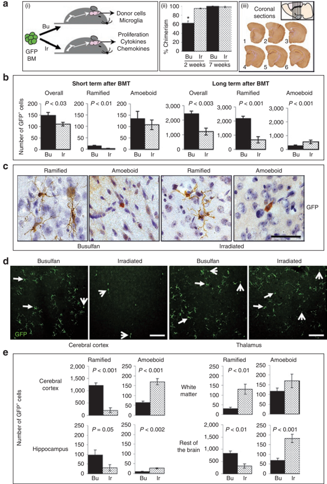
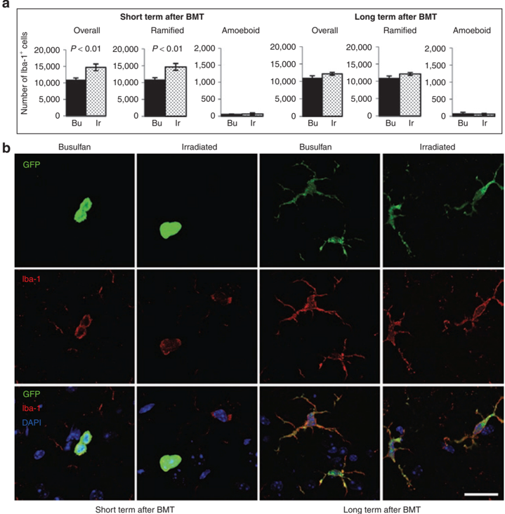
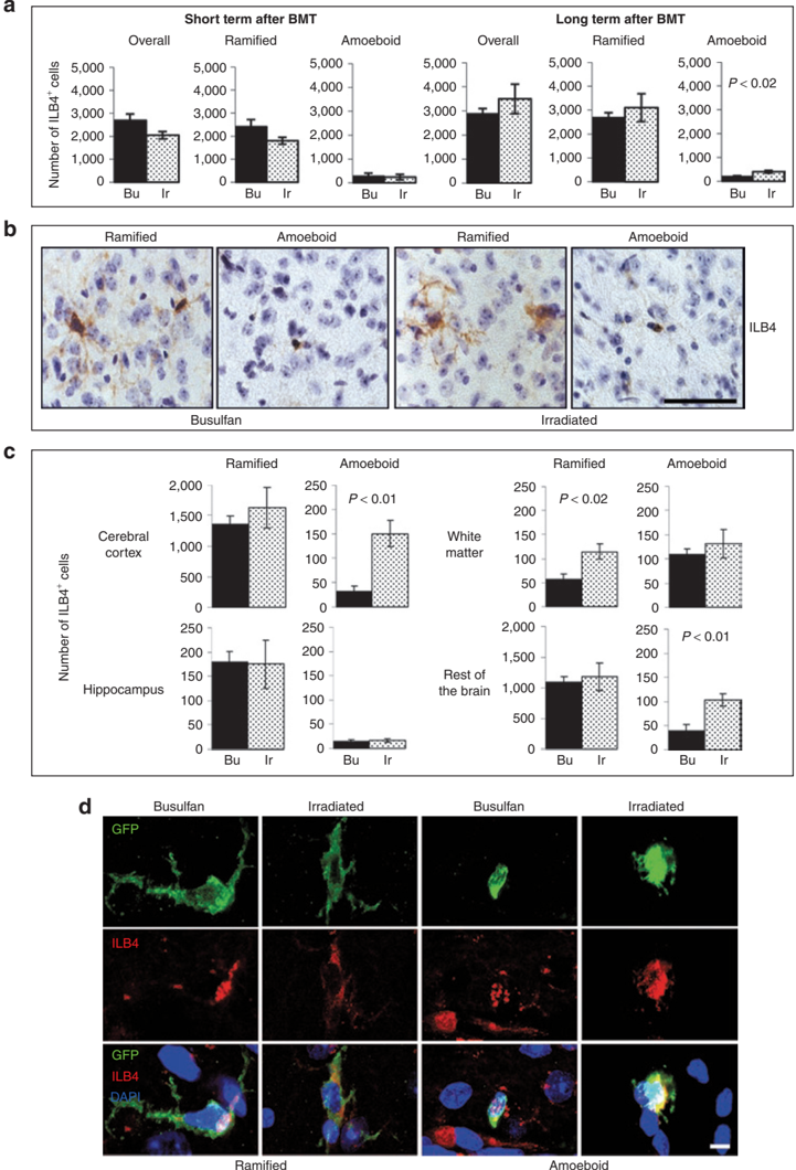
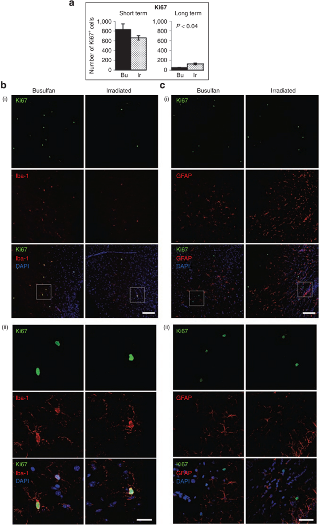
Verification
Claims the paper makes
The claims and statistics of this paper haven’t been checked yet.
Run the check — free, no account needed. It reads the open-access text and checks the sources it cites.
Claims other papers make about this one
Each is checked against this paper’s own text: does it say what the citing paper says it does?
In the citing paper
“The reported efficiencies of microglia replacement following BMT in mice vary greatly (18–23).”
From Treatment of a genetic brain disease by CNS-wide microglia replacement (2022) · cites this paper as [21]
Self-check
Percentages, sums and reported differences re-computed from the paper’s own numbers.
The self-check needs the paper’s full text.
Citations
Cites
p53 is required for radiation-induced apoptosis in mouse thymocytes
Lowe, S. W., Schmitt, E. M., Smith, S. W., Osborne, B. A., & Jacks, T. (1993) · Nature · doi:10.1038/362847a0 · Cited by 2,864
Microglia in the adult brain arise from Ly-6ChiCCR2+ monocytes only under defined host conditions
Mildner, A., Schmidt, H., Nitsche, M., Merkler, D., Hanisch, U.-K., Mack, M., Heikenwalder, M., Brück, W., Priller, J., & Prinz, M. (2007) · Nature Neuroscience · doi:10.1038/nn2015 · Cited by 1,089
The myeloid cells of the central nervous system parenchyma
Ransohoff, R. M., & Cardona, A. E. (2010) · Nature · doi:10.1038/nature09615 · Cited by 804
Bone-marrow-derived microglia: myth or reality?
Soulet, D., & Rivest, S. (2008) · Current Opinion in Pharmacology · doi:10.1016/j.coph.2008.04.002 · Cited by 139
Microgliosis: the questions shape the answers
Ransohoff, R. M. (2007) · Nature Neuroscience · doi:10.1038/nn1207-1507 · Cited by 109
Research works printed but not linked: 38
- Krause, 2001, Multi-organ, multi-lineage engraftment by a single bone marrow-derived stem cell, Cell, 105, 369, 10.1016/S0092-8674(01)00328-2 · DOI
- Ajami, 2007, Local self-renewal can sustain CNS microglia maintenance and function throughout adult life, Nat Neurosci, 10, 1538, 10.1038/nn2014 · DOI · Paper page
- Priller, 2001, Targeting gene-modified hematopoietic cells to the central nervous system: use of green fluorescent protein uncovers microglial engraftment, Nat Med, 7, 1356, 10.1038/nm1201-1356 · DOI · Paper page
- Chen, 2003, Overexpression of monocyte chemoattractant protein 1 in the brain exacerbates ischemic brain injury and is associated with recruitment of inflammatory cells, J Cereb Blood Flow Metab, 23, 748, 10.1097/01.WCB.0000071885.63724.20 · DOI
- Kuziel, 1997, Severe reduction in leukocyte adhesion and monocyte extravasation in mice deficient in CC chemokine receptor 2, Proc Natl Acad Sci USA, 94, 12053, 10.1073/pnas.94.22.12053 · DOI
- Liu, 2007, Origin of dendritic cells in peripheral lymphoid organs of mice, Nat Immunol, 8, 578, 10.1038/ni1462 · DOI
- Diserbo, 2002, Blood-brain barrier permeability after gamma whole-body irradiation: an in vivo microdialysis study, Can J Physiol Pharmacol, 80, 670, 10.1139/y02-070 · DOI
- Li, 2004, Early radiation-induced endothelial cell loss and blood-spinal cord barrier breakdown in the rat spinal cord, Radiat Res, 161, 143, 10.1667/RR3117 · DOI
- Kyrkanides, 2002, Cyclooxygenase-2 modulates brain inflammation-related gene expression in central nervous system radiation injury, Brain Res Mol Brain Res, 104, 159, 10.1016/S0169-328X(02)00353-4 · DOI
- Linard, 2004, Acute induction of inflammatory cytokine expression after gamma-irradiation in the rat: effect of an NF-kappaB inhibitor, Int J Radiat Oncol Biol Phys, 58, 427, 10.1016/j.ijrobp.2003.09.039 · DOI
- Biffi, 2004, Correction of metachromatic leukodystrophy in the mouse model by transplantation of genetically modified hematopoietic stem cells, J Clin Invest, 113, 1118, 10.1172/JCI200419205 · DOI
- Langford-Smith, 2012, Hematopoietic stem cell and gene therapy corrects primary neuropathology and behavior in mucopolysaccharidosis IIIA mice, Mol Ther, 20, 1610, 10.1038/mt.2012.82 · DOI
- Visigalli, 2010, Gene therapy augments the efficacy of hematopoietic cell transplantation and fully corrects mucopolysaccharidosis type I phenotype in the mouse model, Blood, 116, 5130, 10.1182/blood-2010-04-278234 · DOI
- Meng, 2003, Ionizing radiation and busulfan inhibit murine bone marrow cell hematopoietic function via apoptosis-dependent and -independent mechanisms, Exp Hematol, 31, 1348, 10.1016/j.exphem.2003.08.014 · DOI
- Probin, 2006, Busulfan selectively induces cellular senescence but not apoptosis in WI38 fibroblasts via a p53-independent but extracellular signal-regulated kinase-p38 mitogen-activated protein kinase-dependent mechanism, J Pharmacol Exp Ther, 319, 551, 10.1124/jpet.106.107771 · DOI
- Lampron, 2012, Effects of myeloablation, peripheral chimerism, and whole-body irradiation on the entry of bone marrow-derived cells into the brain, Cell Transplant, 21, 1149, 10.3727/096368911X593154 · DOI · Paper page
- Capotondo, 2012, Brain conditioning is instrumental for successful microglia reconstitution following hematopoietic stem cell transplantation, Proc Natl Acad Sci USA, 109, 15018, 10.1073/pnas.1205858109 · DOI · Paper page
- Wake, 2009, Resting microglia directly monitor the functional state of synapses in vivo and determine the fate of ischemic terminals, J Neurosci, 29, 3974, 10.1523/JNEUROSCI.4363-08.2009 · DOI · Paper page
- Kanazawa, 2002, Macrophage/microglia-specific protein Iba1 enhances membrane ruffling and Rac activation via phospholipase C-gamma -dependent pathway, J Biol Chem, 277, 20026, 10.1074/jbc.M109218200 · DOI
- Streit, 1987, Lectin binding by resting and reactive microglia, J Neurocytol, 16, 249, 10.1007/BF01795308 · DOI
- Samlowski, 1989, Peripheral lymph node helper T-cell recovery after syngeneic bone marrow transplantation in mice prepared with either gamma-irradiation or busulfan, Blood, 74, 1436, 10.1182/blood.V74.4.1436.1436 · DOI
- Samlowski, 1987, Recovery of contact hypersensitivity responses following murine bone marrow transplantation: comparison of gamma-irradiation and busulfan as preparative marrow-ablative agents, Blood, 70, 1910, 10.1182/blood.V70.6.1910.1910 · DOI
- Thomas, 2002, Mechanisms of mobilization of hematopoietic progenitors with granulocyte colony-stimulating factor, Curr Opin Hematol, 9, 183, 10.1097/00062752-200205000-00002 · DOI
- Jopling, 2001, A cautionary tale: how to delete mouse haemopoietic stem cells with busulphan, Br J Haematol, 113, 970, 10.1046/j.1365-2141.2001.02825.x · DOI
- Bacigalupo, 2009, Defining the intensity of conditioning regimens: working definitions, Biol Blood Marrow Transplant, 15, 1628, 10.1016/j.bbmt.2009.07.004 · DOI
- Hess, 1996, Phenotype of arylsulfatase A-deficient mice: relationship to human metachromatic leukodystrophy, Proc Natl Acad Sci USA, 93, 14821, 10.1073/pnas.93.25.14821 · DOI
- Kennedy, 1997, Kinetics of central nervous system microglial and macrophage engraftment: analysis using a transgenic bone marrow transplantation model, Blood, 90, 986, 10.1182/blood.V90.3.986 · DOI
- Allan, 2005, Interleukin-1 and neuronal injury, Nat Rev Immunol, 5, 629, 10.1038/nri1664 · DOI · Paper page
- Mihara, 2012, IL-6/IL-6 receptor system and its role in physiological and pathological conditions, Clin Sci, 122, 143, 10.1042/CS20110340 · DOI
- Hassan, 1992, In vivo distribution of [11C]-busulfan in cynomolgus monkey and in the brain of a human patient, Cancer Chemother Pharmacol, 30, 81, 10.1007/BF00686397 · DOI
- Soussain, 2009, CNS complications of radiotherapy and chemotherapy, Lancet, 374, 1639, 10.1016/S0140-6736(09)61299-X · DOI
- Bigger, 2006, Permanent partial phenotypic correction and tolerance in a mouse model of hemophilia B by stem cell gene delivery of human factor IX, Gene Ther, 13, 117, 10.1038/sj.gt.3302638 · DOI
- Siapati, 2007, Murine leukemia following irradiation conditioning for transplantation of lentivirally-modified hematopoietic stem cells, Eur J Haematol, 78, 303, 10.1111/j.1600-0609.2006.00813.x · DOI
- Siapati, 2005, Comparison of HIV- and EIAV-based vectors on their efficiency in transducing murine and human hematopoietic repopulating cells, Mol Ther, 12, 537, 10.1016/j.ymthe.2005.01.022 · DOI
- Okabe, 1997, ‘Green mice' as a source of ubiquitous green cells, FEBS Lett, 407, 313, 10.1016/S0014-5793(97)00313-X · DOI · Paper page
- Malinowska, 2010, Genistein improves neuropathology and corrects behaviour in a mouse model of neurodegenerative metabolic disease, PLoS ONE, 5, e14192, 10.1371/journal.pone.0014192 · DOI
- Canal, 2010, Circadian rhythm and suprachiasmatic nucleus alterations in the mouse model of mucopolysaccharidosis IIIB, Behav Brain Res, 209, 212, 10.1016/j.bbr.2010.01.045 · DOI
- Wilkinson, 2012, Neuropathology in mouse models of mucopolysaccharidosis type I, IIIA and IIIB, PLoS ONE, 7, e35787, 10.1371/journal.pone.0035787 · DOI
Cited by
Microglia and brain macrophages in the molecular age: from origin to neuropsychiatric disease
Prinz, M., & Priller, J. (2014) · Nature Reviews Neuroscience · doi:10.1038/nrn3722 · Cited by 1,329
Shemer, A., Grozovski, J., Tay, T. L., Tao, J., Volaski, A., Süß, P., Ardura-Fabregat, A., Gross-Vered, M., Kim, J.-S., David, E., et al. (2018) · Nature Communications · doi:10.1038/s41467-018-07548-5 · Cited by 1,052
The Semantics of Microglia Activation: Neuroinflammation, Homeostasis, and Stress
Woodburn, S. C., Bollinger, J. L., & Wohleb, E. S. (2021) · Journal of Neuroinflammation · doi:10.1186/s12974-021-02309-6 · Cited by 783
Dodiya, H. B., Forsyth, C. B., Voigt, R. M., Engen, P. A., Patel, J., Shaikh, M., Green, S. J., Naqib, A., Roy, A., Kordower, J. H., et al. (2020) · Neurobiology of Disease · doi:10.1016/j.nbd.2018.12.012 · Cited by 305
Treatment of a genetic brain disease by CNS-wide microglia replacement
Shibuya, Y., Kumar, K. K., Mader, M. M.-D., Yoo, Y., Ayala, L. A., Zhou, M., Mohr, M. A., Neumayer, G., Kumar, I., Yamamoto, R., et al. (2022) · Science Translational Medicine · doi:10.1126/scitranslmed.abl9945 · Cited by 126
Metadata
- Authors
- Wilkinson, F. L., Sergijenko, A., Langford-Smith, K. J., Malinowska, M., Wynn, R. F., & Bigger, B. W.
- Venue
- Molecular Therapy
- Publisher
- Elsevier BV
- Published
- 2013-04
- Volume
- 21
- Issue
- 4
- Pages
- 868-876
- ISSN
- 1525-0016
- Cited by
- 112
- References
- 5
- Language
- en
- Update notices
- erratum (2013-07-01)
About these checks
Registry, retraction, Cites and statistics results are computed, not judged, with one exception: a printed reference that no index lists is read once by a model before it counts as a citation error. Claims are extracted by a model and kept only when their quote appears verbatim in the text; each cited source is then checked, with a model’s help, for whether it says what the claim relies on. Those verdicts can be wrong — read the claim’s quote and the cited source before relying on one.
Statistics and arithmetic are recomputed, not judged.
Confidence bands are shown only for fields calibrated on our annotated test set. None is calibrated yet, so no band is shown.
What counts as a claim ›How we measure confidence ›How we check this ›
Are you an author, or do you see a problem with this page? Request a review
