Cited by 111Cell death mechanisms and regulationNF-κB Signaling PathwaysFerroptosis and cancer prognosis

The DOI matches its Crossref record; 41 of the 41 references it cites were found in a registry or an index.

HS-1371, a novel kinase inhibitor of RIP3-mediated necroptosis

Open access
Park, Han-Hee, Park, Se-Yeon, Mah, Shinmee, Park, Jung-Hee, Hong, Soon-Sun, Hong, Sungwoo, Kim, You-Sun·2018·Experimental & Molecular Medicine

Abstract

Necroptosis is a type of programmed cell death that usually occurs under apoptosis-deficient conditions. Receptor-interacting protein kinase-3 (RIP3, or RIPK3) is a central player in necroptosis, and its kinase activity is essential for downstream necroptotic signaling events. Since RIP3 kinase activity has been associated with various diseases, the development of specific RIP3 inhibitors is an attractive strategy for therapeutic application. In this study, we identified a potent RIP3 inhibitor, HS-1371, by the extensive screening of chemical libraries focused on kinases. HS-1371 directly binds to RIP3 in an ATP-competitive and time-independent manner, providing a mechanism of action. Moreover, the compound inhibited TNF-induced necroptosis but did not inhibit TNF-induced apoptosis, indicating that this novel inhibitor has a specific inhibitory effect on RIP3-mediated necroptosis via the suppression of RIP3 kinase activity. Our results suggest that HS-1371 could serve as a potential preventive or therapeutic agent for diseases involving RIP3 hyperactivation.

Overview

The claims and statistics of this paper haven’t been checked yet.

Run the check — free, no account needed. It reads the open-access text and checks the sources it cites.

A few free checks a day · papers already verified open instantly · results are public

Integrity signals

Registry

Crossref · 10.1038/s12276-018-0152-8

Cites

41 of 41 references verified0 citation errors · 0 not checkable

Statistics

Figures

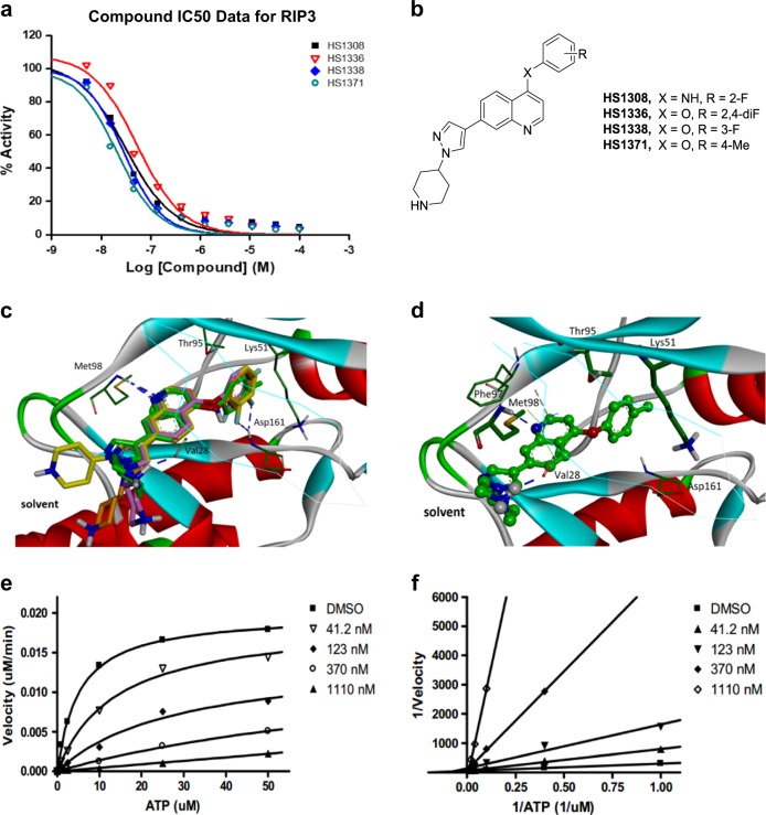
Fig. 1 Identification of HS-1371 as a novel RIP3 kinase inhibitor. a IC50 curves for RIP3 with HS-1308, 1336, 1338, and 1371 treatments. b Chemical structures of HS-1308, 1336, 1338, and 1371. c, d. c Comparative view of the predicted binding conformations of HS-1308, 1336, 1338 and 1371 in the ATP-binding site of RIP3 (PDB ID: 4M66). Carbon atoms of HS-1308, 1336, 1338 and 1371 are colored in yellow, pink, brown, and green, respectively. d Detailed interaction patterns of HS-1371 in the ATP-binding site of RIP3. Hydrogen-bond interactions are represented with blue dotted lines. e, f. e Michaelis–Menten plot and f Lineweaver-Burk Plot for RIP3 with HS-1371
Fig. 1 Identification of HS-1371 as a novel RIP3 kinase inhibitor. a IC50 curves for RIP3 with HS-1308, 1336, 1338, and 1371 treatments. b Chemical structures of HS-1308, 1336, 1338, and 1371. c, d. c Comparative view of the predicted binding conformations of HS-1308, 1336, 1338 and 1371 in the ATP-binding site of RIP3 (PDB ID: 4M66). Carbon atoms of HS-1308, 1336, 1338 and 1371 are colored in yellow, pink, brown, and green, respectively. d Detailed interaction patterns of HS-1371 in the ATP-binding site of RIP3. Hydrogen-bond interactions are represented with blue dotted lines. e, f. e Michaelis–Menten plot and f Lineweaver-Burk Plot for RIP3 with HS-1371CC BY · PubMed Central
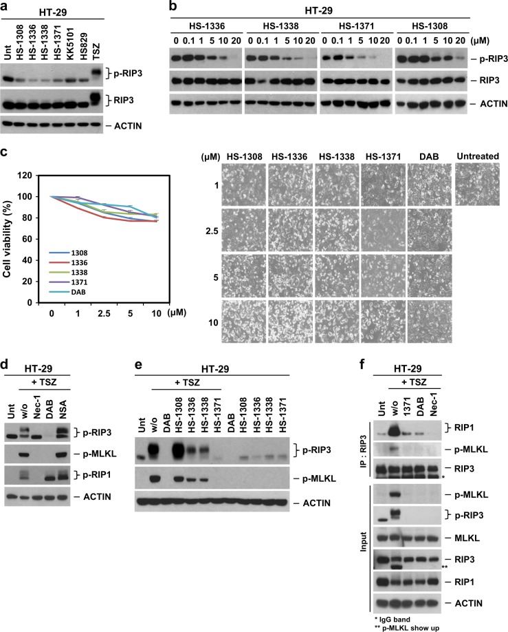
Fig. 2 Novel inhibitors can block RIP3 S227 phosphorylation. a Western blotting showing various kinase inhibitor effects on RIP3 S227 auto-phosphorylation. HT-29 cells were treated with four tested kinase inhibitors (1308, 1336, 1338, and 1371, 10 μM) for 9 h. As a control, KK5101 (Trk alpha inhibitor) and HS829 (IKK beta inhibitor) were also treated at a concentration of 10 μM for 9 h. Prototypical necroptosis stimuli (TNF + zVAD + either cycloheximide or SMAC mimetic; hereafter referred to as TCZ or TSZ) were applied for 6 h. Cell lysates were analyzed with a S227 specific p-RIP3 antibody. b Four tested kinase inhibitors displayed kinase inhibitory effects on basal levels of RIP3 auto-phosphorylation in a dose-dependent manner. HT-29 cells were treated with indicated concentrations for 9 h, and cell lysates were analyzed by immunoblotting. c Four tested kinase inhibitors showed small amounts of cytotoxicity in a dose-dependent manner. HT-29 cells were treated with 4 tested kinase inhibitors for 24 h, and cell viability was analyzed by MTT assay or phase-contrast microscopy. The results are presented as means ± SEM. *P < 0.05, **P < 0.01, ***P < 0.001. d Various necroptosis inhibitors blocked TNF-induced RIP3 phosphorylation. HT-29 cells were pretreated with necrostatin-1 (Nec-1, 40 μM), dabrafenib (DAB, 10 μM) or necrosulfonamide (NSA, 1 μM) for 2 h and then treated with TSZ for 6 h. Cell lysates were analyzed by immunoblotting. e HS-1371 efficiently blocked TNF-induced RIP3 phosphorylation. HT-29 cells were pretreated with four tested kinase inhibitors for 2 h and then treated with TSZ for 6 h. Cell lysates were analyzed by immunoblotting. f HT-29 cells were pretreated with Nec-1 (40 μM), DAB (5 μM) or HS-1371 (5 μM) for 2 h and then treated with TSZ for 4 h. Cell lysates were immunoprecipitated with anti-RIP3 antibody. Immunoprecipitates and total lysates were analyzed by Western blotting
Fig. 2 Novel inhibitors can block RIP3 S227 phosphorylation. a Western blotting showing various kinase inhibitor effects on RIP3 S227 auto-phosphorylation. HT-29 cells were treated with four tested kinase inhibitors (1308, 1336, 1338, and 1371, 10 μM) for 9 h. As a control, KK5101 (Trk alpha inhibitor) and HS829 (IKK beta inhibitor) were also treated at a concentration of 10 μM for 9 h. Prototypical necroptosis stimuli (TNF + zVAD + either cycloheximide or SMAC mimetic; hereafter referred to as TCZ or TSZ) were applied for 6 h. Cell lysates were analyzed with a S227 specific p-RIP3 antibody. b Four tested kinase inhibitors displayed kinase inhibitory effects on basal levels of RIP3 auto-phosphorylation in a dose-dependent manner. HT-29 cells were treated with indicated concentrations for 9 h, and cell lysates were analyzed by immunoblotting. c Four tested kinase inhibitors showed small amounts of cytotoxicity in a dose-dependent manner. HT-29 cells were treated with 4 tested kinase inhibitors for 24 h, and cell viability was analyzed by MTT assay or phase-contrast microscopy. The results are presented as means ± SEM. *P < 0.05, **P < 0.01, ***P < 0.001. d Various necroptosis inhibitors blocked TNF-induced RIP3 phosphorylation. HT-29 cells were pretreated with necrostatin-1 (Nec-1, 40 μM), dabrafenib (DAB, 10 μM) or necrosulfonamide (NSA, 1 μM) for 2 h and then treated with TSZ for 6 h. Cell lysates were analyzed by immunoblotting. e HS-1371 efficiently blocked TNF-induced RIP3 phosphorylation. HT-29 cells were pretreated with four tested kinase inhibitors for 2 h and then treated with TSZ for 6 h. Cell lysates were analyzed by immunoblotting. f HT-29 cells were pretreated with Nec-1 (40 μM), DAB (5 μM) or HS-1371 (5 μM) for 2 h and then treated with TSZ for 4 h. Cell lysates were immunoprecipitated with anti-RIP3 antibody. Immunoprecipitates and total lysates were analyzed by Western blottingCC BY · PubMed Central
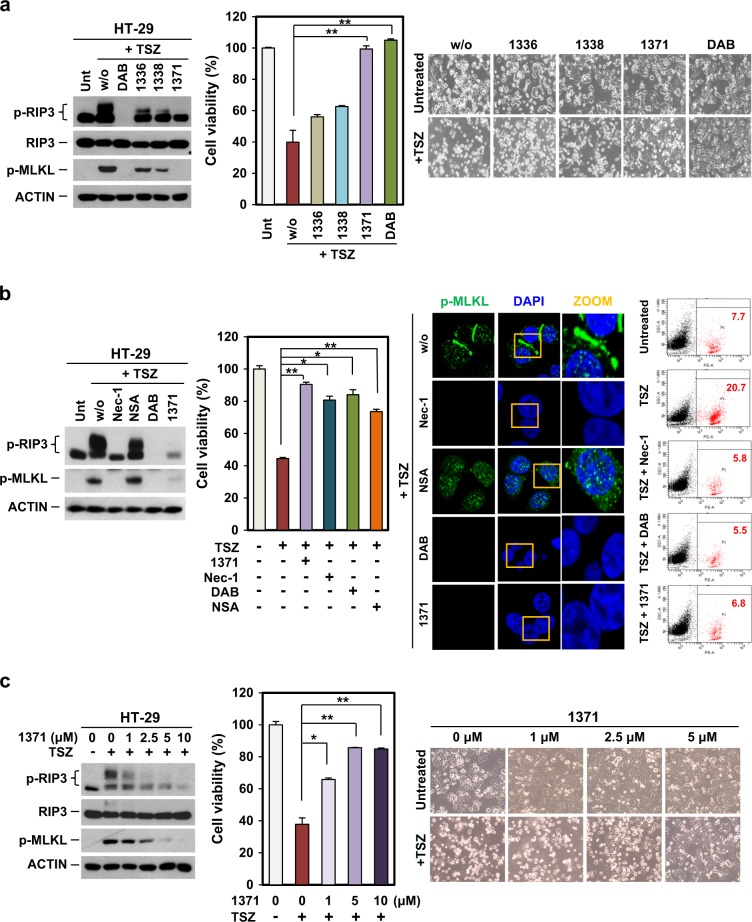
Fig. 3 HS-1371 protects cells from TNF-induced necroptosis. a TNF-induced necroptosis was completely blocked by HS-1371 treatment. HT-29 cells were pretreated with four tested inhibitors for 2 h and then treated with TSZ (6 h for immunoblotting, 24 h for cell death assay). Cell lysates were analyzed by immunoblotting, and cell viability was analyzed by MTT assay or phase-contrast microscopy. The results are presented as the means ± SEM. *P < 0.05, **P < 0.01, ***P < 0.001. b Protection of necroptosis by HS-1371 treatment is similar with dabrafenib, which abolished downstream events. HT-29 cells were pretreated with various necroptosis inhibitors and HS-1371 before TSZ treatment. Cell lysates were analyzed by immunoblotting, and cell viability was analyzed by MTT assay (left panel). Cells were stained with phospho-MLKL antibody and analyzed by confocal fluorescence microscopy (middle panel), and cell death was further analyzed by FACS analysis after PI staining (right panel). c HS-1371 protects cells from TNF-induced necroptosis in a dose-dependent manner. HT-29 cells were pretreated with indicated concentrations of HS-1371 and then treated with TSZ (6 h for immunoblotting, 24 h for cell death assay). Cell lysates were analyzed by immunoblotting, and cell viability was analyzed by MTT assay or phase-contrast microscopy. The results are presented as means ± SEM. *P < 0.05, **P < 0.01, ***P < 0.001
Fig. 3 HS-1371 protects cells from TNF-induced necroptosis. a TNF-induced necroptosis was completely blocked by HS-1371 treatment. HT-29 cells were pretreated with four tested inhibitors for 2 h and then treated with TSZ (6 h for immunoblotting, 24 h for cell death assay). Cell lysates were analyzed by immunoblotting, and cell viability was analyzed by MTT assay or phase-contrast microscopy. The results are presented as the means ± SEM. *P < 0.05, **P < 0.01, ***P < 0.001. b Protection of necroptosis by HS-1371 treatment is similar with dabrafenib, which abolished downstream events. HT-29 cells were pretreated with various necroptosis inhibitors and HS-1371 before TSZ treatment. Cell lysates were analyzed by immunoblotting, and cell viability was analyzed by MTT assay (left panel). Cells were stained with phospho-MLKL antibody and analyzed by confocal fluorescence microscopy (middle panel), and cell death was further analyzed by FACS analysis after PI staining (right panel). c HS-1371 protects cells from TNF-induced necroptosis in a dose-dependent manner. HT-29 cells were pretreated with indicated concentrations of HS-1371 and then treated with TSZ (6 h for immunoblotting, 24 h for cell death assay). Cell lysates were analyzed by immunoblotting, and cell viability was analyzed by MTT assay or phase-contrast microscopy. The results are presented as means ± SEM. *P < 0.05, **P < 0.01, ***P < 0.001CC BY · PubMed Central
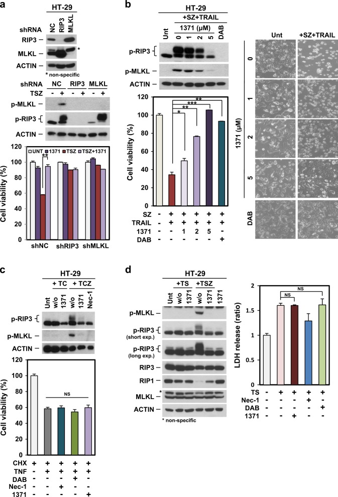
Fig. 4 HS-1371 only rescues cells from RIP3-dependent necroptosis. a Knockdown of RIP3 had no inhibitory effect on HS-1371 in necroptosis. HT-29 cells expressing RIP3 shRNA, MLKL shRNA, or non-silencing control were analyzed by western blotting (upper panel), and these cells were treated with TSZ (6 h for immunoblotting, 24 h for cell death assay). Cell lysates were analyzed by immunoblotting (middle panel), and cell viability was analyzed by MTT assay (bottom panel). The results are presented as means ± SEM. *P < 0.05, **P < 0.01, ***P < 0.001. b HS-1371 protected cells not only from TNF-induced necroptosis but also from TRAIL-induced necroptosis. HT-29 cells were pretreated with indicated concentrations of HS-1371 and then treated with TRAIL + Smac + zVAD (6 h for immunoblotting, 24 h for cell death assay). Cell lysates were analyzed by immunoblotting, and cell viability was analyzed by MTT assay or phase-contrast microscopy. The results are presented as means ± SEM. *P < 0.05, **P < 0.01, ***P < 0.001. c, d HS-1371 had no effect on TNF-induced apoptosis. HT-29 cells were pretreated with HS-1371 (5 μM) for 2 h and then treated with TC or TS (TNF + either cycloheximide or SMAC mimetic, hereafter referred to as TC or TS, 6 h for immunoblotting, 24 h (TC) or 36 h (TS) for cell death assay). Cell lysates were analyzed by immunoblotting, and cell viability was analyzed by MTT assay (TC) or CellTiter-Glo Luminescent Cell Viability Assay Kit (TS). The results are presented as means ± SEM. *P < 0.05, **P < 0.01, ***P < 0.001
Fig. 4 HS-1371 only rescues cells from RIP3-dependent necroptosis. a Knockdown of RIP3 had no inhibitory effect on HS-1371 in necroptosis. HT-29 cells expressing RIP3 shRNA, MLKL shRNA, or non-silencing control were analyzed by western blotting (upper panel), and these cells were treated with TSZ (6 h for immunoblotting, 24 h for cell death assay). Cell lysates were analyzed by immunoblotting (middle panel), and cell viability was analyzed by MTT assay (bottom panel). The results are presented as means ± SEM. *P < 0.05, **P < 0.01, ***P < 0.001. b HS-1371 protected cells not only from TNF-induced necroptosis but also from TRAIL-induced necroptosis. HT-29 cells were pretreated with indicated concentrations of HS-1371 and then treated with TRAIL + Smac + zVAD (6 h for immunoblotting, 24 h for cell death assay). Cell lysates were analyzed by immunoblotting, and cell viability was analyzed by MTT assay or phase-contrast microscopy. The results are presented as means ± SEM. *P < 0.05, **P < 0.01, ***P < 0.001. c, d HS-1371 had no effect on TNF-induced apoptosis. HT-29 cells were pretreated with HS-1371 (5 μM) for 2 h and then treated with TC or TS (TNF + either cycloheximide or SMAC mimetic, hereafter referred to as TC or TS, 6 h for immunoblotting, 24 h (TC) or 36 h (TS) for cell death assay). Cell lysates were analyzed by immunoblotting, and cell viability was analyzed by MTT assay (TC) or CellTiter-Glo Luminescent Cell Viability Assay Kit (TS). The results are presented as means ± SEM. *P < 0.05, **P < 0.01, ***P < 0.001CC BY · PubMed Central
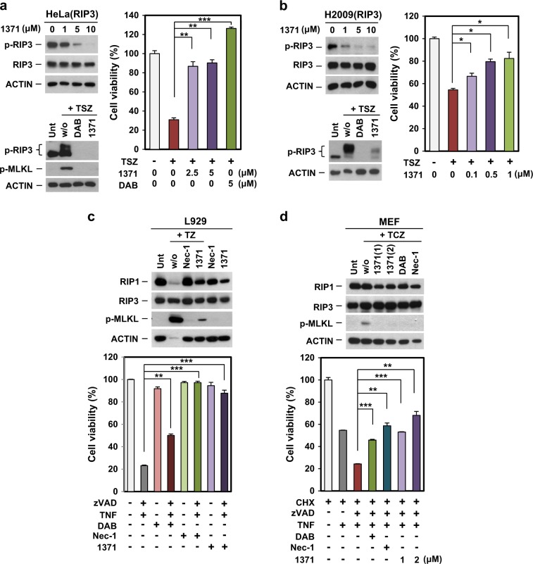
Fig. 5 HS-1371 had an inhibitory effect on RIP3 kinase activity in various cell lines. a, b HeLa (cervical cancer, a) and H2009 (lung cancer, b) cells ectopically expressing RIP3 were treated with indicated concentrations of HS-1371 for 6 h, and cell lysates were analyzed by immunoblotting (left upper panel). Cells were pretreated with DAB or HS-1371 for 2 h and then treated with TSZ (6 h for immunoblotting, 24 h for cell death assay). Cell lysates were analyzed by immunoblotting (left bottom panel), and cell viability was analyzed by MTT assay (right panel). The results are presented as means ± SEM. *P < 0.05, **P < 0.01, ***P < 0.001. c, d HS-1371 had the same effect on mouse cells. L929 (mouse sarcoma, c) and MEF (mouse embryonic fibroblast, d) cells were pretreated with inhibitors and then necroptosis was induced by TNF. Cell lysates were analyzed by immunoblotting (upper panel), and cell viability was analyzed by MTT assay (bottom panel). The results are presented as means ± SEM. *P < 0.05, **P < 0.01, ***P < 0.001
Fig. 5 HS-1371 had an inhibitory effect on RIP3 kinase activity in various cell lines. a, b HeLa (cervical cancer, a) and H2009 (lung cancer, b) cells ectopically expressing RIP3 were treated with indicated concentrations of HS-1371 for 6 h, and cell lysates were analyzed by immunoblotting (left upper panel). Cells were pretreated with DAB or HS-1371 for 2 h and then treated with TSZ (6 h for immunoblotting, 24 h for cell death assay). Cell lysates were analyzed by immunoblotting (left bottom panel), and cell viability was analyzed by MTT assay (right panel). The results are presented as means ± SEM. *P < 0.05, **P < 0.01, ***P < 0.001. c, d HS-1371 had the same effect on mouse cells. L929 (mouse sarcoma, c) and MEF (mouse embryonic fibroblast, d) cells were pretreated with inhibitors and then necroptosis was induced by TNF. Cell lysates were analyzed by immunoblotting (upper panel), and cell viability was analyzed by MTT assay (bottom panel). The results are presented as means ± SEM. *P < 0.05, **P < 0.01, ***P < 0.001CC BY · PubMed Central
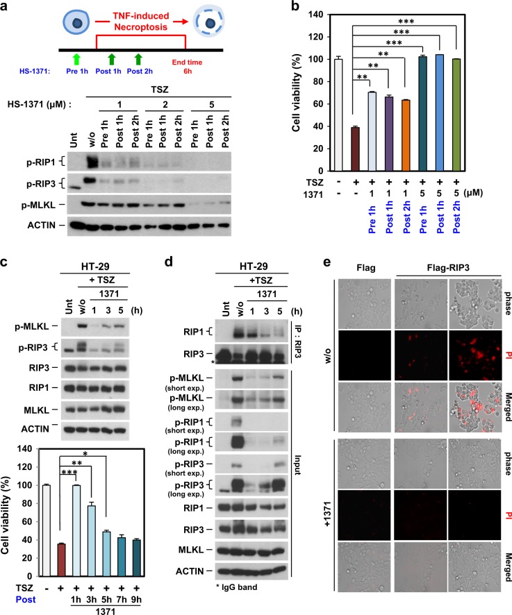
Fig. 6 Possibility of HS-1371 as a drug for necroptosis-related diseases. a, b HT-29 cells were pre- or post-treated with HS-1371 for indicated time points, and RIP3 phosphorylation (a) and cell death (b) were measured by western blotting or MTT assay. Data are presented as means ± SEM. *P < 0.05, **P < 0.01, ***P < 0.001. c HT-29 cells were treated with TSZ and then post-treated with HS-1371 for indicated time points, and then cell lysates were analyzed by immunoblotting (upper panel). Cell viability was analyzed by MTT assay. Data are presented as means ± SEM. *P < 0.05, **P < 0.01, ***P < 0.001. d Cells were treated with the same conditions as (c), and cell lysates were immunoprecipitated with anti-RIP3 antibody. Immunoprecipitates and total lysates were analyzed by Western blotting. e Upregulated RIP3 expression-induced cell death was inhibited by treatment with HS-1371. 293T cells were transfected with Flag vector or Flag-RIP3, and 20 h after transfection, cells were stained with PI. PI-positive cells were analyzed by phase-contrast fluorescence microscopy
Fig. 6 Possibility of HS-1371 as a drug for necroptosis-related diseases. a, b HT-29 cells were pre- or post-treated with HS-1371 for indicated time points, and RIP3 phosphorylation (a) and cell death (b) were measured by western blotting or MTT assay. Data are presented as means ± SEM. *P < 0.05, **P < 0.01, ***P < 0.001. c HT-29 cells were treated with TSZ and then post-treated with HS-1371 for indicated time points, and then cell lysates were analyzed by immunoblotting (upper panel). Cell viability was analyzed by MTT assay. Data are presented as means ± SEM. *P < 0.05, **P < 0.01, ***P < 0.001. d Cells were treated with the same conditions as (c), and cell lysates were immunoprecipitated with anti-RIP3 antibody. Immunoprecipitates and total lysates were analyzed by Western blotting. e Upregulated RIP3 expression-induced cell death was inhibited by treatment with HS-1371. 293T cells were transfected with Flag vector or Flag-RIP3, and 20 h after transfection, cells were stained with PI. PI-positive cells were analyzed by phase-contrast fluorescence microscopyCC BY · PubMed Central

Verification

Claims the paper makes

The claims and statistics of this paper haven’t been checked yet.

Run the check — free, no account needed. It reads the open-access text and checks the sources it cites.

A few free checks a day · papers already verified open instantly · results are public

Claims other papers make about this one

Each is checked against this paper’s own text: does it say what the citing paper says it does?

Self-check

Percentages, sums and reported differences re-computed from the paper’s own numbers.

The self-check needs the paper’s full text.

Citations

Cites

Show all 41 references

Cited by

Sorted by citations

See all 111

Metadata

Authors
Park, H.-H., Park, S.-Y., Mah, S., Park, J.-H., Hong, S.-S., Hong, S., & Kim, Y.-S.
Affiliations
  • Ajou University (KR)
  • Korea Advanced Institute of Science and Technology (KR)
Corresponding author
  • Hong, Soon-Sun
  • Hong, Sungwoo
  • Kim, You-Sun
Venue
Experimental & Molecular Medicine
Published
2018-09
Volume
50
Issue
9
Pages
1-15
ISSN
  • 1226-3613 (ISSN-L)
  • 2092-6413
OpenAlex
W2885340691
Cited by
110
References
41
Citations by year
2026: 8 · 2025: 8 · 2024: 9 · 2023: 13 · 2022: 5 · 2021: 42 · 2020: 19 · 2019: 6
FWCI
6.05
Type
journal article
Language
en
Open access
Locations
  • Experimental & Molecular Medicine (publishedVersion, cc-by)
  • Europe PMC (PubMed Central) (publishedVersion, cc-by)
  • PubMed Central (publishedVersion, cc-by)
  • PubMed (publishedVersion)
  • AJOU Open Repository (Ajou University) (submittedVersion)
  • DOAJ (DOAJ: Directory of Open Access Journals) (submittedVersion, cc-by-sa)
Funders
  • Korea Health Industry Development Institute (HI15C0554)
  • Institute for Basic Science
Topics
  • Life Sciences › Biochemistry, Genetics and Molecular Biology › Molecular Biology › Cell death mechanisms and regulation
  • Life Sciences › Biochemistry, Genetics and Molecular Biology › Cancer Research › NF-κB Signaling Pathways
  • Life Sciences › Biochemistry, Genetics and Molecular Biology › Cancer Research › Ferroptosis and cancer prognosis
Keywords
necroptosis, RIPK3, programmed cell death, kinase inhibition, apoptosis
MeSH terms
Animals, Cell Line, Humans, Necrosis, Phosphorylation, Phosphoserine, Piperidines, Pyrazoles, Quinolines, Tumor Necrosis Factor-alpha, Apoptosis, Cytoprotection, Protein Kinase Inhibitors, Mice, Receptor-Interacting Protein Serine-Threonine Kinases

About these checks

Registry, retraction, Cites and statistics results are computed, not judged, with one exception: a printed reference that no index lists is read once by a model before it counts as a citation error. Claims are extracted by a model and kept only when their quote appears verbatim in the text; each cited source is then checked, with a model’s help, for whether it says what the claim relies on. Those verdicts can be wrong — read the claim’s quote and the cited source before relying on one.

Statistics and arithmetic are recomputed, not judged.

Confidence bands are shown only for fields calibrated on our annotated test set. None is calibrated yet, so no band is shown.

What counts as a claim ›How we measure confidence ›How we check this ›

Are you an author, or do you see a problem with this page? Request a review