

The DOI matches its Crossref record; 40 of the 40 references it cites were found in a registry or an index.
Particulate matter-induced senescence of skin keratinocytes involves oxidative stress-dependent epigenetic modifications
Open accessAbstract
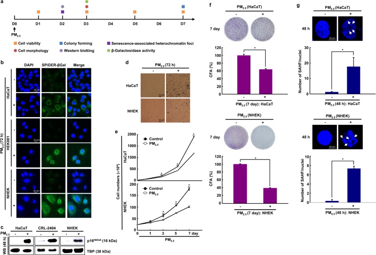
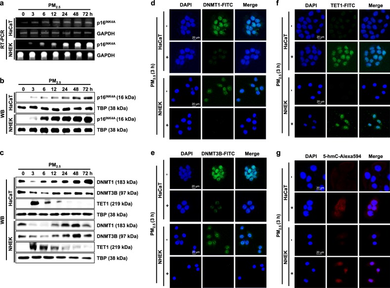
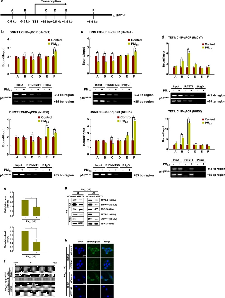
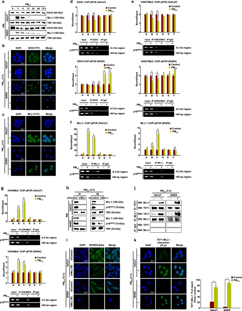
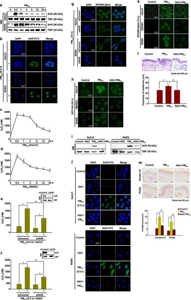
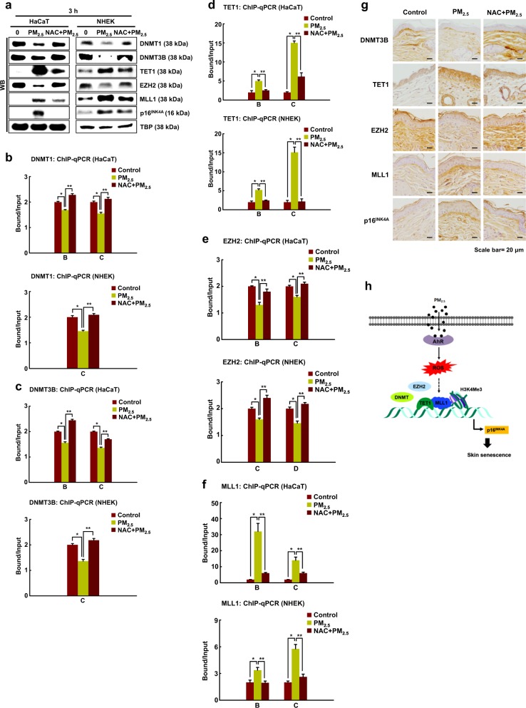
Ambient air particulate matter (PM) induces senescence in human skin cells. However, the underlying mechanisms remain largely unknown. We investigated how epigenetic regulatory mechanisms participate in cellular senescence induced by PM with a diameter <2.5 (PM 2.5) in human keratinocytes and mouse skin tissues. PM 2.5-treated cells exhibited characteristics of cellular senescence. PM 2.5 induced a decrease in DNA methyltransferase (DNMT) expression and an increase in DNA demethylase (ten–eleven translocation; TET) expression, leading to hypomethylation of the p16INK4A promoter region. In addition, PM 2.5 led to a decrease in polycomb EZH2 histone methyltransferase expression, whereas the expression of the epigenetic transcriptional activator MLL1 increased. Furthermore, binding of DNMT1, DNMT3B, and EZH2 to the promoter region of p16INK4A decreased in PM 2.5-treated keratinocytes, whereas TET1 and MLL1 binding increased, leading to decreased histone H3 lysine 27 trimethylation (H3K27Me3) and increased H3K4Me3 in the promoter of p16INK4A. PM 2.5-induced senescence involved aryl hydrocarbon receptor (AhR)-induced reactive oxygen species (ROS) production. ROS scavenging dampened PM 2.5-induced cellular senescence through regulation of DNA and histone methylation. Altogether, our work shows that skin senescence induced by environmental PM 2.5 occurs through ROS-dependent the epigenetic modification of senescence-associated gene expression. Our findings provide information for the design of preventive and therapeutic strategies against skin senescence, particularly in light of the increasing problem of PM 2.5 exposure due to air pollution.
Overview
The claims and statistics of this paper haven’t been checked yet.
Run the check — free, no account needed. It reads the open-access text and checks the sources it cites.
Integrity signals
Registry
Cites
Statistics
Figures
Verification
Claims the paper makes
The claims and statistics of this paper haven’t been checked yet.
Run the check — free, no account needed. It reads the open-access text and checks the sources it cites.
Claims other papers make about this one
Each is checked against this paper’s own text: does it say what the citing paper says it does?
The cited source says what the claim attributes to it.
In the citing paper
“A previous study has also shown that PM2.5 induces cellular senescence in a ROS-dependent manner29.”
From Air Pollution and Skin Diseases (2025) · cites this paper as [29]
Self-check
Percentages, sums and reported differences re-computed from the paper’s own numbers.
The self-check needs the paper’s full text.
Citations
Cites
Methylation-specific PCR: a novel PCR assay for methylation status of CpG islands.
Herman, J. G., Graff, J. R., Myöhänen, S., Nelkin, B. D., & Baylin, S. B. (1996) · Proceedings of the National Academy of Sciences · doi:10.1073/pnas.93.18.9821 · Cited by 5,634
Clearance of p16Ink4a-positive senescent cells delays ageing-associated disorders
Baker, D. J., Wijshake, T., Tchkonia, T., LeBrasseur, N. K., Childs, B. G., van de Sluis, B., Kirkland, J. L., & van Deursen, J. M. (2011) · Nature · doi:10.1038/nature10600 · Cited by 3,670
Tet proteins can convert 5-methylcytosine to 5-formylcytosine and 5-carboxylcytosine
Ito, S., Shen, L., Dai, Q., Wu, S. C., Collins, L. B., Swenberg, J. A., He, C., & Zhang, Y. (2011) · Science · doi:10.1126/science.1210597 · Cited by 3,451
Senescence in Health and Disease
He, S., & Sharpless, N. E. (2017) · Cell · doi:10.1016/j.cell.2017.05.015 · Cited by 1,766
ROS, Cell Senescence, and Novel Molecular Mechanisms in Aging and Age‐Related Diseases
Davalli, P., Mitic, T., Caporali, A., Lauriola, A., & D’Arca, D. (2016) · Oxidative Medicine and Cellular Longevity · doi:10.1155/2016/3565127 · Cited by 1,059
Kim, K. E., Cho, D., & Park, H. J. (2016) · Life Sciences · doi:10.1016/j.lfs.2016.03.039 · Cited by 455
Implications of Genetic and Epigenetic Alterations of CDKN2A (p16 INK4a ) in Cancer
Zhao, R., Choi, B. Y., Lee, M.-H., Bode, A. M., & Dong, Z. (2016) · EBioMedicine · doi:10.1016/j.ebiom.2016.04.017 · Cited by 425
Piao, M. J., Ahn, M. J., Kang, K. A., Ryu, Y. S., Hyun, Y. J., Shilnikova, K., Zhen, A. X., Jeong, J. W., Choi, Y. H., Kang, H. K., et al. (2018) · Archives of Toxicology · doi:10.1007/s00204-018-2197-9 · Cited by 339
Self-citationPollution and skin: from epidemiological and mechanistic studies to clinical implications.
Krutmann, J., Liu, W., Li, L., Pan, X., Crawford, M., Sore, G., & Seite, S. (2014) · Journal of Dermatological Science · doi:10.1016/j.jdermsci.2014.08.008 · Cited by 290
SWI/SNF Mediates Polycomb Eviction and Epigenetic Reprogramming of the INK4b-ARF-INK4a Locus
Kia, S. K., Gorski, M. M., Giannakopoulos, S., & Verrijzer, C. P. (2008) · Molecular and Cellular Biology · doi:10.1128/mcb.02019-07 · Cited by 269
Birch-Machin, M. A., & Bowman, A. (2016) · British Journal of Dermatology · doi:10.1111/bjd.14906 · Cited by 225
Lee, C.-W., Lin, Z.-C., Hu, S. C.-S., Chiang, Y.-C., Hsu, L.-F., Lin, Y.-C., Lee, I.-T., Tsai, M.-H., & Fang, J.-Y. (2016) · Scientific Reports · doi:10.1038/srep27995 · Cited by 188
Detection of Senescence-Associated Heterochromatin Foci (SAHF)
Aird, K. M., & Zhang, R. (2012) · Methods in molecular biology · doi:10.1007/978-1-62703-239-1_12 · Cited by 185
A KRAS-directed transcriptional silencing pathway that mediates the CpG island methylator phenotype
Serra, R. W., Fang, M., Park, S. M., Hutchinson, L., & Green, M. R. (2014) · eLife · doi:10.7554/elife.02313 · Cited by 172
Pan, T.-L., Wang, P.-W., Aljuffali, I. A., Huang, C.-T., Lee, C.-W., & Fang, J.-Y. (2015) · Journal of Dermatological Science · doi:10.1016/j.jdermsci.2015.01.011 · Cited by 158
Ryu, Y. S., Kang, K. A., Piao, M. J., Ahn, M. J., Yi, J. M., Hyun, Y., Kim, S. H., Ko, M. K., Park, C. O., & Hyun, J. W. (2018) · Redox Biology · doi:10.1016/j.redox.2018.101080 · Cited by 155
Self-citationSon, J.-Y., Lee, J.-T., Kim, K., Jung, K., & Bell, M. L. (2012) · Environmental Health Perspectives · doi:10.1289/ehp.1104316 · Cited by 154
Zhou, W. J., Tian, D., He, J., Wang, Y., Zhang, L., Cui, L., Li, J., Zhang, L., Li, L., Shu, Y., et al. (2016) · Oncotarget · doi:10.18632/oncotarget.7842 · Cited by 126
McGarvey, K. M., Greene, E., Fahrner, J. A., Jenuwein, T., & Baylin, S. B. (2007) · Cancer Research · doi:10.1158/0008-5472.can-06-2029 · Cited by 116
Cellular Mechanisms of Oxidative Stress and Action in Melanoma
Venza, M., Visalli, M., Beninati, C., Gaetano, G. V. D., Teti, D. V., & Venza, I. (2015) · Oxidative Medicine and Cellular Longevity · doi:10.1155/2015/481782 · Cited by 110
Reversing wrinkled skin and hair loss in mice by restoring mitochondrial function
Singh, B., Schoeb, T. R., Bajpai, P., Slominski, A. T., & Singh, K. K. (2018) · Cell Death and Disease · doi:10.1038/s41419-018-0765-9 · Cited by 109
Costa, C., Catania, S., Pasquale, R. D., Stancanelli, R., Scribano, G. M., & Melchini, A. (2010) · Toxicology · doi:10.1016/j.tox.2010.02.014 · Cited by 105
Soberanes, S., Gonzalez, A., Urich, D., Chiarella, S. E., Radigan, K. A., Osornio-Vargas, Á. R., Joseph, J., Kalyanaraman, B., Ridge, K. M., Chandel, N. S., et al. (2012) · Scientific Reports · doi:10.1038/srep00275 · Cited by 101
Gao, Z.-X., Song, X., Li, S.-S., Lai, X.-R., Yang, Y., Yang, G., Li, Z., Cui, Y., & Pan, H. (2016) · Investigative Ophthalmology & Visual Science · doi:10.1167/iovs.15-18839 · Cited by 86
Zhou, B. R., Wang, X., Li, F., Wang, Y., Yang, L., Zhen, X., & Tan, W. (2017) · Molecular Medicine Reports · doi:10.3892/mmr.2017.6580 · Cited by 79
The Role of p16INK4a Pathway in Human Epidermal Stem Cell Self-Renewal, Aging and Cancer
D’Arcangelo, D., Tinaburri, L., & Dellambra, E. (2017) · International Journal of Molecular Sciences · doi:10.3390/ijms18071591 · Cited by 77
Zhao, L., Zhang, Y., Gao, Y., Geng, P., Yang, L., Liu, X., Yao, R., Hou, P., Liu, D., Lu, J., et al. (2015) · Cell Death and Differentiation · doi:10.1038/cdd.2015.6 · Cited by 71
DDB1-CUL4 and MLL1 Mediate Oncogene-Induced p16INK4a Activation
Kotake, Y., Zeng, Y., & Xiong, Y. (2009) · Cancer Research · doi:10.1158/0008-5472.can-08-2739 · Cited by 69
Kang, K. A., Piao, M. J., Ryu, Y. S., Kang, H. K., Chang, W. Y., Keum, Y. S., & Hyun, J. W. (2016) · Oncotarget · doi:10.18632/oncotarget.9745 · Cited by 59
Self-citationQiao, Y., Li, Q., Du, H.-Y., Wang, Q.-W., Huang, Y., & Liu, W. (2017) · Biochemical and Biophysical Research Communications · doi:10.1016/j.bbrc.2017.04.160 · Cited by 53
Transcriptional Regulation of the p16 Tumor Suppressor Gene.
Kotake, Y., Naemura, M., Murasaki, C., Inoue, Y., & Okamoto, H. (2015) · PubMed · Cited by 51
Gonçalves, A. C., Cortesão, E., Oliveiros, B., Alves, V., Espadana, A. I., Rito, L., Magalhães, E., Pereira, S., Pereira, A., Costa, N., et al. (2015) · Clinical and Experimental Medicine · doi:10.1007/s10238-015-0357-2 · Cited by 31
DNA Methylation Patterns in Ulcerative Colitis-Associated Cancer: A Systematic Review
Emmett, R. A., Davidson, K. L., Gould, N. J., & Arasaradnam, R. P. (2017) · Epigenomics · doi:10.2217/epi-2017-0025 · Cited by 29
Atorvastatin treatment modulates p16 promoter methylation to regulate p16 expression
Zhu, B., Gong, Y., Yan, G., Wang, D., Wang, Q., Qiao, Y., Hou, J., Liu, B., & Tang, C. (2017) · FEBS Journal · doi:10.1111/febs.14087 · Cited by 27
Bartsch, N., Heidler, J., Vieth, B., Hutzler, C., & Luch, A. (2016) · Journal of Occupational and Environmental Hygiene · doi:10.1080/15459624.2016.1200724 · Cited by 25
Increased P16 DNA Methylation in Mouse Thymic Lymphoma Induced by Irradiation
Song, W., Liu, Y., Liu, Y., Zhang, C., Yuan, B., Zhang, L., & Sun, S. (2014) · PLoS ONE · doi:10.1371/journal.pone.0093850 · Cited by 22
Nikolić, N., Aničić, B., Čarkić, J., Simonović, J., Toljić, B., Tanić, N., Tepavčević, Z., Vukadinović, M., Konstantinović, V. S., & Milašin, J. (2015) · Archives of Oral Biology · doi:10.1016/j.archoralbio.2015.08.011 · Cited by 21
Han, Y., Wang, X.-B., Cui, N., Zhang, S., Wang, C., & Zheng, F. (2016) · Oncotarget · doi:10.18632/oncotarget.12202 · Cited by 15
Abiko, Y., Lin, F.-Y., Lee, H., Puga, A., & Kumagai, Y. (2016) · The Journal of Toxicological Sciences · doi:10.2131/jts.41.775 · Cited by 12
Atorvastatin treatment modulates p16 promoter methylation to regulate p16 expression
Boqian, Zhu, Yaoyao, Gong, M., Gaoliang, Yan, Dong, Wang, Qingjie, Yong, et al. (2017) · FEBS Journal · Cited by 1
Cited by
Sorted by citations
Skin Aging, Cellular Senescence and Natural Polyphenols
Csekes, E., & Račková, L. (2021) · International Journal of Molecular Sciences · doi:10.3390/ijms222312641 · Cited by 352
Skin Senescence: Mechanisms and Impact on Whole-Body Aging
Franco, A. C., Aveleira, C., & Cavadas, C. (2022) · Trends in Molecular Medicine · doi:10.1016/j.molmed.2021.12.003 · Cited by 321
Molecular mechanisms of environmental exposures and human disease
Wu, H., Eckhardt, C. M., & Baccarelli, A. (2023) · Nature Reviews Genetics · doi:10.1038/s41576-022-00569-3 · Cited by 296
Environmentally-Induced (Extrinsic) Skin Aging: Exposomal Factors and Underlying Mechanisms
Krutmann, J., Schikowski, T., Morita, A., & Berneburg, M. (2021) · Journal of Investigative Dermatology · doi:10.1016/j.jid.2020.12.011 · Cited by 253
Liu, H., Cheng, M.-Y., Xun, M., Zhao, Z.-W., Zhang, Y., Tang, W., Cheng, J. J., Ni, J., & Wang, W. (2023) · International Journal of Molecular Sciences · doi:10.3390/ijms24043755 · Cited by 252
Metadata
- Authors
- Ryu, Y. S., Kang, K. A., Piao, M. J., Ahn, M. J., Yi, J. M., Bossis, G., Hyun, Y.-M., Park, C. O., & Hyun, J. W.
- Affiliations
- Jeju National University (KR)
- Inje University (KR)
- Centre National de la Recherche Scientifique (FR)
- Yonsei University (KR)
- Corresponding author
- Hyun, Jin Won
- ORCID
- Venue
- Experimental & Molecular Medicine
- Published
- 2019-09
- Volume
- 51
- Issue
- 9
- Pages
- 1-14
- ISSN
- 1226-3613 (ISSN-L)
- 2092-6413
- PMC
- PMC6802667
- OpenAlex
- W2976086758
- Cited by
- 119
- References
- 40
- Citations by year
- 2026: 10 · 2025: 21 · 2024: 18 · 2023: 14 · 2022: 20 · 2021: 20 · 2020: 16
- FWCI
- 4.63
- Type
- journal article
- Language
- en
- Open access
- gold
- doi.org
- Licence
- Locations
- Experimental & Molecular Medicine (publishedVersion, cc-by)
- PubMed Central (publishedVersion, cc-by)
- PubMed (publishedVersion)
- HAL (submittedVersion, cc-by)
- YUHSpace (Yonsei University Medical Library) (submittedVersion, cc-by-nc-nd)
- Funders
- National Research Foundation of Korea (2017R1A4A1014512)
- Topics
- Life Sciences › Biochemistry, Genetics and Molecular Biology › Molecular Biology › Epigenetics and DNA Methylation
- Life Sciences › Biochemistry, Genetics and Molecular Biology › Cell Biology › Telomeres, Telomerase, and Senescence
- Health Sciences › Medicine › Dermatology › Skin Protection and Aging
- Keywords
- particulate matter, PM2.5, cellular senescence, keratinocytes, epigenetic modifications, DNA methylation, histone methylation, aryl hydrocarbon receptor, reactive oxygen species, oxidative stress, EZH2, MLL1, TET1, air pollution, skin aging
- MeSH terms
- Enhancer of Zeste Homolog 2 Protein, DNA (Cytosine-5-)-Methyltransferase 1, DNA Methyltransferase 3B, Animals, DNA (Cytosine-5-)-Methyltransferases, DNA-Binding Proteins, Humans, Promoter Regions, Genetic, Histone-Lysine N-Methyltransferase, Proto-Oncogene Proteins, Skin, Keratinocytes, Cellular Senescence, Reactive Oxygen Species, Receptors, Aryl Hydrocarbon, Oxidative Stress, DNA Methylation, Cyclin-Dependent Kinase Inhibitor p16, Epigenesis, Genetic, Cell Proliferation, Mice, Myeloid-Lymphoid Leukemia Protein, Particulate Matter
About these checks
Registry, retraction, Cites and statistics results are computed, not judged, with one exception: a printed reference that no index lists is read once by a model before it counts as a citation error. Claims are extracted by a model and kept only when their quote appears verbatim in the text; each cited source is then checked, with a model’s help, for whether it says what the claim relies on. Those verdicts can be wrong — read the claim’s quote and the cited source before relying on one.
Statistics and arithmetic are recomputed, not judged.
Confidence bands are shown only for fields calibrated on our annotated test set. None is calibrated yet, so no band is shown.
What counts as a claim ›How we measure confidence ›How we check this ›
Are you an author, or do you see a problem with this page? Request a review
