

The DOI matches its Crossref record; 183 of the 194 references it cites were found in a registry or an index; 123 of the 205 sources its claims cite were checked for support.
Prefrontal cortex, amygdala, and threat processing: implications for PTSD
Open accessAbstract
Posttraumatic stress disorder can be viewed as a disorder of fear dysregulation. An abundance of research suggests that the prefrontal cortex is central to fear processing—that is, how fears are acquired and strategies to regulate or diminish fear responses. The current review covers foundational research on threat or fear acquisition and extinction in nonhuman animals, healthy humans, and patients with posttraumatic stress disorder, through the lens of the involvement of the prefrontal cortex in these processes. Research harnessing advances in technology to further probe the role of the prefrontal cortex in these processes, such as the use of optogenetics in rodents and brain stimulation in humans, will be highlighted, as well other fear regulation approaches that are relevant to the treatment of posttraumatic stress disorder and involve the prefrontal cortex, namely cognitive regulation and avoidance/active coping. Despite the large body of translational research, many questions remain unanswered and posttraumatic stress disorder remains difficult to treat. We conclude by outlining future research directions related to the role of the prefrontal cortex in fear processing and implications for the treatment of posttraumatic stress disorder.
Overview
CoChat Summary
## TL;DR
This is a narrative review of evidence linking prefrontal cortex circuits with threat learning, extinction, avoidance, and emotion regulation across rodents, healthy humans, and PTSD. The authors argue that altered PFC regulation of amygdala and related subcortical systems helps explain PTSD and may point to better treatments.
## Key findings
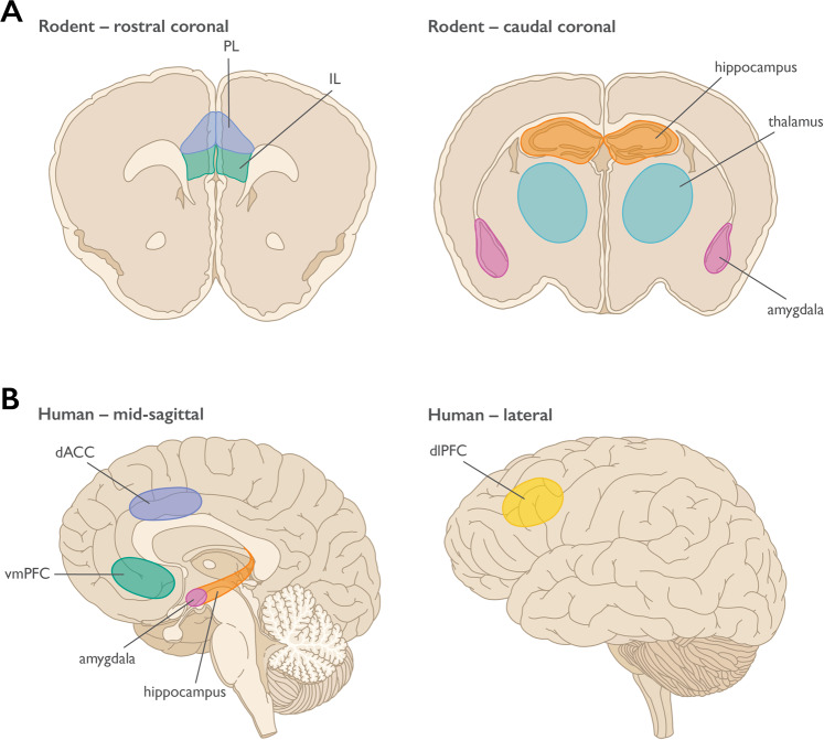
- Rodent work supports a functional distinction in mPFC subregions: PL tends to promote threat expression, while IL supports threat extinction; the review notes this is “extensive, although debated.”
- In humans, vmPFC activity tracks extinction: vmPFC BOLD increases during extinction learning and recall, and vmPFC thickness is positively correlated with extinction recall and learning.
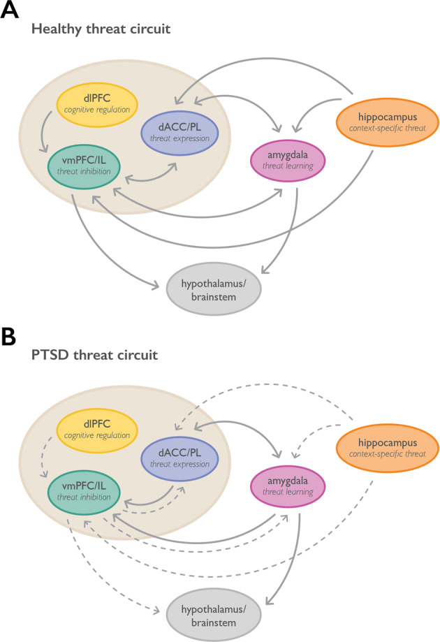
- In PTSD, multiple imaging studies show altered PFC function, including decreased vmPFC activation and increased dACC activation during extinction recall; one cited twin study of 26 male identical twin pairs found diminished mPFC activation during stressful versus neutral script imagery in PTSD.
- Meta-analyses of emotion regulation show that cognitive reappraisal is associated with increased BOLD in dlPFC, vlPFC, and dmPFC, and often decreased amygdala signal, consistent with PFC modulation of threat responses.
## Methods
- Narrative review synthesizing findings from nonhuman animal research, healthy human studies, and PTSD studies on threat learning, extinction, avoidance/active coping, emotion regulation, and stress effects.
- Integrates evidence from lesion, pharmacologic, optogenetic, chemogenetic, fMRI, DTI, meta-analytic, and brain-stimulation studies; the paper does not report a single new sample or experiment.
- Main analysis is qualitative cross-species comparison of PFC subregions and their interactions with the amygdala, hippocampus, and thalamus in threat-related behaviors and PTSD.
## Limitations
- The paper states that many questions remain unanswered and that PTSD remains difficult to treat.
- It notes that much of the human imaging literature has limited sample sizes and that MRI/fMRI evidence is often correlational rather than causal.
- It also points out technical limits of human amygdala fMRI, including coarse spatial resolution that makes discrete subnuclear responses hard to detect; this is a limitation made evident by the text.
Model-written summary. Check it against the paper before relying on it.
Integrity signals
Registry
Cites
Statistics
Figures
Verification
Claims the paper makes
98 Valid · 6 Not valid · 45 Not checked · 30 Own results and interpretations
Some cited sources support this claim and some don’t — read each source’s verdict below.
In the paper
“There is consistent evidence of increases in BOLD signal in the vmPFC during extinction learning [88–90] and recall [89–92] (for review, see [93–95]).”
§ Prefrontal cortex, amygdala, and threat processing: implications for PTSD > Preclinical human threat processing research > Threat extinction
What the sources say
- Not checked — no full text of the sourcecites [88] Gottfried JA, Dolan RJ. Human orbitofrontal cortex mediates extinction learning while accessing conditioned representations of value. Nat Neurosci. 2004;7:1144–52. source
- Not checked — no full text of the sourcecites [89] Milad MR, Wright CI, Orr SP, Pitman RK, Quirk GJ, Rauch SL. Recall of fear extinction in humans activates the ventromedial prefrontal cortex and hippocampus in concert. Biol Psychiatry. 2007;62:446–54. source
- Disputedcites [90] Phelps EA, Delgado MR, Nearing KI, LeDoux JE. Extinction learning in humans: role of the amygdala and vmPFC. Neuron. 2004;43:897–905. sourceExperimental — accuracy not yet measured
The Results (page unavailable) report that subgenual vmPFC BOLD signal to the CS+ was below baseline during both extinction sessions, including the day-2 retention test. The Discussion (page unavailable) says this depression diminished as extinction progressed and that vmPFC responses predicted retention; it does not report the claimed BOLD-signal increase during learning and recall. (LLM, conf=0.86)
Checked by a reasoning model — may be wrong.
- Supportedcites [91] Kalisch R, Korenfeld E, Stephan KE, Weiskopf N, Seymour B, Dolan RJ. Context-dependent human extinction memory is mediated by a ventromedial prefrontal and hippocampal network. J Neurosci. 2006;26:9503–11. source
The Results section (‘VMPFC and anterior hippocampus activation during recall of extinction memory’) reports significantly greater left vmPFC activation in the extinction context during recall on day 2. This paper supports the recall portion of the sentence, where its citation is attached; the extinction-learning portion is cited separately. (LLM, conf=0.92)
- Supportedcites [92] Lonsdorf TB, Haaker J, Kalisch R. Long-term expression of human contextual fear and extinction memories involves amygdala, hippocampus and ventromedial prefrontal cortex: a reinstatement study in two independent samples. Soc Cogn Affect Neurosci. 2014;9:1973–83. source
The Context conditioning results (page not available in our copy) report greater vmPFC hemodynamic responses during the Day 8 recall test in both independent samples. This supports the recall portion of the citing sentence, where this source is cited; the source is not being cited for extinction learning. (LLM, conf=0.90)
boostedstates a cause- Asserted: passes — The authors put it forward as true.
- Checkable: passes — It can be checked against evidence.
- Load-bearing: passes — The paper’s conclusions depend on it.
- Based: passes — The paper gives a reason why it holds.
At least one cited source doesn’t support this claim or points to a different paper, and none confirmed it — see the verdicts below.
In the paper
“Additionally, in a prospective emergency department study following patients longitudinally, Stevens et al. [163] used an unmasked fearful faces design to demonstrate that dorsal ACC activity during presentation of fearful faces predicts ongoing symptom maintenance in the aftermath of acute trauma in civilians.”
§ Prefrontal cortex, amygdala, and threat processing: implications for PTSD > Role of PFC in PTSD > Functional imaging in PFC and PTSD
What the sources say
- Disputedcites [163] Stevens JS, Kim YJ, Galatzer-Levy IR, Reddy R, Ely TD, Nemeroff CB, et al. Amygdala reactivity and anterior cingulate habituation predict posttraumatic stress disorder symptom maintenance after acute civilian trauma. Biol Psychiatry. 2017;81:1023–9. sourceExperimental — accuracy not yet measured
The Results (Associations between fMRI activation and symptom trajectories) state that dACC habituation to fearful stimuli was not associated with symptom slope, and ACC reactivity did not predict it either. The study did use fearful-face stimuli in a longitudinal civilian-trauma cohort, but it was vACC habituation—not dACC activity—that predicted a flatter symptom-recovery slope. (LLM, conf=0.92)
Checked by a reasoning model — may be wrong.
boostedstates an association- Asserted: passes — The authors put it forward as true.
- Checkable: passes — It can be checked against evidence.
- Load-bearing: passes — The paper’s conclusions depend on it.
- Based: passes — The paper gives a reason why it holds.
- Disputed
Every source the paper cites for this claim was checked and supports it.
In the paper
“Additionally, inhibition of PCAF in IL was shown to interfere with threat extinction [62].”
§ Prefrontal cortex, amygdala, and threat processing: implications for PTSD > Nonhuman animal research on threat processing > Molecular pathways in rodent mPFC
What the sources say
- Supportedcites [62] Wei W, Coelho CM, Li X, Marek R, Yan S, Anderson S, et al. p300/CBP-associated factor selectively regulates the extinction of conditioned fear. J Neurosci. 2012;32:11930–41. source
The Results section ('PCAF activity is required for fear extinction but not for fear acquisition') reports that infusing a PCAF inhibitor into the infralimbic prefrontal cortex of mice eliminated the reduction in freezing after extinction training. The authors conclude that PCAF activity there is required for consolidation of fear extinction memory. (LLM, conf=0.94)
plainstates a cause- Asserted: passes — The authors put it forward as true.
- Checkable: passes — It can be checked against evidence.
- Load-bearing: passes — The paper’s conclusions depend on it.
- Based: passes — The paper gives a reason why it holds.
- Supported
Every source the paper cites for this claim was checked and supports it.
In the paper
“These findings suggest that alterations in m6dA deposition are necessary for the formation of threat extinction memories within IL [63].”
§ Prefrontal cortex, amygdala, and threat processing: implications for PTSD > Nonhuman animal research on threat processing > Molecular pathways in rodent mPFC
What the sources say
- Supportedcites [63] Li X, Zhao Q, Wei W, Lin Q, Magnan C, Emami MR, et al. The DNA modification N6-methyl-2’-deoxyadenosine (m6dA) drives activity-induced gene expression and is required for fear extinction. Nat Neurosci. 2019;22:534–44. source
The Results section reports that N6amt1 knockdown in the infralimbic prefrontal cortex (ILPFC) significantly impaired fear extinction memory, while knockdown in the adjacent prelimbic region did not. It concludes that learning-induced m6dA accumulation is critically involved in forming that memory. No page numbers are provided in the supplied passages. (LLM, conf=0.93)
hedgedstates a cause- Asserted: passes — The authors put it forward as true.
- Checkable: passes — It can be checked against evidence.
- Load-bearing: passes — The paper’s conclusions depend on it.
- Based: passes — The paper gives a reason why it holds.
- Supported
Every source the paper cites for this claim was checked and supports it.
In the paper
“Threat conditioning and threat recall are associated with unique, cell-type-specific transcriptional changes that persist for weeks after initial training [57].”
§ Prefrontal cortex, amygdala, and threat processing: implications for PTSD > Nonhuman animal research on threat processing > Molecular pathways in rodent mPFC
What the sources say
- Supportedcites [57] Chen MB, Jiang X, Quake SR, Südhof TC. Persistent transcriptional programmes are associated with remote memory. Nature. 2020;587:437–42. source
The Discussion says fear experience can induce long-lasting gene-expression changes, while recall induces new transcriptional programmes in a different set of neurons. The Memory-associated gene expression section reports subtype-specific programmes, and the study tested recall 16 days after conditioning. Page numbers were not provided in our copy. (LLM, conf=0.92)
plainstates an association- Asserted: passes — The authors put it forward as true.
- Checkable: passes — It can be checked against evidence.
- Load-bearing: unclear — It is unclear whether the paper’s conclusions depend on it.
- Based: passes — The paper gives a reason why it holds.
- Supported
112 more claims · 64 with their cited sources checked
Create a free account to see all 179 claims and to verify the 18 references still open.
- It may state two separate claims as one. 57
- Probably not a substantial claim. 9
Citations
Cites
Emotion regulation: current status and future prospects
Gross, J. J. (2015) · Psychological Inquiry · doi:10.1080/1047840x.2014.940781 · Cited by 5,229 · cited 1×
“Emotion regulation Although extinction and active coping can be investigated across species, humans have the unique ability to use cognitive strategies to alter emotional responses, such as responses to fear provoking stimuli (for review, [117]).”
BackgroundBinder, J. R., Desai, R. H., Graves, W. W., & Conant, L. L. (2009) · Cerebral Cortex · doi:10.1093/cercor/bhp055 · Cited by 4,060
Domingos da Silveira da Luz, A. C., Pereira Dias, G., do Nascimento Bevilaqua, M. C., Cocks, G., Gardino, P. F., Thuret, S., & Nardi, A. E. (2013) · Neuropsychobiology · doi:10.1159/000353269 · Cited by 3,995
Meeting of minds: the medial frontal cortex and social cognition
Amodio, D. M., & Frith, C. D. (2006) · Nature Reviews Neuroscience · doi:10.1038/nrn1884 · Cited by 3,782
Azevedo, R. S. S., de Sousa, J. R., Araujo, M. T. F., Martins Filho, A. J., de Alcantara, B. N., Araujo, F. M. C., Queiroz, M. G. L., Cruz, A. C. R., Vasconcelos, B. H. B., Chiang, J. O., et al. (2018) · Scientific Reports · doi:10.1038/s41598-017-17765-5 · Cited by 3,478
Stress signalling pathways that impair prefrontal cortex structure and function
Arnsten, A. F. T. (2009) · Nature Reviews Neuroscience · doi:10.1038/nrn2648 · Cited by 3,299 · cited 2×
“These changes are proposed to be due to the impact of increased catecholamines, in particular noradrenergic and dopaminergic signaling, on PFC neuronal activity with even relatively mild acute stress exposure [64, 65, 67].”
BackgroundDifferential contribution of amygdala and hippocampus to cued and contextual fear conditioning.
Phillips, R. G., & LeDoux, J. E. (1992) · Behavioral Neuroscience · doi:10.1037/0735-7044.106.2.274 · Cited by 3,292 · cited 1×
“Lesion studies of the hippocampus suggest a critical role in context processing [39].”
BackgroundContributions of the Amygdala to Emotion Processing: From Animal Models to Human Behavior
Phelps, E. A., & LeDoux, J. E. (2005) · Neuron · doi:10.1016/j.neuron.2005.09.025 · Cited by 3,203
Self-citationOngur, D. (2000) · Cerebral Cortex · doi:10.1093/cercor/10.3.206 · Cited by 2,627
The Neurocircuitry of Fear, Stress, and Anxiety Disorders
Shin, L. M., & Liberzon, I. (2010) · Neuropsychopharmacology · doi:10.1038/npp.2009.83 · Cited by 2,083
Cited by
Post-traumatic stress disorder: clinical and translational neuroscience from cells to circuits
Ressler, K. J., Berretta, S., Bolshakov, V. Y., Rosso, I. M., Meloni, E. G., Rauch, S. L., & Carlezon, W. A. (2022) · Nature Reviews Neurology · doi:10.1038/s41582-022-00635-8 · Cited by 481
Self-citationPrecision psychiatry roadmap: towards a biology-informed framework for mental disorders
Kas, M. J., Penninx, B. W. J. H., Knudsen, G. M., Cuthbert, B. N., Falkai, P., Sachs, G. S., Ressler, K. J., Bałkowiec-Iskra, E., Butlen‐Ducuing, F., Leboyer, M., et al. (2025) · Molecular Psychiatry · doi:10.1038/s41380-025-03070-5 · Cited by 116
Self-citationTherapeutic mechanisms of psychedelics and entactogens
Heifets, B. D., & Olson, D. E. (2023) · Neuropsychopharmacology · doi:10.1038/s41386-023-01666-5 · Cited by 79
“Mechanistic preclinical studies have mostly focused on the NAc, involved in prosocial and reward-related behavior [150], and the basolateral amygdala, involved in fear learning and PTSD [151] (Fig.”
BackgroundWen, Z., Raio, C. M., Pace-Schott, E. F., Lazar, S. W., LeDoux, J. E., Phelps, E. A., & Milad, M. R. (2022) · Proceedings of the National Academy of Sciences · doi:10.1073/pnas.2204066119 · Cited by 79
Self-citationHosseini‐Kamkar, N., Farahani, M. V., Nikolic, M., Stewart, K., Goldsmith, S. F., Soltaninejad, M., Rajabli, R., Lowe, C. J., Nicholson, A. A., Morton, J. B., et al. (2023) · JAMA Network Open · doi:10.1001/jamanetworkopen.2023.40018 · Cited by 83
Metadata
- Authors
- Alexandra Kredlow, M., Fenster, R. J., Laurent, E. S., Ressler, K. J., & Phelps, E. A.
- Affiliations
- Harvard University (US)
- Corresponding author
- Ressler, Kerry J.
- Phelps, Elizabeth A.
- Venue
- Neuropsychopharmacology
- Published
- 2021-09-20
- Volume
- 47
- Issue
- 1
- Pages
- 247-259
- ISSN
- 0893-133X (ISSN-L)
- 1740-634X
- PMC
- PMC8617299
- OpenAlex
- W3200528712
- Cited by
- 346
- References
- 194
- Citations by year
- 2026: 83 · 2025: 107 · 2024: 66 · 2023: 60 · 2022: 26 · 2021: 4
- FWCI
- 5.19
- Type
- journal article
- Language
- en
- Open access
- hybrid
- doi.org
- Licence
- cc-by
- creativecommons.org/licenses/by/4.0 (tdm)
- creativecommons.org/licenses/by/4.0 (vor)
- Locations
- Neuropsychopharmacology (publishedVersion, cc-by)
- PubMed Central (publishedVersion, cc-by)
- PubMed (publishedVersion)
- Funders
- National Institutes of Health
- National Institute of Mental Health (P50MH115874, R01MH117292, R01MH108665)
- National Institute on Drug Abuse (R01DA042855)
- Topics
- Social Sciences › Psychology › Clinical Psychology › Posttraumatic Stress Disorder Research
- Life Sciences › Neuroscience › Cognitive Neuroscience › Memory and Neural Mechanisms
- Life Sciences › Neuroscience › Cognitive Neuroscience › Neural and Behavioral Psychology Studies
- Keywords
- post-traumatic stress disorder, prefrontal cortex, amygdala, threat processing, fear acquisition, fear extinction, fear regulation, optogenetics, brain stimulation, cognitive regulation, avoidance, active coping
- MeSH terms
- Amygdala, Animals, Extinction, Psychological, Fear, Humans, Stress Disorders, Post-Traumatic, Prefrontal Cortex
About these checks
Registry, retraction, Cites and statistics results are computed, not judged, with one exception: a printed reference that no index lists is read once by a model before it counts as a citation error. Claims are extracted by a model and kept only when their quote appears verbatim in the text; each cited source is then checked, with a model’s help, for whether it says what the claim relies on. Those verdicts can be wrong — read the claim’s quote and the cited source before relying on one.
The claim verdicts this report counts are experimental: their accuracy is not yet measured.
A language model drew the claims from the paper on 2026-10-09, under version 3 of the claim definition (model deepseek-v4.1-flash, prompt extract-v3).
A second model labelled each claim on 2026-10-09 (model jev-1.13-20260917, definition revision gold-guideline-v0.2).
Cited sources are checked with a language model’s help (gpt-6-sol).
Statistics and arithmetic are recomputed, not judged.
Confidence bands are shown only for fields calibrated on our annotated test set. None is calibrated yet, so no band is shown.
What counts as a claim ›How we measure confidence ›How we check this ›
Are you an author, or do you see a problem with this page? Request a review
How to cite
Cite CoChat for the checks on this page, and the paper for its own findings.
- This page
- CoChat. (2026). Citation check: Prefrontal cortex, amygdala, and threat processing: implications for PTSD. https://app.cochat.ai/papers/10.1038/s41386-021-01155-7
- The paper
- Alexandra Kredlow, M., Fenster, R. J., Laurent, E. S., Ressler, K. J., & Phelps, E. A. (2022). Prefrontal cortex, amygdala, and threat processing: implications for PTSD. Neuropsychopharmacology. https://doi.org/10.1038/s41386-021-01155-7
Data sources
Bibliographic data: Semantic Scholar, OpenAlex, Crossref.
Contains information from the Semantic Scholar Academic Graph Datasets, made available under the ODC Attribution License (ODC-BY 1.0).
Semantic Scholar’s data is described in: Kinney, R., et al. (2023). The Semantic Scholar Open Data Platform. arXiv:2301.10140.
