

Prefrontal cortex, amygdala, and threat processing: implications for PTSD
Open accessThe DOI matches its Crossref record; 183 of the 194 references it cites were found in a registry or an index; 123 of the 205 sources its claims cite were checked for support.
Abstract
Posttraumatic stress disorder can be viewed as a disorder of fear dysregulation. An abundance of research suggests that the prefrontal cortex is central to fear processing—that is, how fears are acquired and strategies to regulate or diminish fear responses. The current review covers foundational research on threat or fear acquisition and extinction in nonhuman animals, healthy humans, and patients with posttraumatic stress disorder, through the lens of the involvement of the prefrontal cortex in these processes. Research harnessing advances in technology to further probe the role of the prefrontal cortex in these processes, such as the use of optogenetics in rodents and brain stimulation in humans, will be highlighted, as well other fear regulation approaches that are relevant to the treatment of posttraumatic stress disorder and involve the prefrontal cortex, namely cognitive regulation and avoidance/active coping. Despite the large body of translational research, many questions remain unanswered and posttraumatic stress disorder remains difficult to treat. We conclude by outlining future research directions related to the role of the prefrontal cortex in fear processing and implications for the treatment of posttraumatic stress disorder.
Overview
CoChat Summary
## TL;DR
This is a narrative review of evidence linking prefrontal cortex circuits with threat learning, extinction, avoidance, and emotion regulation across rodents, healthy humans, and PTSD. The authors argue that altered PFC regulation of amygdala and related subcortical systems helps explain PTSD and may point to better treatments.
## Key findings
- Rodent work supports a functional distinction in mPFC subregions: PL tends to promote threat expression, while IL supports threat extinction; the review notes this is “extensive, although debated.”
- In humans, vmPFC activity tracks extinction: vmPFC BOLD increases during extinction learning and recall, and vmPFC thickness is positively correlated with extinction recall and learning.
- In PTSD, multiple imaging studies show altered PFC function, including decreased vmPFC activation and increased dACC activation during extinction recall; one cited twin study of 26 male identical twin pairs found diminished mPFC activation during stressful versus neutral script imagery in PTSD.
- Meta-analyses of emotion regulation show that cognitive reappraisal is associated with increased BOLD in dlPFC, vlPFC, and dmPFC, and often decreased amygdala signal, consistent with PFC modulation of threat responses.
## Methods
- Narrative review synthesizing findings from nonhuman animal research, healthy human studies, and PTSD studies on threat learning, extinction, avoidance/active coping, emotion regulation, and stress effects.
- Integrates evidence from lesion, pharmacologic, optogenetic, chemogenetic, fMRI, DTI, meta-analytic, and brain-stimulation studies; the paper does not report a single new sample or experiment.
- Main analysis is qualitative cross-species comparison of PFC subregions and their interactions with the amygdala, hippocampus, and thalamus in threat-related behaviors and PTSD.
## Limitations
- The paper states that many questions remain unanswered and that PTSD remains difficult to treat.
- It notes that much of the human imaging literature has limited sample sizes and that MRI/fMRI evidence is often correlational rather than causal.
- It also points out technical limits of human amygdala fMRI, including coarse spatial resolution that makes discrete subnuclear responses hard to detect; this is a limitation made evident by the text.
Model-written summary. Check it against the paper before relying on it.
Integrity signals
Registry
Cites
Statistics
Figures
Verification
Claims the paper makes
98 Valid · 6 Not valid · 45 Not checked · 30 Own results and interpretations
Some cited sources support this claim and some don’t — read each source’s verdict below.
In the paper
“There is consistent evidence of increases in BOLD signal in the vmPFC during extinction learning [88–90] and recall [89–92] (for review, see [93–95]).”
§ Prefrontal cortex, amygdala, and threat processing: implications for PTSD > Preclinical human threat processing research > Threat extinction
What the sources say
- Not checked — no full text of the sourcecites [88] Gottfried JA Dolan RJ Human orbitofrontal cortex mediates extinction learning while accessing conditioned representations of value Nat Neurosci 2004 7 1144 52 15361879 source
- Not checked — no full text of the sourcecites [89] Milad MR Wright CI Orr SP Pitman RK Quirk GJ Rauch SL Recall of fear extinction in humans activates the ventromedial prefrontal cortex and hippocampus in concert Biol Psychiatry 2007 62 446 54 17217927 source
- Disputedcites [90] Phelps EA Delgado MR Nearing KI LeDoux JE Extinction learning in humans: role of the amygdala and vmPFC Neuron. 2004 43 897 905 15363399 sourceExperimental — accuracy not yet measured
The Results (page unavailable) report that subgenual vmPFC BOLD signal to the CS+ was below baseline during both extinction sessions, including the day-2 retention test. The Discussion (page unavailable) says this depression diminished as extinction progressed and that vmPFC responses predicted retention; it does not report the claimed BOLD-signal increase during learning and recall. (LLM, conf=0.86)
Checked by a reasoning model — may be wrong.
- Supportedcites [91] Kalisch R Korenfeld E Stephan KE Weiskopf N Seymour B Dolan RJ Context-dependent human extinction memory is mediated by a ventromedial prefrontal and hippocampal network J Neurosci 2006 26 9503 11 16971534 PMC2634865 source
The Results section (‘VMPFC and anterior hippocampus activation during recall of extinction memory’) reports significantly greater left vmPFC activation in the extinction context during recall on day 2. This paper supports the recall portion of the sentence, where its citation is attached; the extinction-learning portion is cited separately. (LLM, conf=0.92)
- Supportedcites [92] Lonsdorf TB Haaker J Kalisch R Long-term expression of human contextual fear and extinction memories involves amygdala, hippocampus and ventromedial prefrontal cortex: a reinstatement study in two independent samples Soc Cogn Affect Neurosci 2014 9 1973 83 24493848 PMC4249485 source
The Context conditioning results (page not available in our copy) report greater vmPFC hemodynamic responses during the Day 8 recall test in both independent samples. This supports the recall portion of the citing sentence, where this source is cited; the source is not being cited for extinction learning. (LLM, conf=0.90)
boostedstates a cause- Asserted: passes — The authors put it forward as true.
- Checkable: passes — It can be checked against evidence.
- Load-bearing: passes — The paper’s conclusions depend on it.
- Based: passes — The paper gives a reason why it holds.
At least one cited source doesn’t support this claim or points to a different paper, and none confirmed it — see the verdicts below.
In the paper
“Additionally, in a prospective emergency department study following patients longitudinally, Stevens et al. [163] used an unmasked fearful faces design to demonstrate that dorsal ACC activity during presentation of fearful faces predicts ongoing symptom maintenance in the aftermath of acute trauma in civilians.”
§ Prefrontal cortex, amygdala, and threat processing: implications for PTSD > Role of PFC in PTSD > Functional imaging in PFC and PTSD
What the sources say
- Disputedcites [163] Stevens JS Kim YJ Galatzer-Levy IR Reddy R Ely TD Nemeroff CB Amygdala reactivity and anterior cingulate habituation predict posttraumatic stress disorder symptom maintenance after acute civilian trauma Biol Psychiatry 2017 81 1023 9 28117048 PMC5449257 sourceExperimental — accuracy not yet measured
The Results (Associations between fMRI activation and symptom trajectories) state that dACC habituation to fearful stimuli was not associated with symptom slope, and ACC reactivity did not predict it either. The study did use fearful-face stimuli in a longitudinal civilian-trauma cohort, but it was vACC habituation—not dACC activity—that predicted a flatter symptom-recovery slope. (LLM, conf=0.92)
Checked by a reasoning model — may be wrong.
boostedstates an association- Asserted: passes — The authors put it forward as true.
- Checkable: passes — It can be checked against evidence.
- Load-bearing: passes — The paper’s conclusions depend on it.
- Based: passes — The paper gives a reason why it holds.
- Disputed
Every source the paper cites for this claim was checked and supports it.
In the paper
“Additionally, inhibition of PCAF in IL was shown to interfere with threat extinction [62].”
§ Prefrontal cortex, amygdala, and threat processing: implications for PTSD > Nonhuman animal research on threat processing > Molecular pathways in rodent mPFC
What the sources say
- Supportedcites [62] Wei W Coelho CM Li X Marek R Yan S Anderson S p300/CBP-associated factor selectively regulates the extinction of conditioned fear J Neurosci 2012 32 11930 41 22933779 PMC3466419 source
The Results section ('PCAF activity is required for fear extinction but not for fear acquisition') reports that infusing a PCAF inhibitor into the infralimbic prefrontal cortex of mice eliminated the reduction in freezing after extinction training. The authors conclude that PCAF activity there is required for consolidation of fear extinction memory. (LLM, conf=0.94)
plainstates a cause- Asserted: passes — The authors put it forward as true.
- Checkable: passes — It can be checked against evidence.
- Load-bearing: passes — The paper’s conclusions depend on it.
- Based: passes — The paper gives a reason why it holds.
- Supported
Every source the paper cites for this claim was checked and supports it.
In the paper
“These findings suggest that alterations in m6dA deposition are necessary for the formation of threat extinction memories within IL [63].”
§ Prefrontal cortex, amygdala, and threat processing: implications for PTSD > Nonhuman animal research on threat processing > Molecular pathways in rodent mPFC
What the sources say
- Supportedcites [63] Li X Zhao Q Wei W Lin Q Magnan C Emami MR The DNA modification N6-methyl-2’-deoxyadenosine (m6dA) drives activity-induced gene expression and is required for fear extinction Nat Neurosci 2019 22 534 44 30778148 PMC6462436 source
The Results section reports that N6amt1 knockdown in the infralimbic prefrontal cortex (ILPFC) significantly impaired fear extinction memory, while knockdown in the adjacent prelimbic region did not. It concludes that learning-induced m6dA accumulation is critically involved in forming that memory. No page numbers are provided in the supplied passages. (LLM, conf=0.93)
hedgedstates a cause- Asserted: passes — The authors put it forward as true.
- Checkable: passes — It can be checked against evidence.
- Load-bearing: passes — The paper’s conclusions depend on it.
- Based: passes — The paper gives a reason why it holds.
- Supported
Every source the paper cites for this claim was checked and supports it.
In the paper
“Threat conditioning and threat recall are associated with unique, cell-type-specific transcriptional changes that persist for weeks after initial training [57].”
§ Prefrontal cortex, amygdala, and threat processing: implications for PTSD > Nonhuman animal research on threat processing > Molecular pathways in rodent mPFC
What the sources say
- Supportedcites [57] Chen MB Jiang X Quake SR Südhof TC Persistent transcriptional programmes are associated with remote memory Nature. 2020 587 437 42 33177708 PMC9097329 source
The Discussion says fear experience can induce long-lasting gene-expression changes, while recall induces new transcriptional programmes in a different set of neurons. The Memory-associated gene expression section reports subtype-specific programmes, and the study tested recall 16 days after conditioning. Page numbers were not provided in our copy. (LLM, conf=0.92)
plainstates an association- Asserted: passes — The authors put it forward as true.
- Checkable: passes — It can be checked against evidence.
- Load-bearing: unclear — It is unclear whether the paper’s conclusions depend on it.
- Based: passes — The paper gives a reason why it holds.
- Supported
112 more claims · 64 with their cited sources checked
Create a free account to see all 179 claims and to verify the 18 references still open.
- It may state two separate claims as one. 57
- Probably not a substantial claim. 9
Citations
Cites
Emotion regulation: current status and future prospects
Gross, J. J. (2015) · Psychological Inquiry · doi:10.1080/1047840x.2014.940781 · Cited by 5,229 · cited 1×
“Emotion regulation Although extinction and active coping can be investigated across species, humans have the unique ability to use cognitive strategies to alter emotional responses, such as responses to fear provoking stimuli (for review, [117]).”
BackgroundBinder, J. R., Desai, R. H., Graves, W. W., & Conant, L. L. (2009) · Cerebral Cortex · doi:10.1093/cercor/bhp055 · Cited by 4,060
Domingos da Silveira da Luz, A. C., Pereira Dias, G., do Nascimento Bevilaqua, M. C., Cocks, G., Gardino, P. F., Thuret, S., & Nardi, A. E. (2013) · Neuropsychobiology · doi:10.1159/000353269 · Cited by 3,995
Meeting of minds: the medial frontal cortex and social cognition
Amodio, D. M., & Frith, C. D. (2006) · Nature Reviews Neuroscience · doi:10.1038/nrn1884 · Cited by 3,782
Azevedo, R. S. S., de Sousa, J. R., Araujo, M. T. F., Martins Filho, A. J., de Alcantara, B. N., Araujo, F. M. C., Queiroz, M. G. L., Cruz, A. C. R., Vasconcelos, B. H. B., Chiang, J. O., et al. (2018) · Scientific Reports · doi:10.1038/s41598-017-17765-5 · Cited by 3,478
Stress signalling pathways that impair prefrontal cortex structure and function
Arnsten, A. F. T. (2009) · Nature Reviews Neuroscience · doi:10.1038/nrn2648 · Cited by 3,299 · cited 2×
“These changes are proposed to be due to the impact of increased catecholamines, in particular noradrenergic and dopaminergic signaling, on PFC neuronal activity with even relatively mild acute stress exposure [64, 65, 67].”
BackgroundDifferential contribution of amygdala and hippocampus to cued and contextual fear conditioning.
Phillips, R. G., & LeDoux, J. E. (1992) · Behavioral Neuroscience · doi:10.1037/0735-7044.106.2.274 · Cited by 3,292 · cited 1×
“Lesion studies of the hippocampus suggest a critical role in context processing [39].”
BackgroundContributions of the Amygdala to Emotion Processing: From Animal Models to Human Behavior
Phelps, E. A., & LeDoux, J. E. (2005) · Neuron · doi:10.1016/j.neuron.2005.09.025 · Cited by 3,203
Self-citationOngur, D. (2000) · Cerebral Cortex · doi:10.1093/cercor/10.3.206 · Cited by 2,627
The Neurocircuitry of Fear, Stress, and Anxiety Disorders
Shin, L. M., & Liberzon, I. (2010) · Neuropsychopharmacology · doi:10.1038/npp.2009.83 · Cited by 2,083
The prefontral cortex and cognitive control
Miller, E. K. (2000) · Nature Reviews Neuroscience · doi:10.1038/35036228 · Cited by 2,016
Cognitive Reappraisal of Emotion: A Meta-Analysis of Human Neuroimaging Studies
Buhle, J. T., Silvers, J. A., Wager, T. D., Lopez, R., Onyemekwu, C., Kober, H., Weber, J., & Ochsner, K. N. (2014) · Cerebral Cortex · doi:10.1093/cercor/bht154 · Cited by 1,896 · cited 7×
“[121] meta-analysis involved cognitive reappraisal of negativelyvalenced photos.”
BackgroundResultInfluentialNeuroimaging studies of working memory:
Wager, T. D., & Smith, E. E. (2003) · Cognitive, Affective, & Behavioral Neuroscience · doi:10.3758/cabn.3.4.255 · Cited by 1,889
Phelps, E. A., Delgado, M. R., Nearing, K. I., & LeDoux, J. E. (2004) · Neuron · doi:10.1016/j.neuron.2004.08.042 · Cited by 1,753 · cited 8×
“Specifically, greater thickness was associated with smaller SCR to the conditioned stimulus during extinction recall, suggestive of better extinction recall (see also [97]).”
BackgroundResultInfluentialSelf-citationLeDoux, J., Iwata, J., Cicchetti, P., & Reis, D. (1988) · The Journal of Neuroscience · doi:10.1523/jneurosci.08-07-02517.1988 · Cited by 1,580 · cited 1×
“For thirty years, the amygdala has been implicated in both threat learning [48] and threat extinction [49] processes.”
BackgroundDifferential projections of the infralimbic and prelimbic cortex in the rat
Vertes, R. P. (2004) · Synapse · doi:10.1002/syn.10279 · Cited by 1,550 · cited 2×
“Although there is some overlap in projection patterns, IL projects most heavily to lateral septum, bed nucleus of the stria terminalis, amygdala, hypothalamus, and brainstem, while PL sends more projections to insular cortex, nucleus accumbens, thalamus, and raphe nuclei [29].”
BackgroundFear Extinction as a Model for Translational Neuroscience: Ten Years of Progress
Milad, M. R., & Quirk, G. J. (2012) · Annual Review of Psychology · doi:10.1146/annurev.psych.121208.131631 · Cited by 1,466 · cited 1×
“The vmPFC is proposed to be the homologue for the IL in rodents [87] and serves to inhibit threat responses produced by the amygdala.”
BackgroundLaBar, K. S., Gatenby, J. C., Gore, J. C., LeDoux, J. E., & Phelps, E. A. (1998) · Neuron · doi:10.1016/s0896-6273(00)80475-4 · Cited by 1,402 · cited 2×
“Consistent with these early patient studies, functional magnetic resonance imaging (fMRI) studies soon followed that showed increased blood oxygenation level dependent (BOLD) signal in the amygdala to a conditioned stimulus (relative to stimulus never paired with shock) [75, 76], and the magnitude of this BOLD respons…”
BackgroundResultSelf-citationBechara, A., Tranel, D., Damasio, H., Adolphs, R., Rockland, C., & Damasio, A. R. (1995) · Science · doi:10.1126/science.7652558 · Cited by 1,400
Rauch, S. L., Shin, L. M., & Phelps, E. A. (2006) · Biological Psychiatry · doi:10.1016/j.biopsych.2006.06.004 · Cited by 1,397
Self-citationNeurobiological Basis of Failure to Recall Extinction Memory in Posttraumatic Stress Disorder
Milad, M. R., Pitman, R. K., Ellis, C. B., Gold, A. L., Shin, L. M., Lasko, N. B., Zeidan, M. A., Handwerger, K., Orr, S. P., & Rauch, S. L. (2009) · Biological Psychiatry · doi:10.1016/j.biopsych.2009.06.026 · Cited by 1,339 · cited 1×
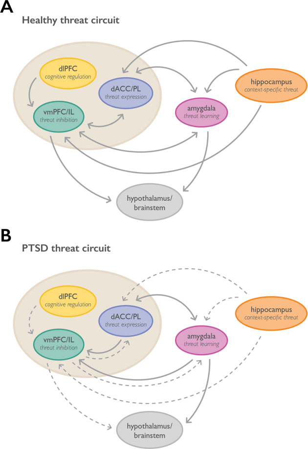
“They found that during extinction recall there was decreased activation of vmPFC and greater activation in dACC in PTSD relative to healthy trauma-exposed participants, which was associated with impaired physiological measures of extinction recall [165].”
BackgroundMilad, M. R., Wright, C. I., Orr, S. P., Pitman, R. K., Quirk, G. J., & Rauch, S. L. (2007) · Biological Psychiatry · doi:10.1016/j.biopsych.2006.10.011 · Cited by 1,197 · cited 10×
“Connectivity analyses have demonstrated functional coupling between the mPFC and amygdala during extinction learning [107], and vmPFC and amygdala during extinction recall [89, 108].”
BackgroundResultInfluentialSierra-Mercado, D., Padilla-Coreano, N., & Quirk, G. J. (2011) · Neuropsychopharmacology · doi:10.1038/npp.2010.184 · Cited by 1,190 · cited 1×
“Pharmacological inactivation of PL and IL with agents such as the GABA agonist muscimol further suggested opposing roles for these structures in threat conditioning and threat extinction, respectively [33].”
BackgroundJohnstone, T., van Reekum, C. M., Urry, H. L., Kalin, N. H., & Davidson, R. J. (2007) · The Journal of Neuroscience · doi:10.1523/jneurosci.2063-07.2007 · Cited by 1,051 · cited 1×
“For example, the nature of vmPFC involvement during cognitive reappraisal has been observed to vary as a function of psychiatric symptoms [138, 139].”
BackgroundUsing Neuroscience to Help Understand Fear and Anxiety: A Two-System Framework
LeDoux, J. E., & Pine, D. S. (2016) · American Journal of Psychiatry · doi:10.1176/appi.ajp.2016.16030353 · Cited by 1,035 · cited 1×
“However, in line with the two-system view of fear and anxiety [17] and in order to not make assumptions about emotional states, the term “threat” will be used when referring to the behavioral, psychophysiological, or neural outcomes of conditioning research.”
BackgroundSimmonds, D. J., Pekar, J. J., & Mostofsky, S. H. (2008) · Neuropsychologia · doi:10.1016/j.neuropsychologia.2007.07.015 · Cited by 1,015 · cited 1×
“The vlPFC is hypothesized to support choosing and inhibiting appraisals of stimuli [121, 124, 125] or potentially may signal salience and the need to reappraise [122].”
BackgroundThe role of ventromedial prefrontal cortex in the recovery of extinguished fear
Quirk, G. J., Russo, G. K., Barron, J. L., & Lebron, K. (2000) · The Journal of Neuroscience · doi:10.1523/jneurosci.20-16-06225.2000 · Cited by 957 · cited 1×
“supported this result when they [32] performed electrolytic lesions of the rat vmPFC and assessed threat extinction memory.”
ResultNeural network of cognitive emotion regulation—an ALE meta-analysis and MACM analysis
Kohn, N., Eickhoff, S. B., Scheller, M., Laird, A. R., Fox, P. T., & Habel, U. (2014) · NeuroImage · doi:10.1016/j.neuroimage.2013.11.001 · Cited by 956 · cited 3×
“The vlPFC is hypothesized to support choosing and inhibiting appraisals of stimuli [121, 124, 125] or potentially may signal salience and the need to reappraise [122].”
BackgroundResultBrain systems mediating aversive conditioning: an event-related fMRI study
Büchel, C., Morris, J., Dolan, R. J., & Friston, K. J. (1998) · Neuron · doi:10.1016/s0896-6273(00)80476-6 · Cited by 951 · cited 1×
“Consistent with these early patient studies, functional magnetic resonance imaging (fMRI) studies soon followed that showed increased blood oxygenation level dependent (BOLD) signal in the amygdala to a conditioned stimulus (relative to stimulus never paired with shock) [75, 76], and the magnitude of this BOLD respons…”
ResultExtinction of emotional learning: contribution of medial prefrontal cortex
Morgan, M. A., Romanski, L. M., & LeDoux, J. E. (1993) · Neuroscience Letters · doi:10.1016/0304-3940(93)90241-c · Cited by 897
Activation of the left amygdala to a cognitive representation of fear
Phelps, E. A., O'Connor, K. J., Gatenby, J. C., Gore, J. C., Grillon, C., & Davis, M. (2001) · Nature Neuroscience · doi:10.1038/86110 · Cited by 895 · cited 1×
“For instructed learning, the amygdala BOLD response is left-lateralized [83], and only patients with left, but not right, amygdala damage show impaired physiological evidence of threat learning, perhaps because of the verbally mediated nature of this learning [84].”
BackgroundSelf-citationHeidbreder, C. A., & Groenewegen, H. J. (2003) · Neuroscience & Biobehavioral Reviews · doi:10.1016/j.neubiorev.2003.09.003 · Cited by 882 · cited 2×
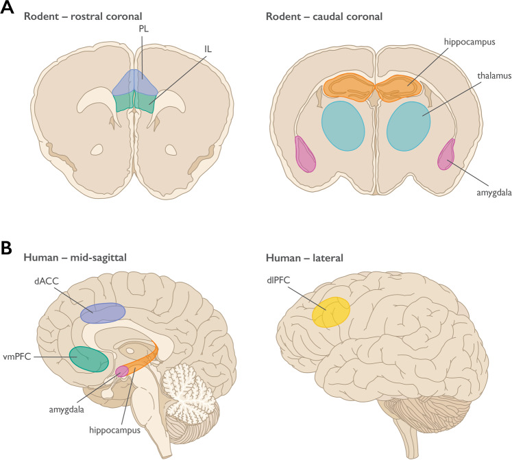
“The rodent mPFC is generally considered to consist of the medial precentral area (Fr2), the dorsal anterior cingulate cortex (dACC), prelimbic cortex (PL), and infralimbic cortex (IL) [24] (see Fig.”
BackgroundPrefrontal–hippocampal interactions in episodic memory
Eichenbaum, H. (2017) · Nature Reviews Neuroscience · doi:10.1038/nrn.2017.74 · Cited by 870 · cited 1×
“The vHPC sends dense direct projections to the mPFC from CA1, but also bidirectional disynaptic indirect connections to the mPFC through the reuniens nucleus of the thalamus and the perirhinal cortex [38].”
BackgroundGhashghaei, H. T., Hilgetag, C. C., & Barbas, H. (2007) · NeuroImage · doi:10.1016/j.neuroimage.2006.09.046 · Cited by 845
Ghashghaei, H. T., & Barbas, H. (2002) · Neuroscience · doi:10.1016/s0306-4522(02)00446-3 · Cited by 785
Response Variation following Trauma: A Translational Neuroscience Approach to Understanding PTSD
Yehuda, R., & LeDoux, J. (2007) · Neuron · doi:10.1016/j.neuron.2007.09.006 · Cited by 783
Fear and the brain: where have we been, and where are we going?
LeDoux, J. (1998) · Biological Psychiatry · doi:10.1016/s0006-3223(98)00282-0 · Cited by 782
Morgan, M. A., & LeDoux, J. E. (1995) · Behavioral Neuroscience · doi:10.1037/0735-7044.109.4.681 · Cited by 717 · cited 1×
“A follow-up study demonstrated that more dorsal areas of the mPFC affected threat learning, while more ventral mPFC was required for threat extinction [31].”
BackgroundNeural circuitry underlying the regulation of conditioned fear and its relation to extinction
Delgado, M. R., Nearing, K. I., LeDoux, J. E., & Phelps, E. A. (2008) · Neuron · doi:10.1016/j.neuron.2008.06.029 · Cited by 716 · cited 4×
“[131] involved cognitive reappraisal of threat conditioned stimuli, whereas most of the studies included in the Buhle et al.”
BackgroundMethodResultInfluentialSelf-citationDifferential Control of Learning and Anxiety along the Dorsoventral Axis of the Dentate Gyrus
Kheirbek, M. A., Drew, L. J., Burghardt, N. S., Costantini, D. O., Tannenholz, L., Ahmari, S. E., Zeng, H., Fenton, A. A., & Hen, R. (2013) · Neuron · doi:10.1016/j.neuron.2012.12.038 · Cited by 713 · cited 1×
“The hippocampus itself appears to have a dorsal-ventral functional gradient, with the dorsal hippocampus encoding context more specifically, while the ventral hippocampus (vHPC) includes affective information as well [37].”
BackgroundNeural signatures of human fear conditioning: an updated and extended meta-analysis of fMRI studies
Fullana, M. A., Harrison, B. J., Soriano-Mas, C., Vervliet, B., Cardoner, N., Àvila-Parcet, A., & Radua, J. (2016) · Molecular Psychiatry · doi:10.1038/mp.2015.88 · Cited by 702 · cited 1×
“The challenges of using BOLD imaging to study the human amygdala is reflected in recent meta-analyses of fMRI threat learning studies, which fail to find BOLD changes in the amygdala [78], in spite of its critical role in threat learning in rodent models and patient studies.”
BackgroundNievergelt, C. M., Maihofer, A. X., Klengel, T., Atkinson, E. G., Chen, C.-Y., Choi, K. W., Coleman, J. R. I., Dalvie, S., Duncan, L. E., Gelernter, J., et al. (2019) · Nature Communications · doi:10.1038/s41467-019-12576-w · Cited by 635
Self-citationA Neural Circuit for Memory Specificity and Generalization
Xu, W., & Südhof, T. C. (2013) · Science · doi:10.1126/science.1229534 · Cited by 634 · cited 1×
“The reuniens nucleus of the thalamus coordinates oscillatory synchrony between the mPFC and the vHPC, which is necessary for proper contextual representation of threat memories [46, 53].”
BackgroundImpaired fear conditioning following unilateral temporal lobectomy in humans
LaBar, K., LeDoux, J., Spencer, D., & Phelps, E. (1995) · The Journal of Neuroscience · doi:10.1523/jneurosci.15-10-06846.1995 · Cited by 631 · cited 1×
“Relative to healthy controls, both bilateral [72] and unilateral [73] amygdala damage resulted in impaired conditioned responses, as measured by the skin conductance response (SCR).”
BackgroundSelf-citationOlsson, A., & Phelps, E. A. (2007) · Nature Neuroscience · doi:10.1038/nn1968 · Cited by 622 · cited 1×
“Consistent with Pavlovian threat conditioning, fMRI studies of both instructed and observational threat learning show activation in the amygdala, dACC, and insula [82].”
BackgroundSelf-citationBredy, T. W., Wu, H., Crego, C., Zellhoefer, J., Sun, Y. E., & Barad, M. (2007) · Learning & Memory · doi:10.1101/lm.500907 · Cited by 548 · cited 1×
“In the IL, threat extinction is associated with acetylation of histones near the Bdnf locus [61], changes to the p300/ CBP complex (PCAF) [62], as well as deposition of DNA-modification marks such as 5-hydroxymethylcytosine and N6-methyl-2’deoxyadenosine (m6dA) near loci of activity-dependent genes such as Bdnf.”
BackgroundLong-range connectivity defines behavioral specificity of amygdala neurons
Senn, V., Wolff, S. B. E., Herry, C., Grenier, F., Ehrlich, I., Gründemann, J., Fadok, J. P., Müller, C., Letzkus, J. J., & Lüthi, A. (2014) · Neuron · doi:10.1016/j.neuron.2013.11.006 · Cited by 522 · cited 1×
“There is evidence to suggest that there is a dorso-ventral topographic segregation of BLA input to the mPFC; more dorsal projections (to PL) encode threatstimulating information while more ventral projections (to IL) encode threat extinction-related information [51].”
BackgroundThe Role of the Medial Prefrontal Cortex in the Conditioning and Extinction of Fear
Giustino, T. F., & Maren, S. (2015) · Frontiers in Behavioral Neuroscience · doi:10.3389/fnbeh.2015.00298 · Cited by 519 · cited 2×
“Decades of basic research on the mPFC in rodents indicate that it plays a key role in the expression and storage of the Pavlovian threat response and the establishment of threat-related extinction memories [21].”
BackgroundA meta-analysis of D-cycloserine and the facilitation of fear extinction and exposure therapy
Norberg, M. M., Krystal, J. H., & Tolin, D. F. (2008) · Biological Psychiatry · doi:10.1016/j.biopsych.2008.01.012 · Cited by 510 · cited 1×
“From rodent research showing that intra-amygdala infusion of the NMDA receptor agonist d-cycloserine, which enhances NMDAdependent plasticity, facilitates extinction learning, and successful translation of this work to humans (see [104] for review), we know that the human amygdala plays a role in extinction learning.”
BackgroundProjections from neocortex mediate top-down control of memory retrieval
Rajasethupathy, P., Sankaran, S., Marshel, J. H., Kim, C. K., Ferenczi, E., Lee, S. Y., Berndt, A., Ramakrishnan, C., Jaffe, A., Lo, M., et al. (2015) · Nature · doi:10.1038/nature15389 · Cited by 509 · cited 1×
“In addition, there may be more routes of information flow from the PFC to the hippocampus, including direct routes from the nearby anterior cingulate [47].”
BackgroundKalisch, R., Korenfeld, E., Stephan, K. E., Weiskopf, N., Seymour, B., & Dolan, R. J. (2006) · The Journal of Neuroscience · doi:10.1523/jneurosci.2021-06.2006 · Cited by 493 · cited 1×
“Brain imaging studies of the contextual modulation of extinction typically manipulate the visual background during extinction and report hippocampal activation during extinction recall [89, 91, 92].”
BackgroundA temporal shift in the circuits mediating retrieval of fear memory
Do-Monte, F. H., Quiñones-Laracuente, K., & Quirk, G. J. (2015) · Nature · doi:10.1038/nature14030 · Cited by 491 · cited 1×
“The paraventricular nucleus of the thalamus plays a crucial role in the encoding of threat memories over time [54] and appears to be necessary in shifting the temporal nature of how the mPFC encodes threat memories [55].”
BackgroundA Role for the Human Dorsal Anterior Cingulate Cortex in Fear Expression
Milad, M. R., Quirk, G. J., Pitman, R. K., Orr, S. P., Fischl, B., & Rauch, S. L. (2007) · Biological Psychiatry · doi:10.1016/j.biopsych.2007.04.032 · Cited by 485 · cited 2×
“The dACC is a prefrontal region that is proposed to be the human homolog of the PL cortex in rodents [80].”
BackgroundConsolidation of Fear Extinction Requires Protein Synthesis in the Medial Prefrontal Cortex
Santini, E., Ge, H., Ren, K., de Ortiz, S. P., & Quirk, G. J. (2004) · The Journal of Neuroscience · doi:10.1523/jneurosci.0786-04.2004 · Cited by 479 · cited 1×
“This suggests that translation of new protein is necessary for the formation of a novel threat extinction memory [56].”
BackgroundInduction of Fear Extinction with Hippocampal-Infralimbic BDNF
Peters, J., Dieppa-Perea, L. M., Melendez, L. M., & Quirk, G. J. (2010) · Science · doi:10.1126/science.1186909 · Cited by 462 · cited 1×
“Expression of Bdnf in PL is necessary for consolidation of cued threat conditioning [59], while infusion of Bdnf into IL after threat acquisition is sufficient to diminish threat responses in the absence of extinction training [60].”
BackgroundIzquierdo, A., Wellman, C. L., & Holmes, A. (2006) · The Journal of Neuroscience · doi:10.1523/jneurosci.0474-06.2006 · Cited by 459 · cited 2×
“Studies in animal models have shown that acute stress leads to changes in neuronal signaling that impair function in the dlPFC [64] and IL cortex [66].”
BackgroundMorawetz, C., Bode, S., Derntl, B., & Heekeren, H. R. (2017) · Neuroscience & Biobehavioral Reviews · doi:10.1016/j.neubiorev.2016.11.014 · Cited by 458 · cited 1×
“The most recent meta-analysis of fMRI studies of emotion regulation [118] found that all strategies aimed at downregulating negative emotions were collectively associated with increased BOLD signal in the following areas: ventrolateral prefrontal cortex (vlPFC), dorsolateral prefrontal cortex (dlPFC), and dorsomedial…”
BackgroundHippocampal Inactivation Disrupts Contextual Retrieval of Fear Memory after Extinction
Corcoran, K. A., & Maren, S. (2001) · The Journal of Neuroscience · doi:10.1523/jneurosci.21-05-01720.2001 · Cited by 455 · cited 2×
“, [40]), research suggests that the hippocampus is involved in contextual modulation of extinction and works in concert with the PFC during contextual extinction learning.”
BackgroundEmotion regulation: quantitative meta-analysis of functional activation and deactivation
Frank, D. W., Dewitt, M., Hudgens-Haney, M., Schaeffer, D. J., Ball, B. H., Schwarz, N. F., Hussein, A. A., Smart, L. M., & Sabatinelli, D. (2014) · Neuroscience & Biobehavioral Reviews · doi:10.1016/j.neubiorev.2014.06.010 · Cited by 454
Two different lateral amygdala cell populations contribute to the initiation and storage of memory
Repa, J. C., Muller, J., Apergis, J., Desrochers, T. M., Zhou, Y., & LeDoux, J. E. (2001) · Nature Neuroscience · doi:10.1038/89512 · Cited by 439 · cited 1×
“However, there is electrophysiological evidence in rodents showing that a subset of lateral nucleus amygdala neurons respond preferentially during initial learning [77], and there is greater responding overall at this time.”
BackgroundHow the Neurocircuitry and Genetics of Fear Inhibition May Inform Our Understanding of PTSD
Jovanovic, T., & Ressler, K. J. (2010) · American Journal of Psychiatry · doi:10.1176/appi.ajp.2009.09071074 · Cited by 437
Self-citationDuncan, L. E., Ratanatharathorn, A., Aiello, A. E., Almli, L. M., Amstadter, A. B., Ashley-Koch, A. E., Baker, D. G., Beckham, J. C., Bierut, L. J., Bisson, J., et al. (2018) · Molecular Psychiatry · doi:10.1038/mp.2017.77 · Cited by 425
Self-citationFear is only as deep as the mind allows
Diekhof, E. K., Geier, K., Falkai, P., & Gruber, O. (2011) · NeuroImage · doi:10.1016/j.neuroimage.2011.05.073 · Cited by 413 · cited 2×
“, the dlPFC, vlPFC, and dmPFC) influence the amygdala: (1) that they engage the vmPFC which then modulates the amygdala, similar to the neurocircuitry of extinction [93, 131, 132], and (2) that they modulate lateral temporal areas associated with semantic and perceptual representations, which then indirectly influence…”
BackgroundRobbins, T. W. (2007) · Philosophical Transactions of the Royal Society B: Biological Sciences · doi:10.1098/rstb.2007.2097 · Cited by 413 · cited 1×
“The vlPFC is hypothesized to support choosing and inhibiting appraisals of stimuli [121, 124, 125] or potentially may signal salience and the need to reappraise [122].”
BackgroundThickness of ventromedial prefrontal cortex in humans is correlated with extinction memory
Milad, M. R., Quinn, B. T., Pitman, R. K., Orr, S. P., Fischl, B., & Rauch, S. L. (2005) · Proceedings of the National Academy of Sciences · doi:10.1073/pnas.0502441102 · Cited by 412 · cited 1×
“[96] found vmPFC thickness to be positively correlated with extinction recall.”
BackgroundRevisiting the Role of Infralimbic Cortex in Fear Extinction with Optogenetics
Do-Monte, F. H., Manzano-Nieves, G., Quiñones-Laracuente, K., Ramos-Medina, L., & Quirk, G. J. (2015) · The Journal of Neuroscience · doi:10.1523/jneurosci.3137-14.2015 · Cited by 385 · cited 1×
“These data suggest that IL neurons are necessary for encoding threat extinction memories but may not be necessary for threat extinction memory storage or retention [36].”
BackgroundArnsten, A. F. T., Raskind, M. A., Taylor, F. B., & Connor, D. F. (2015) · Neurobiology of Stress · doi:10.1016/j.ynstr.2014.10.002 · Cited by 381 · cited 1×
“These changes are proposed to be due to the impact of increased catecholamines, in particular noradrenergic and dopaminergic signaling, on PFC neuronal activity with even relatively mild acute stress exposure [64, 65, 67].”
BackgroundHopper, J. W., Frewen, P. A., van der Kolk, B. A., & Lanius, R. A. (2007) · Journal of Traumatic Stress · doi:10.1002/jts.20284 · Cited by 376
Bromet, E., Sonnega, A., & Kessler, R. C. (1998) · American Journal of Epidemiology · doi:10.1093/oxfordjournals.aje.a009457 · Cited by 370
The epidemiology of posttraumatic stress disorder: what is the extent of the problem?
Breslau, N. (2001) · PubMed · Cited by 368
Synaptic Encoding of Fear Extinction in mPFC-amygdala Circuits
Cho, J.-H., Deisseroth, K., & Bolshakov, V. Y. (2013) · Neuron · doi:10.1016/j.neuron.2013.09.025 · Cited by 366 · cited 1×
“However, these results have not been universally reproduced [34, 35].”
Gottfried, J. A., & Dolan, R. J. (2004) · Nature Neuroscience · doi:10.1038/nn1314 · Cited by 353 · cited 1×
“Human imaging studies, however, have been less consistent with finding changes in BOLD signal in the amygdala during extinction [76, 88, 90, 95, 105].”
BackgroundLearning fears by observing others: the neural systems of social fear transmission
Olsson, A., Nearing, K. I., & Phelps, E. A. (2007) · Social Cognitive and Affective Neuroscience · doi:10.1093/scan/nsm005 · Cited by 349 · cited 7×
“Specifically, greater thickness was associated with smaller SCR to the conditioned stimulus during extinction recall, suggestive of better extinction recall (see also [97]).”
BackgroundSelf-citationThe birth, death and resurrection of avoidance: a reconceptualization of a troubled paradigm
LeDoux, J. E., Moscarello, J., Sears, R., & Campese, V (2017) · Molecular Psychiatry · doi:10.1038/mp.2016.166 · Cited by 348 · cited 1×
“However, in order for the animal to produce an avoidance action, conditioned freezing must be inhibited which requires the IL cortex, much like in the expression of extinction (see [111] for a review).”
BackgroundCircuit-Based Corticostriatal Homologies Between Rat and Primate
Heilbronner, S. R., Rodriguez-Romaguera, J., Quirk, G. J., Groenewegen, H. J., & Haber, S. N. (2016) · Biological Psychiatry · doi:10.1016/j.biopsych.2016.05.012 · Cited by 325 · cited 1×
“Cross-species comparisons can be more easily made with respect to connectivity patterns [23].”
BackgroundDörfel, D., Lamke, J.-P., Hummel, F., Wagner, U., Erk, S., & Walter, H. (2014) · NeuroImage · doi:10.1016/j.neuroimage.2014.06.051 · Cited by 322 · cited 1×
“[120] found that some emotion regulation strategies are associated with reduced activity in the amygdala, whereas others are not.”
BackgroundAmygdala inputs to prefrontal cortex guide behavior amid conflicting cues of reward and punishment
Burgos-Robles, A., Kimchi, E. Y., Izadmehr, E. M., Porzenheim, M. J., Ramos-Guasp, W. A., Nieh, E. H., Felix-Ortiz, A. C., Namburi, P., Leppla, C. A., Presbrey, K. N., et al. (2017) · Nature Neuroscience · doi:10.1038/nn.4553 · Cited by 318 · cited 1×
“The BLA sends bidirectional projections to the mPFC [50].”
BackgroundOrsini, C. A., Kim, J. H., Knapska, E., & Maren, S. (2011) · The Journal of Neuroscience · doi:10.1523/jneurosci.4095-11.2011 · Cited by 315 · cited 1×
“Inhibition of double-projecting vHPC neurons to the mPFC and basolateral amygdala (BLA) interferes with contextual threat recall [42] and disconnection of the vHPC from the mPFC interferes with renewal of threat memories, a context-dependent process [43].”
BackgroundAcute and sustained effects of cognitive emotion regulation in major depression
Erk, S., Mikschl, A., Stier, S., Ciaramidaro, A., Gapp, V., Weber, B., & Walter, H. (2010) · The Journal of Neuroscience · doi:10.1523/jneurosci.1856-10.2010 · Cited by 313 · cited 1×
“For example, the nature of vmPFC involvement during cognitive reappraisal has been observed to vary as a function of psychiatric symptoms [138, 139].”
BackgroundDistinct hippocampal engrams control extinction and relapse of fear memory
Lacagnina, A. F., Brockway, E. T., Crovetti, C. R., Shue, F., McCarty, M. J., Sattler, K. P., Lim, S. C., Santos, S. L., Denny, C. A., & Drew, M. R. (2019) · Nature Neuroscience · doi:10.1038/s41593-019-0361-z · Cited by 311 · cited 1×
“Activity-tagging coupled with optogenetic inhibition suggests that threat conditioning and extinction memories exist in separate populations of neurons within the hippocampus [44], and the hippocampus may influence mPFC activity through feed-forward inhibition mechanisms through parvalbumin interneurons [45].”
BackgroundFear extinction in the human brain: A meta-analysis of fMRI studies in healthy participants
Fullana, M. A., Albajes-Eizagirre, A., Soriano-Mas, C., Vervliet, B., Cardoner, N., Benet, O., Radua, J., & Harrison, B. J. (2018) · Neuroscience & Biobehavioral Reviews · doi:10.1016/j.neubiorev.2018.03.002 · Cited by 308 · cited 1×
“Human imaging studies, however, have been less consistent with finding changes in BOLD signal in the amygdala during extinction [76, 88, 90, 95, 105].”
BackgroundCognitive emotion regulation fails the stress test
Raio, C. M., Orederu, T. A., Palazzolo, L., Shurick, A. A., & Phelps, E. A. (2013) · Proceedings of the National Academy of Sciences · doi:10.1073/pnas.1305706110 · Cited by 302 · cited 1×
“In addition, mild acute stress in humans impairs the efficacy of previously acquired cognitive reappraisal strategies in reducing conditioned threat and subjective fear [146] and results in enhanced spontaneous recovery following extinction training [147].”
BackgroundSelf-citationOverlapping neural systems mediating extinction, reversal and regulation of fear
Schiller, D., & Delgado, M. R. (2010) · Trends in Cognitive Sciences · doi:10.1016/j.tics.2010.04.002 · Cited by 294 · cited 2×
“Further, connectivity analyses [131, 132], a metaanalysis conducted by Diekhof et al.”
BackgroundMethodAltered Processing of Contextual Information during Fear Extinction in PTSD: An fMRI Study
Rougemont-Bücking, A., Linnman, C., Zeffiro, T. A., Zeidan, M. A., Lebron-Milad, K., Rodriguez-Romaguera, J., Rauch, S. L., Pitman, R. K., & Milad, M. R. (2011) · CNS Neuroscience & Therapeutics · doi:10.1111/j.1755-5949.2010.00152.x · Cited by 292 · cited 1×
“Increased dACC activation during threat expression and extinction and decreased vmPFC activation with extinction recall were replicated in another study by this group examining context modulation in PTSD [166].”
ResultThe role of social cognition in emotion
Olsson, A., & Ochsner, K. N. (2008) · Trends in Cognitive Sciences · doi:10.1016/j.tics.2007.11.010 · Cited by 289
Corticoamygdala Transfer of Socially Derived Information Gates Observational Learning
Allsop, S. A., Wichmann, R., Mills, F., Burgos-Robles, A., Chang, C.-J., Felix-Ortiz, A. C., Vienne, A., Beyeler, A., Izadmehr, E. M., Glober, G., et al. (2018) · Cell · doi:10.1016/j.cell.2018.04.004 · Cited by 288 · cited 1×
“For the purposes of this review, we will focus on rodent PL and IL, although recent work has implicated dACC in observational threat pathways, which may be relevant to PTSD from witnessed trauma [25].”
Active Avoidance Learning Requires Prefrontal Suppression of Amygdala-Mediated Defensive Reactions
Moscarello, J. M., & LeDoux, J. E. (2013) · The Journal of Neuroscience · doi:10.1523/jneurosci.2596-12.2013 · Cited by 277 · cited 2×
“One benefit of avoidance learning over extinction for controlling threat reactions is that avoidance learning results in a persistent reduction in the passive conditioned response, even when the avoidance action is no longer available [112].”
BackgroundMcLaughlin, K. A., Koenen, K. C., Friedman, M. J., Ruscio, A. M., Karam, E. G., Shahly, V., Stein, D. J., Hill, E. D., Petukhova, M., Alonso, J., et al. (2015) · Biological Psychiatry · doi:10.1016/j.biopsych.2014.03.028 · Cited by 275
Krystal, J. H., Davis, L. L., Neylan, T. C., A. Raskind, M., Schnurr, P. P., Stein, M. B., Vessicchio, J., Shiner, B., Gleason, T. D., & Huang, G. D. (2017) · Biological Psychiatry · doi:10.1016/j.biopsych.2017.03.007 · Cited by 265 · cited 1×
“In contrast to the promise of current scientific approaches, in the clinic PTSD remains very difficult to treat [15, 16].”
ResultSleep and REM sleep disturbance in the pathophysiology of PTSD: the role of extinction memory
Pace-Schott, E. F., Germain, A., & Milad, M. R. (2015) · Biology of Mood & Anxiety Disorders · doi:10.1186/s13587-015-0018-9 · Cited by 255 · cited 1×
“Because of evidence for sleep’s modulation of extinction learning across species, it has been suggested that disruptions of sleep following acute trauma, or predating the traumatic experience, may contribute to the etiology or perpetuation of PTSD [110].”
BackgroundBerboth, S., & Morawetz, C. (2021) · Neuropsychologia · doi:10.1016/j.neuropsychologia.2021.107767 · Cited by 254 · cited 1×
“There are many possible reasons for these differences in findings (see [121, 136, 137] for discussion), however, one possibility is that it has to do with the type of cognitive reappraisal procedure used.”
BackgroundAmygdala and hippocampal activity during acquisition and extinction of human fear conditioning
Knight, D. C., Smith, C. N., Cheng, D. T., Stein, E. A., & Helmstetter, F. J. (2004) · Cognitive, Affective, & Behavioral Neuroscience · doi:10.3758/cabn.4.3.317 · Cited by 249 · cited 1×
“Human imaging studies, however, have been less consistent with finding changes in BOLD signal in the amygdala during extinction [76, 88, 90, 95, 105].”
BackgroundStress-Induced Dendritic Remodeling in the Prefrontal Cortex is Circuit Specific
Shansky, R. M., Hamo, C., Hof, P. R., McEwen, B. S., & Morrison, J. H. (2009) · Cerebral Cortex · doi:10.1093/cercor/bhp003 · Cited by 248 · cited 1×
“In rodents, chronic stress also impacts neural activity in both PL and IL cortex [70] and leads to structural changes in IL cortex [71].”
BackgroundFear-potentiated startle in posttraumatic stress disorder
Morgan, C. A., Grillon, C., Southwick, S. M., Davis, M., & Charney, D. S. (1995) · Biological Psychiatry · doi:10.1016/0006-3223(94)00321-s · Cited by 242
Morey, R. A., Haswell, C. C., Hooper, S. R., & De Bellis, M. D. (2016) · Neuropsychopharmacology · doi:10.1038/npp.2015.205 · Cited by 237 · cited 1×
“Similarly, in a study of over 100 participants, it was found that maltreated youth with PTSD demonstrated decreased right vmPFC volumes compared to both maltreated youth without PTSD and nonmaltreated healthy controls [152].”
BackgroundMarek, R., Jin, J., Goode, T. D., Giustino, T. F., Wang, Q., Acca, G. M., Holehonnur, R., Ploski, J. E., Fitzgerald, P. J., Lynagh, T., et al. (2018) · Nature Neuroscience · doi:10.1038/s41593-018-0073-9 · Cited by 226 · cited 1×
“Activity-tagging coupled with optogenetic inhibition suggests that threat conditioning and extinction memories exist in separate populations of neurons within the hippocampus [44], and the hippocampus may influence mPFC activity through feed-forward inhibition mechanisms through parvalbumin interneurons [45].”
BackgroundTranscranial Direct Current Stimulation Enhances Cognitive Control During Emotion Regulation
Feeser, M., Prehn, K., Kazzer, P., Mungee, A., & Bajbouj, M. (2014) · Brain Stimulation · doi:10.1016/j.brs.2013.08.006 · Cited by 225
Funayama, E. S., Grillon, C., Davis, M., & Phelps, E. A. (2001) · Journal of Cognitive Neuroscience · doi:10.1162/08989290152541395 · Cited by 225 · cited 1×
“For instructed learning, the amygdala BOLD response is left-lateralized [83], and only patients with left, but not right, amygdala damage show impaired physiological evidence of threat learning, perhaps because of the verbally mediated nature of this learning [84].”
BackgroundSelf-citationKing, A. P., Block, S. R., Sripada, R. K., Rauch, S., Giardino, N., Favorite, T., Angstadt, M., Kessler, D., Welsh, R., & Liberzon, I. (2016) · Depression and Anxiety · doi:10.1002/da.22481 · Cited by 225 · cited 1×
“[169] found that posterior cingulate cortex (PCC)-dlPFC functional connectivity was correlated with improvement in PTSD avoidance and hyperarousal symptoms.”
BackgroundMultiple large-scale neural networks underlying emotion regulation
Morawetz, C., Riedel, M. C., Salo, T., Berboth, S., Eickhoff, S. B., Laird, A. R., & Kohn, N. (2020) · Neuroscience & Biobehavioral Reviews · doi:10.1016/j.neubiorev.2020.07.001 · Cited by 225 · cited 1×
“There are many possible reasons for these differences in findings (see [121, 136, 137] for discussion), however, one possibility is that it has to do with the type of cognitive reappraisal procedure used.”
BackgroundJeong, T. Y., Kim, H., Choi, S.-J., Watanabe, K., Taniguchi, T., Yee, K. J., Kim, Y.-S., & Jung, S. (2019) · Nature Communications · doi:10.1038/s41467-019-11751-3 · Cited by 219
Effects of sleep on memory for conditioned fear and fear extinction.
Pace-Schott, E. F., Germain, A., & Milad, M. R. (2015) · Psychological Bulletin · doi:10.1037/bul0000014 · Cited by 218 · cited 1×
“Which of these competing memory representations is selectively strengthened depends on contextual factors such a recency of learning and replay [109].”
BackgroundPrelimbic cortical BDNF is required for memory of learned fear but not extinction or innate fear
Choi, D. C., Maguschak, K. A., Ye, K., Jang, S.-W., Myers, K. M., & Ressler, K. J. (2010) · Proceedings of the National Academy of Sciences · doi:10.1073/pnas.0909359107 · Cited by 212 · cited 1×
“Expression of Bdnf in PL is necessary for consolidation of cued threat conditioning [59], while infusion of Bdnf into IL after threat acquisition is sufficient to diminish threat responses in the absence of extinction training [60].”
BackgroundSelf-citationDepartment of Veterans Affairs Cooperative Studies Program (#575B) and Million Veteran Program, Gelernter, J., Sun, N., Polimanti, R., Pietrzak, R., Levey, D. F., Bryois, J., Lu, Q., Hu, Y., Li, B., et al. (2019) · Nature Neuroscience · doi:10.1038/s41593-019-0447-7 · Cited by 200
Going and stopping: dichotomies in behavioral control by the prefrontal cortex
Gourley, S. L., & Taylor, J. R. (2016) · Nature Neuroscience · doi:10.1038/nn.4275 · Cited by 195 · cited 2×
“However, there are other data to suggest that, more generally, PL encodes for the learning of rules, while IL allows for rule reversal [18].”
BackgroundVan De Werd, H. J. J. M., Rajkowska, G., Evers, P., & Uylings, H. B. M. (2010) · Brain Structure and Function · doi:10.1007/s00429-010-0247-z · Cited by 191 · cited 1×
“Histologically in the mouse, PL and IL differ in the thickness of layer II/III and the prominence of layer separations between superficial II/III and layer V; however, this boundary is not easily demarcated [26, 27].”
BackgroundProtection from extinction in human fear conditioning
Lovibond, P. F., Davis, N. R., & O'Flaherty, A. S. (2000) · Behaviour Research and Therapy · doi:10.1016/s0005-7967(99)00121-7 · Cited by 189 · cited 1×
“Simply providing the option of an action to avoid the unconditioned stimulus yields no lasting reduction of conditioned responses when the avoidance action is no longer available, and in fact can increase them by preventing extinction learning (called “protection from extinction”, [116]).”
BackgroundReinstatement of Conditioned Fear in Humans Is Context Dependent and Impaired in Amnesia.
LaBar, K. S., & Phelps, E. A. (2005) · Behavioral Neuroscience · doi:10.1037/0735-7044.119.3.677 · Cited by 184 · cited 2×
“However, these patients were able to verbally report the contingency between the conditioned stimulus and shock after the procedure, which was impaired in patients with hippocampal damage whose amygdala was intact [72, 74].”
BackgroundSelf-citationStevens, J. S., Kim, Y. J., Galatzer-Levy, I. R., Reddy, R., Ely, T. D., Nemeroff, C. B., Hudak, L. A., Jovanovic, T., Rothbaum, B. O., & Ressler, K. J. (2017) · Biological Psychiatry · doi:10.1016/j.biopsych.2016.11.015 · Cited by 177 · cited 1×
“[163] used an unmasked fearful faces design to demonstrate that dorsal ACC activity during presentation of fearful faces predicts ongoing symptom maintenance in the aftermath of acute trauma in civilians.”
MethodSelf-citationBromis, K., Calem, M., Reinders, A. A. T. S., Williams, S. C. R., & Kempton, M. J. (2018) · American Journal of Psychiatry · doi:10.1176/appi.ajp.2018.17111199 · Cited by 177 · cited 1×
“A recent meta-analysis confirmed, using voxel-based morphometry, that there were prominent volumetric reductions in the mPFC, including the ACC, when examining over 80 different MRI studies in PTSD compared to depression [159].”
MethodLang, S., Kroll, A., Lipinski, S. J., Wessa, M., Ridder, S., Christmann, C., Schad, L. R., & Flor, H. (2009) · European Journal of Neuroscience · doi:10.1111/j.1460-9568.2009.06624.x · Cited by 171 · cited 2×
“Connectivity analyses have demonstrated functional coupling between the mPFC and amygdala during extinction learning [107], and vmPFC and amygdala during extinction recall [89, 108].”
The Neurobiology and Pharmacotherapy of Posttraumatic Stress Disorder
Abdallah, C. G., Averill, L. A., Akiki, T. J., Raza, M., Averill, C. L., Gomaa, H., Adikey, A., & Krystal, J. H. (2019) · Annual Review of Pharmacology and Toxicology · doi:10.1146/annurev-pharmtox-010818-021701 · Cited by 167 · cited 1×
“In contrast to the promise of current scientific approaches, in the clinic PTSD remains very difficult to treat [15, 16].”
ResultChildhood abuse and reduced cortical thickness in brain regions involved in emotional processing
Gold, A. L., Sheridan, M. A., Peverill, M., Busso, D. S., Lambert, H. K., Alves, S., Pine, D. S., & McLaughlin, K. A. (2016) · Journal of Child Psychology and Psychiatry · doi:10.1111/jcpp.12630 · Cited by 164 · cited 1×
“In humans, experimental studies of chronic stress are not possible due to ethical concerns; however, there is evidence that a history of childhood abuse is correlated with reduced gray matter volume in PFC regions, including the vmPFC and orbital frontal cortex [145].”
BackgroundNicholson, A. A., Friston, K. J., Zeidman, P., Harricharan, S., McKinnon, M. C., Densmore, M., Neufeld, R. W. J., Théberge, J., Corrigan, F., Jetly, R., et al. (2017) · Human Brain Mapping · doi:10.1002/hbm.23748 · Cited by 159
Prefrontal Cortex Stimulation Enhances Fear Extinction Memory in Humans
Raij, T., Nummenmaa, A., Marin, M.-F., Porter, D., Furtak, S., Setsompop, K., & Milad, M. R. (2018) · Biological Psychiatry · doi:10.1016/j.biopsych.2017.10.022 · Cited by 156 · cited 1×
“[102] found that TMS, during extinction learning, to an area of the frontal cortex functionally connected to the vmPFC (i.”
BackgroundRole of the Amygdala in Fear Extinction Measured with Potentiated Startle
DAVIS, M., WALKER, D. L., & MYERS, K. M. (2003) · Annals of the New York Academy of Sciences · doi:10.1111/j.1749-6632.2003.tb07084.x · Cited by 152
Cato, M. A., Crosson, B., Gökçay, D., Soltysik, D., Wierenga, C., Gopinath, K., Himes, N., Belanger, H., Bauer, R. M., Fischler, I. S., et al. (2004) · Journal of Cognitive Neuroscience · doi:10.1162/089892904322984481 · Cited by 149
PTSD Psychotherapy Outcome Predicted by Brain Activation During Emotional Reactivity and Regulation
Fonzo, G. A., Goodkind, M. S., Oathes, D. J., Zaiko, Y. V., Harvey, M., Peng, K. K., Weiss, M. E., Thompson, A. L., Zack, S. E., Lindley, S. E., et al. (2017) · American Journal of Psychiatry · doi:10.1176/appi.ajp.2017.16091072 · Cited by 149 · cited 1×
“[167] reported on fMRI task-based assessment while completing three tasks assessing emotional reactivity and regulation prior to prolonged exposure psychotherapy for PTSD.”
BackgroundJovanovic, T., Ely, T., Fani, N., Glover, E. M., Gutman, D., Tone, E. B., Norrholm, S. D., Bradley, B., & Ressler, K. J. (2013) · Cortex · doi:10.1016/j.cortex.2012.08.011 · Cited by 146 · cited 1×
“[174] found lower vmPFC activation during a simple stop signal (or “Go/Nogo”) task related to behavioral inhibition in civilians with PTSD.”
BackgroundSelf-citationHerringa, R., Phillips, M., Almeida, J., Insana, S., & Germain, A. (2012) · Psychiatry Research: Neuroimaging · doi:10.1016/j.pscychresns.2012.02.005 · Cited by 143 · cited 1×
“A study of 85 veterans suggested decreased structural volumes of vmPFC and ACC in those with PTSD compared to controls [153] and a smaller study of 28 veterans found smaller subgenual cingulate volumes compared to controls, in addition to a number of other limbic region structural abnormalities [154].”
ResultHartley, C. A., Fischl, B., & Phelps, E. A. (2011) · Cerebral Cortex · doi:10.1093/cercor/bhq253 · Cited by 143 · cited 2×
“Specifically, greater thickness was associated with smaller SCR to the conditioned stimulus during extinction recall, suggestive of better extinction recall (see also [97]).”
BackgroundSelf-citationPrefrontal projections to the thalamic nucleus reuniens mediate fear extinction
Ramanathan, K. R., Jin, J., Giustino, T. F., Payne, M. R., & Maren, S. (2018) · Nature Communications · doi:10.1038/s41467-018-06970-z · Cited by 140 · cited 2×
“In return, the mPFC appears to suppress expression of erroneous contexts in a “topdown” manner through a disynaptic pathway through the reuniens nucleus of the thalamus [46].”
BackgroundAdmon, R., Leykin, D., Lubin, G., Engert, V., Andrews, J., Pruessner, J., & Hendler, T. (2013) · Human Brain Mapping · doi:10.1002/hbm.22100 · Cited by 138 · cited 1×
“[148] used diffusion tensor imaging (DTI) pre- and post-military service, reporting that soldiers with decreased hippocampal structural connectivity with the vmPFC had a more maladaptive response to stressful military service.”
MethodKim, W. B., & Cho, J.-H. (2017) · The Journal of Neuroscience · doi:10.1523/jneurosci.3579-16.2017 · Cited by 131 · cited 1×
“Inhibition of double-projecting vHPC neurons to the mPFC and basolateral amygdala (BLA) interferes with contextual threat recall [42] and disconnection of the vHPC from the mPFC interferes with renewal of threat memories, a context-dependent process [43].”
BackgroundActive avoidance: Neural mechanisms and attenuation of pavlovian conditioned responding
Boeke, E. A., Moscarello, J. M., LeDoux, J. E., Phelps, E. A., & Hartley, C. A. (2017) · The Journal of Neuroscience · doi:10.1523/jneurosci.3261-16.2017 · Cited by 131 · cited 2×
“Consistent with the circuity of avoidance learning detailed in rodent models, trial-by-trial avoidance learning yields increased BOLD activation in the vmPFC and ventral striatum, relative to standard extinction [114], suggesting the brain mechanisms of active coping are preserved across species.”
BackgroundSelf-citationHelpman, L., Marin, M.-F., Papini, S., Zhu, X., Sullivan, G. M., Schneier, F., Neria, M., Shvil, E., Malaga Aragon, M. J., Markowitz, J. C., et al. (2016) · NeuroImage: Clinical · doi:10.1016/j.nicl.2016.10.007 · Cited by 121 · cited 1×
“[173] examined threat conditioning and extinction in an fMRI task before and after a course of prolonged exposure treatment for PTSD.”
BackgroundSelective Effects of Psychotherapy on Frontopolar Cortical Function in PTSD
Fonzo, G. A., Goodkind, M. S., Oathes, D. J., Zaiko, Y. V., Harvey, M., Peng, K. K., Weiss, M. E., Thompson, A. L., Zack, S. E., Mills-Finnerty, C. E., et al. (2017) · American Journal of Psychiatry · doi:10.1176/appi.ajp.2017.16091073 · Cited by 120 · cited 1×
“Further analysis of this study suggested that psychotherapy increased lateral frontopolar cortex activity and connectivity with the vmPFC [168].”
BackgroundKillgore, W. D. S., Britton, J. C., Schwab, Z. J., Price, L. M., Weiner, M. R., Gold, A. L., Rosso, I. M., Simon, N. M., Pollack, M. H., & Rauch, S. L. (2014) · Depression and Anxiety · doi:10.1002/da.22156 · Cited by 119 · cited 1×
“[162] used a masked emotional probe task and compared adults with anxiety or PTSD to healthy controls.”
MethodChronic stress alters neural activity in medial prefrontal cortex during retrieval of extinction
Wilber, A. A., Walker, A. G., Southwood, C. J., Farrell, M. R., Lin, G. L., Rebec, G. V., & Wellman, C. L. (2011) · Neuroscience · doi:10.1016/j.neuroscience.2010.10.070 · Cited by 115 · cited 2×
“In rodents, chronic stress also impacts neural activity in both PL and IL cortex [70] and leads to structural changes in IL cortex [71].”
BackgroundAbnormal Structure of Fear Circuitry in Pediatric Post-Traumatic Stress Disorder
Keding, T. J., & Herringa, R. J. (2015) · Neuropsychopharmacology · doi:10.1038/npp.2014.239 · Cited by 115 · cited 1×
“In development, with a study of ~50 youth, Keding and Herringa [151] found that those with PTSD had reduced gray matter volume in anterior vmPFC, which inversely correlated with PTSD duration.”
BackgroundLebois, L. A. M., Li, M., Baker, J. T., Wolff, J. D., Wang, D., Lambros, A. M., Grinspoon, E., Winternitz, S., Ren, J., Gönenç, A., et al. (2021) · American Journal of Psychiatry · doi:10.1176/appi.ajp.2020.19060647 · Cited by 115 · cited 1×
“[172], and may benefit from distinct approaches to treatment and recovery.”
BackgroundSelf-citationPersistent transcriptional programmes are associated with remote memory
Chen, M. B., Jiang, X., Quake, S. R., & Südhof, T. C. (2020) · Nature · doi:10.1038/s41586-020-2905-5 · Cited by 111 · cited 1×
“Threat conditioning and threat recall are associated with unique, cell-type-specific transcriptional changes that persist for weeks after initial training [57].”
BackgroundStressor controllability modulates fear extinction in humans
Hartley, C. A., Gorun, A., Reddan, M. C., Ramirez, F., & Phelps, E. A. (2014) · Neurobiology of Learning and Memory · doi:10.1016/j.nlm.2013.12.003 · Cited by 110 · cited 2×
“In humans, there is evidence that both avoidance learning and history with escapable shock can persistently reduce conditioned threat actions as measured with SCR, even when no avoidance action is available [114, 115].”
BackgroundSelf-citationGiustino, T. F., Ramanathan, K. R., Totty, M. S., Miles, O. W., & Maren, S. (2020) · The Journal of Neuroscience · doi:10.1523/jneurosci.1092-19.2019 · Cited by 110 · cited 1×
“the amygdala was recently shown to be necessary to produce the immediate extinction deficit, an impairment in extinction learning that occurs soon after fear learning and is thought to be related to the stress of the fear learning process [68].”
BackgroundFalls, W. A., & Davis, M. (1995) · Behavioral Neuroscience · doi:10.1037/0735-7044.109.3.379 · Cited by 110 · cited 1×
“For thirty years, the amygdala has been implicated in both threat learning [48] and threat extinction [49] processes.”
BackgroundBechara, A. (1997) · Neurocase · doi:10.1093/neucas/3.4.267-c · Cited by 109 · cited 2×
“However, these patients were able to verbally report the contingency between the conditioned stimulus and shock after the procedure, which was impaired in patients with hippocampal damage whose amygdala was intact [72, 74].”
BackgroundEndocannabinoid Signaling Collapse Mediates Stress-Induced Amygdalo-Cortical Strengthening
Marcus, D. J., Bedse, G., Gaulden, A. D., Ryan, J. D., Kondev, V., Winters, N. D., Rosas-Vidal, L. E., Altemus, M., Mackie, K., Lee, F. S., et al. (2020) · Neuron · doi:10.1016/j.neuron.2019.12.024 · Cited by 104 · cited 1×
“Synaptic connections between PL neurons and BLA inputs also strengthen in response to stress, in part through endocannabinoid-mediated mechanisms [52].”
BackgroundNoradrenergic blockade stabilizes prefrontal activity and enables fear extinction under stress
Fitzgerald, P. J., Giustino, T. F., Seemann, J. R., & Maren, S. (2015) · Proceedings of the National Academy of Sciences · doi:10.1073/pnas.1500682112 · Cited by 103 · cited 1×
“These changes are proposed to be due to the impact of increased catecholamines, in particular noradrenergic and dopaminergic signaling, on PFC neuronal activity with even relatively mild acute stress exposure [64, 65, 67].”
BackgroundPrefrontal and Auditory Input to Intercalated Neurons of the Amygdala
Strobel, C., Marek, R., Gooch, H. M., Sullivan, R. K. P., & Sah, P. (2015) · Cell Reports · doi:10.1016/j.celrep.2015.02.008 · Cited by 103 · cited 1×
“Projections to the amygdala are bidirectional, although there are differences in the projection patterns of PL and IL to the amygdaloid complex, and there is some controversy about whether PL and IL synapse onto functionally different cell types [28, 29].”
BackgroundA time-dependent role of midline thalamic nuclei in the retrieval of fear memory
Padilla-Coreano, N., Do-Monte, F. H., & Quirk, G. J. (2012) · Neuropharmacology · doi:10.1016/j.neuropharm.2011.08.037 · Cited by 100 · cited 1×
“The paraventricular nucleus of the thalamus plays a crucial role in the encoding of threat memories over time [54] and appears to be necessary in shifting the temporal nature of how the mPFC encodes threat memories [55].”
BackgroundCritical role of the right VLPFC in emotional regulation of social exclusion: a tDCS study
He, Z., Lin, Y., Xia, L., Liu, Z., Zhang, D., & Elliott, R. (2018) · Social Cognitive and Affective Neuroscience · doi:10.1093/scan/nsy026 · Cited by 100
Menz, M. M., Rihm, J. S., & Büchel, C. (2016) · The Journal of Neuroscience · doi:10.1523/jneurosci.3083-15.2016 · Cited by 99
p300/CBP-Associated Factor Selectively Regulates the Extinction of Conditioned Fear
Wei, W., Coelho, C. M., Li, X., Marek, R., Yan, S., Anderson, S., Meyers, D., Mukherjee, C., Sbardella, G., Castellano, S., et al. (2012) · The Journal of Neuroscience · doi:10.1523/jneurosci.0178-12.2012 · Cited by 93 · cited 3×
“Additionally, inhibition of PCAF in IL was shown to interfere with threat extinction [62].”
BackgroundVicarious Fear Learning Depends on Empathic Appraisals and Trait Empathy
Olsson, A., McMahon, K., Papenberg, G., Zaki, J., Bolger, N., & Ochsner, K. N. (2016) · Psychological Science · doi:10.1177/0956797615604124 · Cited by 92 · cited 1×
“In addition, during observational learning, activation in a rostral mPFC region that has been implicated in mentalizing about others is correlated with the strength of the learned threat response as measured by SCR [85], and learning is stronger with greater empathy for the person being observed [86].”
BackgroundAcute stress impairs the retrieval of extinction memory in humans
Raio, C. M., Brignoni-Perez, E., Goldman, R., & Phelps, E. A. (2014) · Neurobiology of Learning and Memory · doi:10.1016/j.nlm.2014.01.015 · Cited by 90 · cited 1×
“In addition, mild acute stress in humans impairs the efficacy of previously acquired cognitive reappraisal strategies in reducing conditioned threat and subjective fear [146] and results in enhanced spontaneous recovery following extinction training [147].”
BackgroundSelf-citationLonsdorf, T. B., Haaker, J., & Kalisch, R. (2014) · Social Cognitive and Affective Neuroscience · doi:10.1093/scan/nsu018 · Cited by 89 · cited 1×
“Brain imaging studies of the contextual modulation of extinction typically manipulate the visual background during extinction and report hippocampal activation during extinction recall [89, 91, 92].”
BackgroundModulation of fear extinction processes using transcranial electrical stimulation
Abend, R., Jalon, I., Gurevitch, G., Sar-El, R., Shechner, T., Pine, D. S., Hendler, T., & Bar‐Haim, Y. (2016) · Translational Psychiatry · doi:10.1038/tp.2016.197 · Cited by 86
Can Transcranial Direct Current Stimulation Augment Extinction of Conditioned Fear?
van 't Wout, M., Mariano, T. Y., Garnaat, S. L., Reddy, M. K., Rasmussen, S. A., & Greenberg, B. D. (2016) · Brain Stimulation · doi:10.1016/j.brs.2016.03.004 · Cited by 81
Fani, N., King, T. Z., Shin, J., Srivastava, A., Brewster, R. C., Jovanovic, T., Bradley, B., & Ressler, K. J. (2016) · Depression and Anxiety · doi:10.1002/da.22483 · Cited by 80 · cited 1×
“[156] found that civilians with PTSD had decreased structural connectivity via the cingulum bundle, which supports the hippocampus-dACC pathway.”
BackgroundSelf-citationKoch, S. B. J., van Zuiden, M., Nawijn, L., Frijling, J. L., Veltman, D. J., & Olff, M. (2017) · Journal of Psychiatry and Neuroscience · doi:10.1503/jpn.160129 · Cited by 80 · cited 1×
“[155] used DTI to show decreased integrity of the uncinate fasciculus tract, connecting the vmPFC to multiple subcortical, limbic regions including the amygdala, in patients with PTSD.”
MethodRoberts, A. C., & Clarke, H. F. (2019) · Proceedings of the National Academy of Sciences · doi:10.1073/pnas.1902288116 · Cited by 79
Marin, M.-F., Song, H., VanElzakker, M. B., Staples-Bradley, L. K., Linnman, C., Pace-Schott, E. F., Lasko, N. B., Shin, L. M., & Milad, M. R. (2016) · American Journal of Psychiatry · doi:10.1176/appi.ajp.2015.14111460 · Cited by 74 · cited 1×
“[164] found that dACC resting activity positively correlated with PTSD symptom severity and predicted increased dACC activations during extinction recall.”
BackgroundLi, X., Zhao, Q., Wei, W., Lin, Q., Magnan, C., Emami, M. R., Wearick-Silva, L. E., Viola, T. W., Marshall, P. R., Yin, J., et al. (2019) · Nature Neuroscience · doi:10.1038/s41593-019-0339-x · Cited by 71 · cited 2×
“These findings suggest that alterations in m6dA deposition are necessary for the formation of threat extinction memories within IL [63].”
BackgroundGarcia, R., Chang, C.-H., & Maren, S. (2006) · Learning & Memory · doi:10.1101/lm.60406 · Cited by 70 · cited 1×
“However, these results have not been universally reproduced [34, 35].”
Augmentation of Fear Extinction by Transcranial Direct Current Stimulation (tDCS)
Dittert, N., Hüttner, S., Polak, T., & Herrmann, M. J. (2018) · Frontiers in Behavioral Neuroscience · doi:10.3389/fnbeh.2018.00076 · Cited by 63 · cited 1×
“[100] administered tDCS via bitemporal electrodes aimed at the vmPFC prior to and during extinction and found that tDCS, relative to sham, stimulation resulted in faster early extinction learning.”
MethodEvidence for disrupted gray matter structural connectivity in posttraumatic stress disorder
Mueller, S. G., Ng, P., Neylan, T., Mackin, S., Wolkowitz, O., Mellon, S., Yan, X., Flory, J., Yehuda, R., Marmar, C. R., et al. (2015) · Psychiatry Research: Neuroimaging · doi:10.1016/j.pscychresns.2015.09.006 · Cited by 61 · cited 1×
“A study of 85 veterans suggested decreased structural volumes of vmPFC and ACC in those with PTSD compared to controls [153] and a smaller study of 28 veterans found smaller subgenual cingulate volumes compared to controls, in addition to a number of other limbic region structural abnormalities [154].”
ResultMarques, L. M., Morello, L. Y. N., & Boggio, P. S. (2018) · Scientific Reports · doi:10.1038/s41598-018-33711-5 · Cited by 59 · cited 1×
“While some studies have found that tDCS over the dlPFC or vlPFC enhances cognitive reappraisal [141–143], others have not [143, 144].”
BackgroundBaratta, M. V., Lucero, T. R., Amat, J., Watkins, L. R., & Maier, S. F. (2008) · Learning & Memory · doi:10.1101/lm.800308 · Cited by 58 · cited 1×
“Mirroring these findings, studies have shown that previous history with escapable shock results in a lasting reduction of the conditioned response, and this effect is eliminated with IL inactivation [113].”
BackgroundComparison of (stereotactic) parcellations in mouse prefrontal cortex
Van De Werd, H. J. J. M., & Uylings, H. B. M. (2014) · Brain Structure and Function · doi:10.1007/s00429-013-0630-7 · Cited by 56 · cited 1×
“Histologically in the mouse, PL and IL differ in the thickness of layer II/III and the prominence of layer separations between superficial II/III and layer V; however, this boundary is not easily demarcated [26, 27].”
BackgroundPrefrontal-Amygdala Connectivity and State Anxiety during Fear Extinction Recall in Adolescents
Ganella, D. E., Barendse, M. E. A., Kim, J. H., & Whittle, S. (2017) · Frontiers in Human Neuroscience · doi:10.3389/fnhum.2017.00587 · Cited by 56 · cited 1×
“Connectivity analyses have demonstrated functional coupling between the mPFC and amygdala during extinction learning [107], and vmPFC and amygdala during extinction recall [89, 108].”
BackgroundDecreased Premotor Cortex Volume in Victims of Urban Violence with Posttraumatic Stress Disorder
Rocha-Rego, V., Pereira, M. G., Oliveira, L., Mendlowicz, M. V., Fiszman, A., Marques-Portella, C., Berger, W., Chu, C., Joffily, M., Moll, J., et al. (2012) · PLoS ONE · doi:10.1371/journal.pone.0042560 · Cited by 55 · cited 1×
“[149] found significant reductions in gray matter volume in the ventral premotor cortex and in the pregenual ACC as a function of civilian violence.”
BackgroundDahlgren, M. K., Laifer, L. M., VanElzakker, M. B., Offringa, R., Hughes, K. C., Staples-Bradley, L. K., Dubois, S. J., Lasko, N. B., Hinojosa, C. A., Orr, S. P., et al. (2018) · Psychological Medicine · doi:10.1017/s003329171700263x · Cited by 55 · cited 1×
“For example, in a case-control twin study with 26 male identical twin pairs (12 PTSD; 14 non-PTSD) discordant for PTSD and combat, script-driven imagery fMRI revealed diminished activation in the mPFC during stressful versus neutral imagery in PTSD patients relative to others [161].”
BackgroundJo, Y. S., Namboodiri, V. M. K., Stuber, G. D., & Zweifel, L. S. (2020) · Nature Communications · doi:10.1038/s41467-020-14393-y · Cited by 53 · cited 1×
“Activity of CRFexpressing neurons within the CeA was also recently shown to contribute to this phenomenon [69].”
BackgroundDynamic Neural Interactions Supporting the Cognitive Reappraisal of Emotion
Steward, T., Davey, C. G., Jamieson, A. J., Stephanou, K., Soriano-Mas, C., Felmingham, K. L., & Harrison, B. J. (2021) · Cerebral Cortex · doi:10.1093/cercor/bhaa268 · Cited by 52 · cited 1×
“in 2011, and more recent dynamic causal modeling of cognitive reappraisal data [135], also support this hypothesis.”
ResultCrosson, B., Cato, M. A., Sadek, J. R., Gökçay, D., Bauer, R. M., Fischler, I. S., Maron, L., Gopinath, K., Auerbach, E. J., Browd, S. R., et al. (2002) · Journal of the International Neuropsychological Society · doi:10.1017/s1355617702801394 · Cited by 49
Workgroup, V. M.-A. M., Clausen, A. N., Clarke, E., Phillips, R. D., Haswell, C., & Morey, R. A. (2020) · Neuropsychopharmacology · doi:10.1038/s41386-019-0539-9 · Cited by 49 · cited 1×
“[150] found that higher combat exposure uniquely related to lower cortical thickness in the left prefrontal lobe; and that, overall, combat exposure, PTSD, and head injuries differentially relate to alterations in cortical thickness.”
BackgroundKroes, M. C. W., Dunsmoor, J. E., Hakimi, M., Oosterwaal, S., NYU PROSPEC collaboration, Meager, M. R., & Phelps, E. A. (2019) · Social Cognitive and Affective Neuroscience · doi:10.1093/scan/nsz039 · Cited by 46 · cited 1×
“Although there do not seem to be structural changes related to the successful cognitive reappraisal, there is one lesion study [140] that supports a role of the dlPFC in cognitive reappraisal.”
BackgroundSelf-citationPARP-1 activity is required for the reconsolidation and extinction of contextual fear memory
Inaba, H., Tsukagoshi, A., & Kida, S. (2015) · Molecular Brain · doi:10.1186/s13041-015-0153-7 · Cited by 45 · cited 1×
“Threat extinction also requires transcriptional processes within IL: injection of an inhibitor of PARP-1, a gene involved in ADP-ribosylaton that is necessary for transcription, into mPFC impairs contextual threat extinction [58].”
BackgroundWoon, E. P., Sequeira, M. K., Barbee, B. R., & Gourley, S. L. (2020) · Journal of Neuroscience Research · doi:10.1002/jnr.24567 · Cited by 42 · cited 1×
“NONHUMAN ANIMAL RESEARCH ON THREAT PROCESSING The medial prefrontal cortex (mPFC) of the rodent regulates a balance between goal-oriented and habitual behaviors [18, 19].”
BackgroundStevens, J. S., Ely, T. D., Sawamura, T., Guzman, D., Bradley, B., Ressler, K. J., & Jovanovic, T. (2016) · Depression and Anxiety · doi:10.1002/da.22506 · Cited by 40
Self-citationZimmermann, K. S., Li, C.-C., Rainnie, D. G., Ressler, K. J., & Gourley, S. L. (2018) · Neuropsychopharmacology · doi:10.1038/npp.2017.219 · Cited by 38
Self-citationZimmermann, K. S., Li, C.-C., Rainnie, D. G., Ressler, K. J., & Gourley, S. L. (2018) · Neuropsychopharmacology · doi:10.1038/npp.2017.139 · Cited by 37 · cited 1×
“Together, these findings indicate that cortical volumes in PTSD patients are smaller in prefrontal regulatory regions, consistent with preclinical work suggesting a critical role for orbital frontal PFC regions in recovery and extinction of threat behaviors [160].”
ResultSelf-citationNeural correlates of individual differences in fear learning
MacNamara, A., Rabinak, C. A., Fitzgerald, D. A., Zhou, X. J., Shankman, S. A., Milad, M. R., & Phan, K. L. (2015) · Behavioural Brain Research · doi:10.1016/j.bbr.2015.03.035 · Cited by 35 · cited 1×
“In contrast to difficulties in detecting BOLD changes in the amygdala during threat learning, meta-analyses and individual studies reliably show activation in a number of other brain regions, including the insula cortex, which is linked to physiological arousal responses [79], the striatum, and the dACC (e.”
ResultThe effect of cathodal tDCS on fear extinction: A cross-measures study
Ganho-Ávila, A., Gonçalves, Ó. F., Guiomar, R. N. R. M., Boggio, P. S., Asthana, M. K., Krypotos, A., & Almeida, J. (2019) · PLoS ONE · doi:10.1371/journal.pone.0221282 · Cited by 34
Winkelmann, T., Grimm, O., Pohlack, S. T., Nees, F., Cacciaglia, R., Dinu-Biringer, R., Steiger, F., Wicking, M., Ruttorf, M., Schad, L. R., et al. (2016) · Brain Structure and Function · doi:10.1007/s00429-015-1013-z · Cited by 33 · cited 1×
“[98] examined the relationship between vmPFC thickness and extinction learning, rather than recall, and found similar results.”
ResultIt's time: A commentary on fear extinction in the human brain using fMRI
Morriss, J., Hoare, S., & van Reekum, C. M. (2018) · Neuroscience & Biobehavioral Reviews · doi:10.1016/j.neubiorev.2018.06.025 · Cited by 32 · cited 1×
“Much like with threat acquisition, it may be that the involvement of the amygdala in extinction is more subtle and difficult to detect using standard fMRI techniques [106].”
MethodHarnett, N. G., Stevens, J. S., Rooij, S. J. H., Ely, T. D., Michopoulos, V., Hudak, L., Jovanovic, T., Rothbaum, B. O., Ressler, K. J., & Fani, N. (2021) · Depression and Anxiety · doi:10.1002/da.23104 · Cited by 31 · cited 1×
“Furthermore, as the traumatized participants were followed over time, increased gray matter volume of the vmPFC was also associated with reduced trauma-related symptoms over the 12 months following trauma [157].”
BackgroundSelf-citationHallock, H. L., Quillian, H. M., Maynard, K. R., Mai, Y., Chen, H.-Y., Hamersky, G. R., Shin, J. H., Maher, B. J., Jaffe, A. E., & Martinowich, K. (2020) · Biological Psychiatry · doi:10.1016/j.biopsych.2020.04.014 · Cited by 25 · cited 1×
“Reversible inactivation of the dorsal hippocampus, through either pharmacologic or chemogenetic means, interferes with context-specific information of a threat memory [40, 41].”
BackgroundStein, M. B., Yuh, E., Jain, S., Okonkwo, D. O., Mac Donald, C. L., Levin, H., Giacino, J. T., Dikmen, S., Vassar, M. J., Diaz-Arrastia, R., et al. (2021) · Biological Psychiatry: Cognitive Neuroscience and Neuroimaging · doi:10.1016/j.bpsc.2020.10.008 · Cited by 24 · cited 1×
“In another study recruiting patients after trauma exposure with mild traumatic brain injury, smaller cortical volumes of superior frontal cortex and rostral and caudal cingulate at 2 weeks after trauma exposure contributed to the prediction of increased likelihood of 3-month PTSD diagnosis in multivariable models inco…”
BackgroundHaswell, C. C., Hooper, S. R., Bellis, M. D. D., & Morey, R. A. (2020) · Carolina Digital Repository (University of North Carolina at Chapel Hill) · doi:10.17615/9zc8-2t34 · Cited by 11
Transcranial direct current stimulation effects on cognitive reappraisal: An unexpected result?
Vieira, L., Marques, D., Melo, L., Marques, R. C., Monte-Silva, K., & Cantilino, A. (2020) · Brain Stimulation · doi:10.1016/j.brs.2020.02.010 · Cited by 7
[Clinical and radiological diagnosis of arthropathia deformans of the temporomandibular joint].
Stiebitz, R., Leber, J., & Hj, M. (1980) · PubMed · Cited by 4
The effect of cathodal tDCS on fear extinction: a cross-measures studyPreprint
Ganho-Ávila, A., Gonçalves, Ó. F., Guiomar, R. N. R. M., Boggio, P. S., Asthana, M. K., Krypotos, A., & Almeida, J. (2019) · bioRxiv (Cold Spring Harbor Laboratory) · doi:10.1101/561803 · Cited by 3
Cited by
Post-traumatic stress disorder: clinical and translational neuroscience from cells to circuits
Ressler, K. J., Berretta, S., Bolshakov, V. Y., Rosso, I. M., Meloni, E. G., Rauch, S. L., & Carlezon, W. A. (2022) · Nature Reviews Neurology · doi:10.1038/s41582-022-00635-8 · Cited by 481
Self-citationPrecision psychiatry roadmap: towards a biology-informed framework for mental disorders
Kas, M. J., Penninx, B. W. J. H., Knudsen, G. M., Cuthbert, B. N., Falkai, P., Sachs, G. S., Ressler, K. J., Bałkowiec-Iskra, E., Butlen‐Ducuing, F., Leboyer, M., et al. (2025) · Molecular Psychiatry · doi:10.1038/s41380-025-03070-5 · Cited by 123
Self-citationTherapeutic mechanisms of psychedelics and entactogens
Heifets, B. D., & Olson, D. E. (2023) · Neuropsychopharmacology · doi:10.1038/s41386-023-01666-5 · Cited by 79
“Mechanistic preclinical studies have mostly focused on the NAc, involved in prosocial and reward-related behavior [150], and the basolateral amygdala, involved in fear learning and PTSD [151] (Fig.”
BackgroundWen, Z., Raio, C. M., Pace-Schott, E. F., Lazar, S. W., LeDoux, J. E., Phelps, E. A., & Milad, M. R. (2022) · Proceedings of the National Academy of Sciences · doi:10.1073/pnas.2204066119 · Cited by 80
Self-citationHosseini‐Kamkar, N., Farahani, M. V., Nikolic, M., Stewart, K., Goldsmith, S. F., Soltaninejad, M., Rajabli, R., Lowe, C. J., Nicholson, A. A., Morton, J. B., et al. (2023) · JAMA Network Open · doi:10.1001/jamanetworkopen.2023.40018 · Cited by 83
Metadata
- Authors
- Alexandra Kredlow, M., Fenster, R. J., Laurent, E. S., Ressler, K. J., & Phelps, E. A.
- Affiliations
- Harvard University (US)
- Corresponding author
- Ressler, Kerry J.
- Phelps, Elizabeth A.
- Venue
- Neuropsychopharmacology
- Published
- 2021-09-20
- Volume
- 47
- Issue
- 1
- Pages
- 247-259
- ISSN
- 0893-133X (ISSN-L)
- 1740-634X
- PMC
- PMC8617299
- OpenAlex
- W3200528712
- Cited by
- 346
- References
- 194
- Citations by year
- 2026: 83 · 2025: 107 · 2024: 66 · 2023: 60 · 2022: 26 · 2021: 4
- FWCI
- 5.19
- Type
- journal article
- Language
- en
- Open access
- hybrid
- doi.org
- Licence
- cc-by
- creativecommons.org/licenses/by/4.0 (tdm)
- creativecommons.org/licenses/by/4.0 (vor)
- Locations
- Neuropsychopharmacology (publishedVersion, cc-by)
- PubMed Central (publishedVersion, cc-by)
- PubMed (publishedVersion)
- Funders
- National Institutes of Health
- National Institute of Mental Health (P50MH115874, R01MH117292, R01MH108665)
- National Institute on Drug Abuse (R01DA042855)
- Topics
- Social Sciences › Psychology › Clinical Psychology › Posttraumatic Stress Disorder Research
- Life Sciences › Neuroscience › Cognitive Neuroscience › Memory and Neural Mechanisms
- Life Sciences › Neuroscience › Cognitive Neuroscience › Neural and Behavioral Psychology Studies
- Keywords
- post-traumatic stress disorder, prefrontal cortex, amygdala, threat processing, fear acquisition, fear extinction, fear regulation, optogenetics, brain stimulation, cognitive regulation, avoidance, active coping
- MeSH terms
- Amygdala, Animals, Extinction, Psychological, Fear, Humans, Stress Disorders, Post-Traumatic, Prefrontal Cortex
About these checks
Registry, retraction, Cites and statistics results are computed, not judged, with one exception: a printed reference that no index lists is read once by a model before it counts as a citation error. Claims are extracted by a model and kept only when their quote appears verbatim in the text; each cited source is then checked, with a model’s help, for whether it says what the claim relies on. Those verdicts can be wrong — read the claim’s quote and the cited source before relying on one.
The claim verdicts this report counts are experimental: their accuracy is not yet measured.
A language model drew the claims from the paper on 2026-10-09, under version 3 of the claim definition (model deepseek-v4.1-flash, prompt extract-v3).
A second model labelled each claim on 2026-10-09 (model jev-1.13-20260917, definition revision gold-guideline-v0.2).
Cited sources are checked with a language model’s help (gpt-6-sol).
Statistics and arithmetic are recomputed, not judged.
Confidence bands are shown only for fields calibrated on our annotated test set. None is calibrated yet, so no band is shown.
What counts as a claim ›How we measure confidence ›How we check this ›
Are you an author, or do you see a problem with this page? Request a review
Data sources
Bibliographic data: Semantic Scholar, OpenAlex, Crossref.
Contains information from the Semantic Scholar Academic Graph Datasets, made available under the ODC Attribution License (ODC-BY 1.0).
Semantic Scholar’s data is described in: Kinney, R., et al. (2023). The Semantic Scholar Open Data Platform. arXiv:2301.10140.
