

The DOI matches its Crossref record; 2 of the 2 references it cites were found in a registry or an index.
Exosome‐mediated uptake of mast cell tryptase into the nucleus of melanoma cells: a novel axis for regulating tumor cell proliferation and gene expression
Abstract
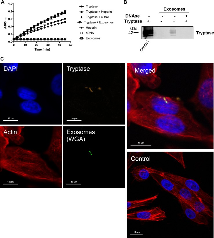
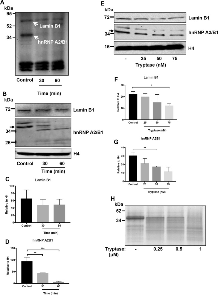
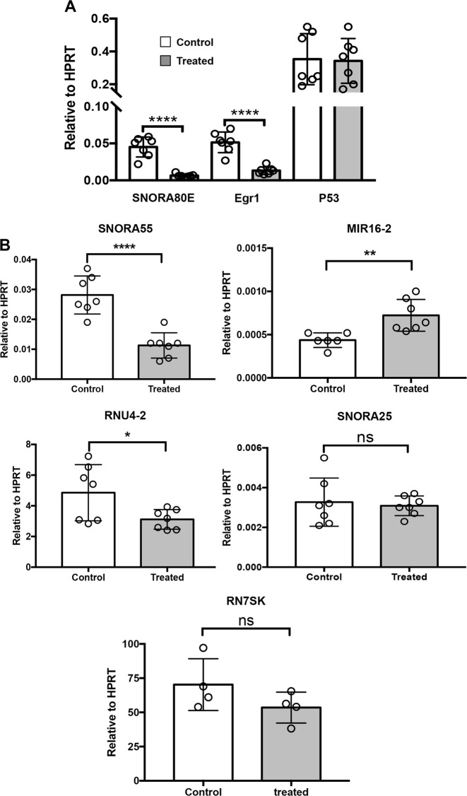
It is well established that mast cell accumulation accompanies most malignancies. However, the knowledge of how mast cells functionally impact on tumors is still rudimentary. Here we addressed this issue and show that mast cells have anti-proliferative activity on melanoma cells and that this effect is dependent on tryptase, a tetrameric protease stored in mast cell granules. Mechanistically, tryptase was found to be endocytosed by melanoma cells as cargo of DNA-coated exosomes released from melanoma cells, followed by transport to the nucleus. In the nucleus, tryptase executed clipping of histone 3 and degradation of Lamin B1, accompanied by extensive nuclear remodeling. Moreover, tryptase degraded hnRNP A2/B1, a protein involved in mRNA stabilization and interaction with non-coding RNAs. This was followed by downregulated expression of the oncogene EGR1 and of multiple non-coding RNAs, including oncogenic species. Altogether, these findings establish a new principle for regulation of tumor cell proliferation.
Overview
The claims and statistics of this paper haven’t been checked yet.
Run the check — free, no account needed. It reads the open-access text and checks the sources it cites.
Integrity signals
Registry
Cites
Statistics
Figures
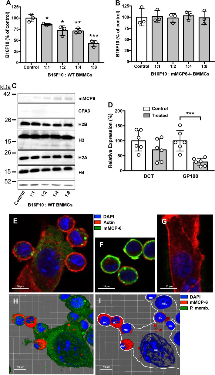
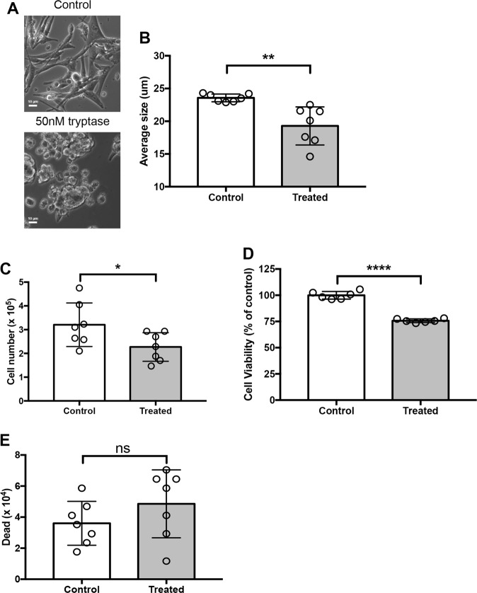
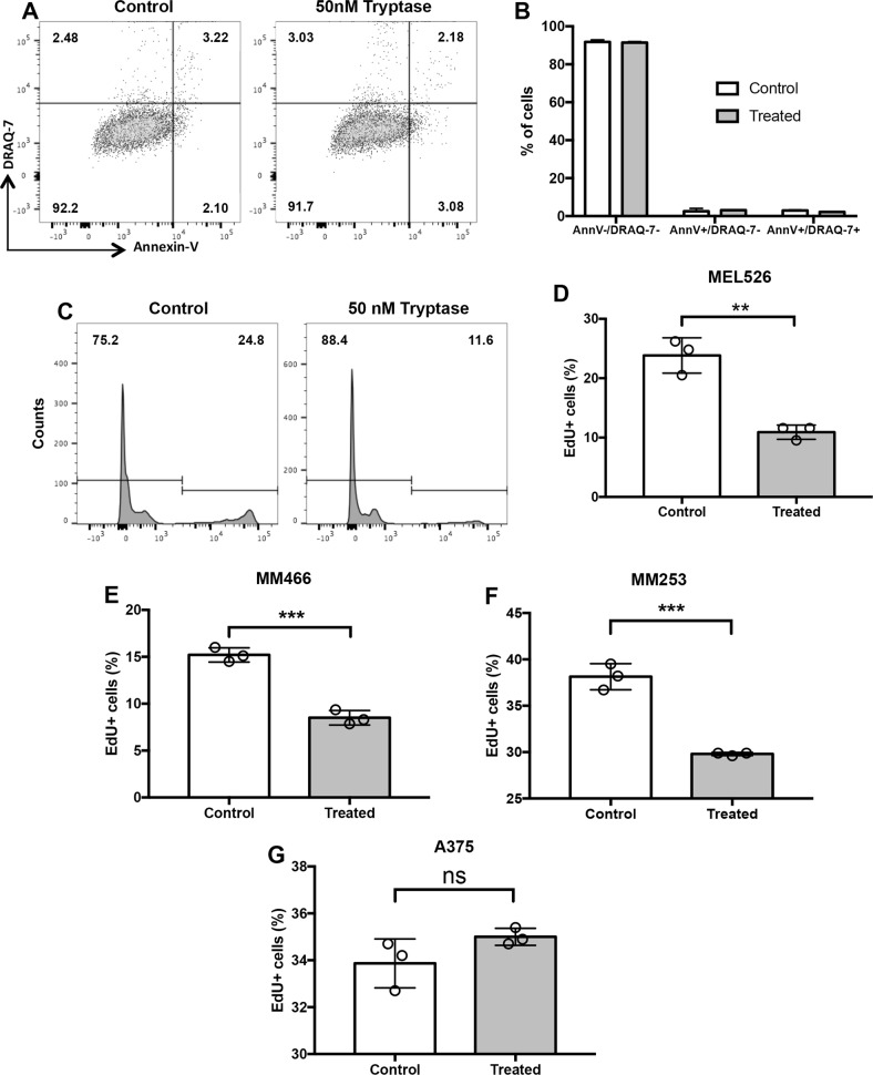
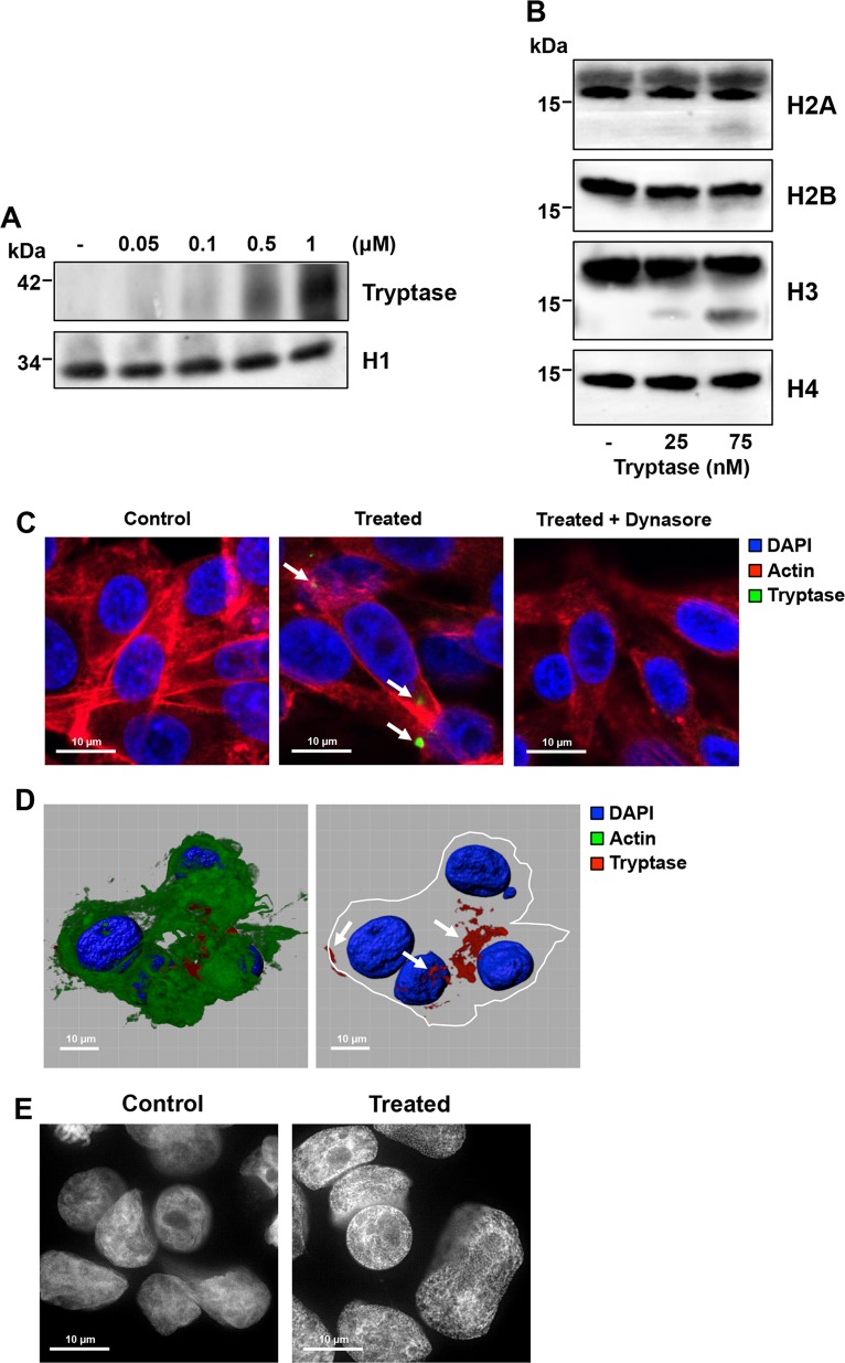
Verification
Claims the paper makes
The claims and statistics of this paper haven’t been checked yet.
Run the check — free, no account needed. It reads the open-access text and checks the sources it cites.
Claims other papers make about this one
Each is checked against this paper’s own text: does it say what the citing paper says it does?
In the citing paper
“This led to cleavage of histone H3 and lamin B1 and extensive nuclear remodeling, ultimately leading to reduced tumor growth.”
From Mast cell ontogeny: From fetal development to life‐long health and disease (2023) · cites this paper as [214]
In the citing paper
“Indeed, it was later shown that melanoma cells can take up tryptase via exosome trafficking. 214”
From Mast cell ontogeny: From fetal development to life‐long health and disease (2023) · cites this paper as [214]
In the citing paper
“For example, mast cell‐derived histamine and TNF‐α regulate cytokine production by tumor cells, and tryptase can inhibit melanoma proliferation. 214”
From Mast cell ontogeny: From fetal development to life‐long health and disease (2023) · cites this paper as [214]
Self-check
Percentages, sums and reported differences re-computed from the paper’s own numbers.
The self-check needs the paper’s full text.
Citations
Cites
Inflammatory mast cells up-regulate angiogenesis during squamous epithelial carcinogenesis
Coussens, L. M., Raymond, W. W., Bergers, G., Laig-Webster, M., Behrendtsen, O., Werb, Z., Caughey, G. H., & Hanahan, D. (1999) · Genes & Development · doi:10.1101/gad.13.11.1382 · Cited by 946
Palucka, A. K., & Coussens, L. M. (2016) · Cell · doi:10.1016/j.cell.2016.01.049 · Cited by 843
Research works printed but not linked: 48
- Metcalfe, D. D., Baram, D. & Mekori, Y. A. Mast cells. Physiol. Rev. 77, 1033–1079 (1997). · DOI
- Gurish, M. F. & Austen, K. F. Developmental origin and functional specialization of mast cell subsets. Immunity 37, 25–33 (2012). · DOI
- Galli, S. J., Nakae, S. & Tsai, M. Mast cells in the development of adaptive immune responses. Nat. Immunol. 6, 135–142 (2005). · DOI
- Ribatti, D. & Crivellato, E. The controversial role of mast cells in tumor growth. Int. Rev. Cell Mol. Biol. 275, 89–131 (2009). · DOI
- Dalton, D. K. & Noelle, R. J. The roles of mast cells in anticancer immunity. Cancer Immunol. Immunother. 61, 1511–1520 (2012). · DOI
- Marichal, T., Tsai, M. & Galli, S. J. Mast cells: potential positive and negative roles in tumor biology. Cancer Immunol. Res. 1, 269–279 (2013). · DOI
- Theoharides, T. C. & Conti, P. Mast cells: the JEKYLL and HYDE of tumor growth. Trends Immunol. 25, 235–241 (2004). · DOI
- Oldford, S. A. & Marshall, J. S. Mast cells as targets for immunotherapy of solid tumors. Mol. Immunol. 63, 113–124 (2015). · DOI
- Rabenhorst, A. et al. Mast cells play a protumorigenic role in primary cutaneous lymphoma. Blood 120, 2042–2054 (2012). · DOI
- Soucek, L. et al. Mast cells are required for angiogenesis and macroscopic expansion of Myc-induced pancreatic islet tumors. Nat. Med. 13, 1211–1218 (2007). · DOI
- Wernersson, S. & Pejler, G. Mast cell granules: armed for battle. Nat. Rev. Immunol. 14, 478–494 (2014). · DOI · Paper page
- Pejler, G., Rönnberg, E., Waern, I. & Wernersson, S. Mast cell proteases: multifaceted regulators of inflammatory disease. Blood 115, 4981–4990 (2010). · DOI
- Pejler, G., Åbrink, M., Ringvall, M. & Wernersson, S. Mast cell proteases. Adv. Immunol. 95, 167–255 (2007). · DOI
- Galli, S. J. et al. Approaches for analyzing the roles of mast cells and their proteases in vivo. Adv. Immunol. 126, 45–127 (2015). · DOI · Paper page
- Grujic, M. et al. The combined action of mast cell chymase, tryptase and carboxypeptidase A3 protects against melanoma colonization of the lung. Oncotarget 8, 25066–25079 (2017). · DOI
- Siiskonen, H. et al. Low numbers of tryptase+ and chymase+ mast cells associated with reduced survival and advanced tumor stage in melanoma. Melanoma Res. 25, 479–485 (2015). · DOI
- Stieglitz, D. et al. BMP6-induced modulation of the tumor micro-milieu. Oncogene 38, 609–621 (2019). · DOI
- Thakurdas, S. M. et al. The mast cell-restricted tryptase mMCP-6 has a critical immunoprotective role in bacterial infections. J. Biol. Chem. 282, 20809–20815 (2007). · DOI
- Shin, K. et al. Mouse mast cell tryptase mMCP-6 is a critical link between adaptive and innate immunity in the chronic phase of Trichinella spiralis infection. J. Immunol. 180, 4885–4891 (2008). · DOI
- Waern, I. et al. Mast cells limit extracellular levels of IL-13 via a serglycin proteoglycan-serine protease axis. Biol. Chem. 393, 1555–1567 (2012). · DOI
- Melo, F. R. et al. Proteolytic histone modification by mast cell tryptase, a serglycin proteoglycan-dependent secretory granule protease. J. Biol. Chem. 289, 7682–7690 (2014). · DOI
- Melo, F. R. et al. Tryptase-catalyzed core histone truncation: a novel epigenetic regulatory mechanism in mast cells. J. Allergy Clin. Immunol. 140, 474–485 (2017). · DOI
- Akers, I. A. et al. Mast cell tryptase stimulates human lung fibroblast proliferation via protease-activated receptor-2. Am. J. Physiol. Lung Cell Mol. Physiol. 278, L193–L201 (2000). · DOI
- Berger, P., Tunon-De-Lara, J. M., Savineau, J. P. & Marthan, R. Selected contribution: tryptase-induced PAR-2-mediated Ca(2+) signaling in human airway smooth muscle cells. J. Appl. Physiol. 91, 995–1003 (2001). · DOI
- Corvera, C. U. et al. Mast cell tryptase regulates rat colonic myocytes through proteinase- activated receptor 2. J. Clin. Invest. 100, 1383–1393 (1997). · DOI
- Hallgren, J. & Pejler, G. Biology of mast cell tryptase. An inflammatory mediator. FEBS J. 273, 1871–1895 (2006). · DOI
- Mori, S. et al. Nafamostat mesilate is an extremely potent inhibitor of human tryptase. J. Pharm. Sci. 92, 420–423 (2003). · DOI
- Sharma, P. et al. Immunoaffinity-based isolation of melanoma cell-derived exosomes from plasma of patients with melanoma. J. Extracell. Vesicles 7, 1435138 (2018). · DOI
- Meehan, K. & Vella, L. J. The contribution of tumour-derived exosomes to the hallmarks of cancer. Crit. Rev. Clin. Lab Sci. 53, 121–131 (2016). · DOI
- Åbrink, M., Grujic, M. & Pejler, G. Serglycin is essential for maturation of mast cell secretory granule. J. Biol. Chem. 279, 40897–40905 (2004). · DOI
- Hallgren, J., Karlson, U., Poorafshar, M., Hellman, L. & Pejler, G. Mechanism for activation of mouse mast cell tryptase: dependence on heparin and acidic pH for formation of active tetramers of mouse mast cell protease 6. Biochemistry 39, 13068–13077 (2000). · DOI
- Hallgren, J., Spillmann, D. & Pejler, G. Structural requirements and mechanism for heparin-induced activation of a recombinant mouse mast cell tryptase, mouse mast cell protease-6: formation of active tryptase monomers in the presence of low molecular weight heparin. J. Biol. Chem. 276, 42774–42781 (2001). · DOI
- Thakur, B. K. et al. Double-stranded DNA in exosomes: a novel biomarker in cancer detection. Cell Res. 24, 766–769 (2014). · DOI
- Hanahan, D. & Weinberg, R. A. Hallmarks of cancer: the next generation. Cell 144, 646–674 (2011). · DOI · Paper page
- Coussens, L. M., Zitvogel, L. & Palucka, A. K. Neutralizing tumor-promoting chronic inflammation: a magic bullet? Science 339, 286–291 (2013). · DOI
- Pereira, P. J. et al. Human beta-tryptase is a ring-like tetramer with active sites facing a central pore. Nature 392, 306–311 (1998). · DOI
- Sommerhoff, C. P. et al. The structure of the human betaII-tryptase tetramer: fo(u)r better or worse. Proc. Natl Acad. Sci. USA 96, 10984–10991 (1999). · DOI
- Camps, J., Erdos, M. R. & Ried, T. The role of lamin B1 for the maintenance of nuclear structure and function. Nucleus 6, 8–14 (2015). · DOI · Paper page
- Jean-Philippe, J., Paz, S. & Caputi, M. hnRNP A1: the Swiss army knife of gene expression. Int. J. Mol. Sci. 14, 18999–19024 (2013). · DOI
- Okugawa, Y. et al. Clinical significance of SNORA42 as an oncogene and a prognostic biomarker in colorectal cancer. Gut 66, 107–117 (2017). · DOI
- Mei, Y. P. et al. Small nucleolar RNA 42 acts as an oncogene in lung tumorigenesis. Oncogene 31, 2794–2804 (2012). · DOI
- Crea, F. et al. Integrated analysis of the prostate cancer small-nucleolar transcriptome reveals SNORA55 as a driver of prostate cancer progression. Mol. Oncol. 10, 693–703 (2016). · DOI
- Kuhlmann, J. D. et al. Increased level of circulating U2 small nuclear RNA fragments indicates metastasis in melanoma patients. Clin. Chem. Lab Med. 53, 605–611 (2015). · DOI
- Tian, R. et al. Differential expression of miR16 in glioblastoma and glioblastoma stem cells: their correlation with proliferation, differentiation, metastasis and prognosis. Oncogene 36, 5861–5873 (2017). · DOI
- Zhao, D. Y., Jacobs, K. M., Hallahan, D. E. & Thotala, D. Silencing Egr1 attenuates radiation-induced apoptosis in normal tissues while killing cancer cells and delaying tumor growth. Mol. Cancer Ther. 14, 2343–2352 (2015). · DOI
- Kouzarides, T. Chromatin modifications and their function. Cell 128, 693–705 (2007). · DOI · Paper page
- Rönnberg, E. & Pejler, G. Serglycin: the master of the mast cell. Methods Mol. Biol. 836, 201–217 (2012). · DOI
- Paivandy, A., Eriksson, J., Melo, F. R., Sellin, M. E. & Pejler, G. Lysosomotropic challenge of mast cells causes intra-granular reactive oxygen species production. Cell Death Discov. 5, 95 (2019). · DOI
Cited by
Sorted by citations
Mast cell ontogeny: From fetal development to life‐long health and disease
Chia, S. L., Kapoor, S., Carvalho, C., Bajénoff, M., & Gentek, R. (2023) · Immunological Reviews · doi:10.1111/imr.13191 · Cited by 60
Metadata
- Authors
- Rabelo Melo, F., Santosh Martin, S., Sommerhoff, C. P., & Pejler, G.
- Venue
- Cell Death & Disease
- Published
- 2019-09-10
- Volume
- 10
- Issue
- 9
- ISSN
- 2041-4889
- PMC
- PMC6736983
- Cited by
- 41
- References
- 2
- Language
- en
- Funders
- Cancerfonden
- Barncancerfonden
- Knut och Alice Wallenbergs Stiftelse
About these checks
Registry, retraction, Cites and statistics results are computed, not judged, with one exception: a printed reference that no index lists is read once by a model before it counts as a citation error. Claims are extracted by a model and kept only when their quote appears verbatim in the text; each cited source is then checked, with a model’s help, for whether it says what the claim relies on. Those verdicts can be wrong — read the claim’s quote and the cited source before relying on one.
Statistics and arithmetic are recomputed, not judged.
Confidence bands are shown only for fields calibrated on our annotated test set. None is calibrated yet, so no band is shown.
What counts as a claim ›How we measure confidence ›How we check this ›
Are you an author, or do you see a problem with this page? Request a review
