

The DOI matches its Crossref record; 5 of the 5 references it cites were found in a registry or an index.
Targeting JNK pathway promotes human hematopoietic stem cell expansion
Abstract
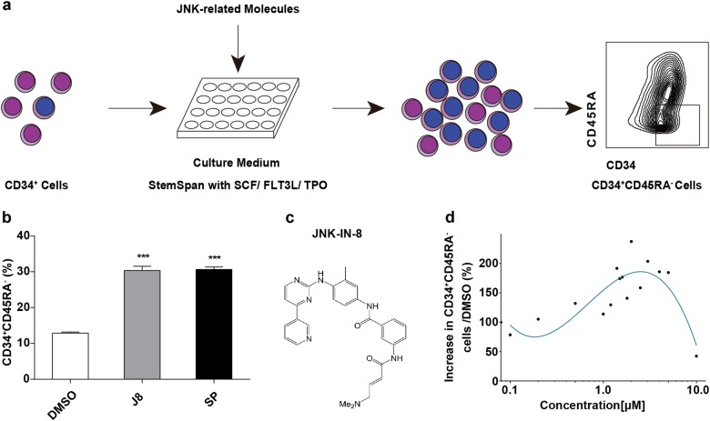
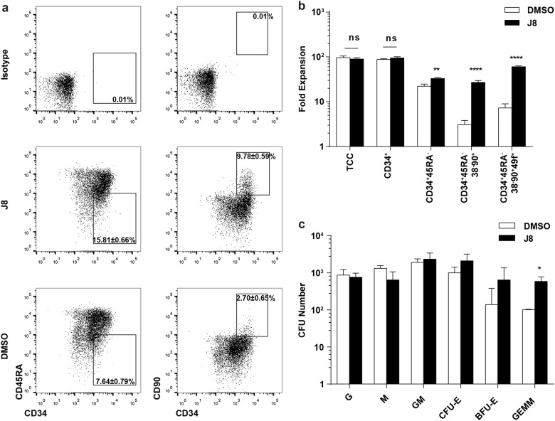
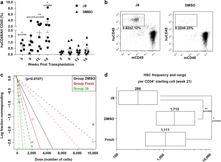
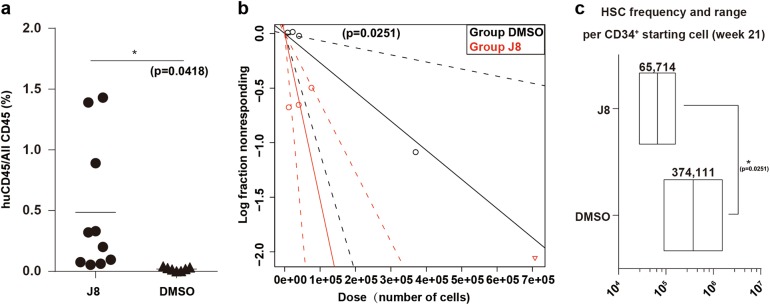
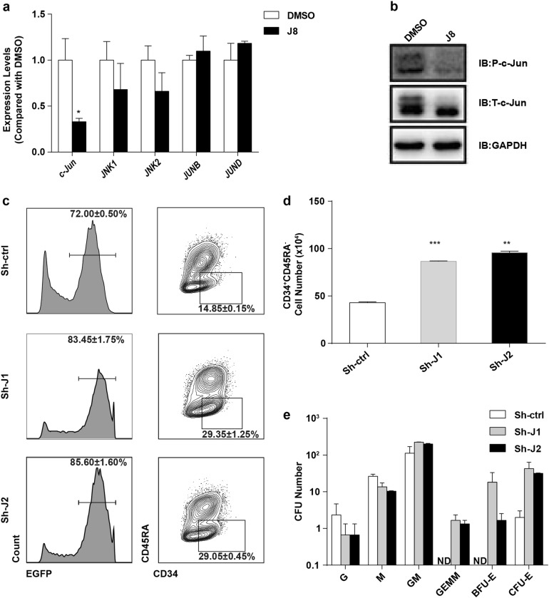
The limited number of human hematopoietic stem cells (HSCs) has restrained their widespread clinical application. Despite great efforts in recent years, the in vitro expansion of HSCs remains a challenge due to incomplete understanding of the signaling networks underlying HSC self-renewal. Here, we show that culturing human cord blood (CB) CD34 + cells with JNK-IN-8, an inhibitor of the JNK signaling pathway, can enhance the self-renewal of HSCs with a 3.88-fold increase in cell number. These cultured CD34 + cells repopulated recipient mice for 21 weeks and can form secondary engraftment that lasted for more than 21 weeks. Knockdown of c-Jun , a major downstream target in the JNK pathway, promoted the expansion of hematopoietic stem and progenitor cells (HSPCs). Our findings demonstrate a critical role of the JNK pathway in regulating HSC expansion, provide new insights into HSC self-renewal mechanism, and may lead to improved clinical application of HSCs.
Overview
The claims and statistics of this paper haven’t been checked yet.
Run the check — free, no account needed. It reads the open-access text and checks the sources it cites.
Integrity signals
Registry
Cites
Statistics
Figures
Verification
Claims the paper makes
The claims and statistics of this paper haven’t been checked yet.
Run the check — free, no account needed. It reads the open-access text and checks the sources it cites.
Claims other papers make about this one
Each is checked against this paper’s own text: does it say what the citing paper says it does?
In the citing paper
“Further, TFs known to drive terminal hematopoietic differentiation (ELF4, PU.1, CEBPB, JUN)38–41 are among the most methylated binding sites in Tet2-KO LSKs upon IL1β stimulation.”
From Clonal hematopoiesis related TET2 loss-of-function impedes IL1β-mediated epigenetic reprogramming in hematopoietic stem and progenitor cells (2023) · cites this paper as [41]
In the citing paper
“Similarly, IL1β promoted hypermethylation of CEBPB binding sites, known to drive HSCs towards terminal differentiation, and promoted hypermethylation in genes driving proliferation, such as Cdk438–41.”
From Clonal hematopoiesis related TET2 loss-of-function impedes IL1β-mediated epigenetic reprogramming in hematopoietic stem and progenitor cells (2023) · cites this paper as [41]
Self-check
Percentages, sums and reported differences re-computed from the paper’s own numbers.
The self-check needs the paper’s full text.
Citations
Cites
AP-1 as a regulator of cell life and death
Shaulian, E., & Karin, M. (2002) · Nature Cell Biology · doi:10.1038/ncb0502-e131 · Cited by 2,961
A Differentiation Checkpoint Limits Hematopoietic Stem Cell Self-Renewal in Response to DNA Damage
Wang, J., Sun, Q., Morita, Y., Jiang, H., Groß, A., Lechel, A., Hildner, K., Guachalla, L. M., Gompf, A., Hartmann, D., et al. (2012) · Cell · doi:10.1016/j.cell.2012.01.040 · Cited by 343
Lineage-specific enhancers activate self-renewal genes in macrophages and embryonic stem cells
Soucie, E. L., Weng, Z., Geirsdóttir, L., Molawi, K., Maurizio, J., Fenouil, R., Mossadegh-Keller, N., Gimenez, G., VanHille, L., Beniazza, M., et al. (2016) · Science · doi:10.1126/science.aad5510 · Cited by 263
c-Jun Is a JNK-independent Coactivator of the PU.1 Transcription Factor
Behre, G., Whitmarsh, A. J., Coghlan, M. P., Hoang, T., Carpenter, C. L., Zhang, D.-E., Davis, R. J., & Tenen, D. G. (1999) · Journal of Biological Chemistry · doi:10.1074/jbc.274.8.4939 · Cited by 180
Wojtowicz, E. E., Lechman, E. R., Hermans, K. G., Schoof, E. M., Wienholds, E., Isserlin, R., van Veelen, P. A., Broekhuis, M. J. C., Janssen, G. M. C., Trotman-Grant, A., et al. (2016) · Cell stem cell · doi:10.1016/j.stem.2016.06.008 · Cited by 60
Research works printed but not linked: 32
- Passweg, J. R. et al. Use of haploidentical stem cell transplantation continues to increase: the 2015 European society for blood and marrow transplant activity survey report. Bone Marrow Transplant. 52, 811–817 (2017). · DOI
- Passweg, J. R. et al. Hematopoietic stem cell transplantation in Europe 2014: more than 40 000 transplants annually. Bone Marrow Transplant. 51, 786–792 (2016). · DOI
- Oran, B. & Shpall, E. Umbilical cord blood transplantation: a maturing technology. Hematol. Am. Soc. Hematol. Educ. Program 2012, 215–222 (2012). · DOI
- Ballen, K. K., Gluckman, E. & Broxmeyer, H. E. Umbilical cord blood transplantation: the first 25 years and beyond. Blood 122, 491–498 (2013). · DOI
- Pineault, N. & Abu-Khader, A. Advances in umbilical cord blood stem cell expansion and clinical translation. Exp. Hematol. 43, 498–513 (2015). · DOI
- Trowbridge, J. J., Xenocostas, A., Moon, R. T. & Bhatia, M. Glycogen synthase kinase-3 is an in vivo regulator of hematopoietic stem cell repopulation. Nat. Med. 12, 89–98 (2006). · DOI
- Huang, J., Nguyen-McCarty, M., Hexner, E. O., Danet-Desnoyers, G. & Klein, P. S. Maintenance of hematopoietic stem cells through regulation of Wnt and mTOR pathways. Nat. Med. 18, 1778–1785 (2012). · DOI
- Boitano, A. E. et al. Aryl hydrocarbon receptor antagonists promote the expansion of human hematopoietic stem cells. Science 329, 1345–1348 (2010). · DOI
- Fares, I. et al. Cord blood expansion. Pyrimidoindole derivatives are agonists of human hematopoietic stem cell self-renewal. Science 345, 1509–1512 (2014). · DOI
- Wagner, JE.Jr et al. Phase I/II trial of StemRegenin-1 expanded umbilical cord blood hematopoietic stem cells supports testing as a stand-alone graft. Cell. Stem. Cell. 18, 144–155 (2016). · DOI
- Fares, I. et al. EPCR expression marks UM171-expanded CD34(+) cord blood stem cells. Blood 129, 3344–3351 (2017). · DOI
- Gafni, O. et al. Derivation of novel human ground state naive pluripotent stem cells. Nature 504, 282–286 (2013). · DOI
- Fang, R. et al. Generation of naive induced pluripotent stem cells from rhesus monkey fibroblasts. Cell. Stem. Cell. 15, 488–497 (2014). · DOI
- Yang, J. et al. Establishment of mouse expanded potential stem cells. Nature 550, 393–397 (2017). · DOI
- Santaguida, M. et al. JunB protects against myeloid malignancies by limiting hematopoietic stem cell proliferation and differentiation without affecting self-renewal. Cancer Cell. 15, 341–352 (2009). · DOI
- Passegue, E., Wagner, E. F. & Weissman, I. L. JunB deficiency leads to a myeloproliferative disorder arising from hematopoietic stem cells. Cell 119, 431–443 (2004). · DOI
- Zhang, T. et al. Discovery of potent and selective covalent inhibitors of JNK. Chem. & Biol. 19, 140–154 (2012). · DOI
- Majeti, R., Park, C. Y. & Weissman, I. L. Identification of a hierarchy of multipotent hematopoietic progenitors in human cord blood. Cell. Stem. Cell. 1, 635–645 (2007). · DOI
- Notta, F. et al. Isolation of single human hematopoietic stem cells capable of long-term multilineage engraftment. Science 333, 218–221 (2011). · DOI · Paper page
- Zeke, A., Misheva, M., Remenyi, A. & Bogoyevitch, M. A. JNK signaling: regulation and functions based on complex protein-protein partnerships. Microbiol. Mol. Biol. Rev.: MMBR 80, 793–835 (2016). · DOI
- Cai, D. H. et al. C/EBP alpha:AP-1 leucine zipper heterodimers bind novel DNA elements, activate the PU.1 promoter and direct monocyte lineage commitment more potently than C/EBP alpha homodimers or AP-1. Oncogene 27, 2772–2779 (2008). · DOI · Paper page
- Frelin, C. et al. GATA-3 regulates the self-renewal of long-term hematopoietic stem cells. Nat. Immunol. 14, 1037–1044 (2013). · DOI
- Okada, S., Fukuda, T., Inada, K. & Tokuhisa, T. Prolonged expression of c-fos suppresses cell cycle entry of dormant hematopoietic stem cells. Blood 93, 816–825 (1999). · DOI
- Deneault, E. et al. A functional screen to identify novel effectors of hematopoietic stem cell activity. Cell 137, 369–379 (2009). · DOI
- Guo, B., Huang, X., Lee, M. R., Lee, S. A. & Broxmeyer, H. E. Antagonism of PPAR-gamma signaling expands human hematopoietic stem and progenitor cells by enhancing glycolysis. Nat. Med. 24, 360–367 (2018). · DOI
- Huang, X., Guo, B., Liu, S., Wan, J. & Broxmeyer, H. E. Neutralizing negative epigenetic regulation by HDAC5 enhances human haematopoietic stem cell homing and engraftment. Nat. Commun. 9, 2741 (2018). · DOI
- Cheung, A. M. S. et al. Analysis of the clonal growth and differentiation dynamics of primitive barcoded human cord blood cells in NSG mice. Blood 122, 3129–3137 (2013). · DOI
- Hu, Y. & Smyth, G. K. ELDA: extreme limiting dilution analysis for comparing depleted and enriched populations in stem cell and other assays. J. Immunol. Methods 347, 70–78 (2009). · DOI
- Trapnell, C., Pachter, L. & Salzberg, S. L. TopHat: discovering splice junctions with RNA-Seq. Bioinforma. (Oxf., Engl.) 25, 1105–1111 (2009). · DOI
- Langmead, B. & Salzberg, S. L. Fast gapped-read alignment with Bowtie 2. Nat. Methods 9, 357–U354 (2012). · DOI · Paper page
- Li, H. et al. The sequence Alignment/Map format and SAMtools. Bioinforma. (Oxf., Engl.) 25, 2078–2079 (2009). · DOI · Paper page
- Trapnell, C. et al. Transcript assembly and quantification by RNA-Seq reveals unannotated transcripts and isoform switching during cell differentiation. Nat. Biotechnol. 28, 511–U174 (2010). · DOI · Paper page
Cited by
McClatchy, J., Strogantsev, R., Wolfe, E., Lin, H. Y., Mohammadhosseini, M., Davis, B. A., Eden, C., Goldman, D., Fleming, W. H., Conley, P., et al. (2023) · Nature Communications · doi:10.1038/s41467-023-43697-y · Cited by 95
Raghav, P. K., & Gangenahalli, G. (2021) · International Journal of Nanomedicine · doi:10.2147/ijn.s303235 · Cited by 13
Metadata
- Authors
- Xiao, X., Lai, W., Xie, H., Liu, Y., Guo, W., Liu, Y., Li, Y., Li, Y., Zhang, J., Chen, W., Shi, M., Shang, L., Yin, M., Wang, C., & Deng, H.
- Venue
- Cell Discovery
- Published
- 2019-01-08
- Volume
- 5
- Issue
- 1
- ISSN
- 2056-5968
- PMC
- PMC6323118
- Cited by
- 51
- References
- 5
- Language
- en
About these checks
Registry, retraction, Cites and statistics results are computed, not judged, with one exception: a printed reference that no index lists is read once by a model before it counts as a citation error. Claims are extracted by a model and kept only when their quote appears verbatim in the text; each cited source is then checked, with a model’s help, for whether it says what the claim relies on. Those verdicts can be wrong — read the claim’s quote and the cited source before relying on one.
Statistics and arithmetic are recomputed, not judged.
Confidence bands are shown only for fields calibrated on our annotated test set. None is calibrated yet, so no band is shown.
What counts as a claim ›How we measure confidence ›How we check this ›
Are you an author, or do you see a problem with this page? Request a review
