

The DOI matches its Crossref record; 13 of the 13 references it cites were found in a registry or an index.
A novel receptor-binding domain (RBD)-based mRNA vaccine against SARS-CoV-2
Open accessAbstract
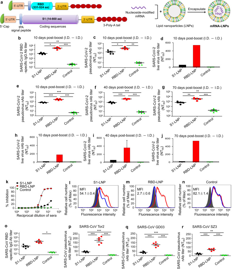
The pandemic of coronavirus disease 2019 (COVID-19) caused by severe acute respiratory syndrome coronavirus 2 (SARS-CoV-2) highlights the need to develop effective and safe vaccines. Similar to SARS-CoV, SARS-CoV-2 recognizes angiotensin-converting enzyme 2 (ACE2) as receptor for host cell entry. 1 , 2 SARS-CoV-2 spike (S) protein consists of S1, including receptor-binding domain (RBD), and S2 subunits. 3 , 4 We previously demonstrated that RBDs of SARS-CoV and MERS-CoV serve as important targets for the development of effective vaccines. 5 , 6
Overview
The claims and statistics of this paper haven’t been checked yet.
Run the check — free, no account needed. It reads the open-access text and checks the sources it cites.
Integrity signals
Registry
Cites
Statistics
Figures
Verification
Claims the paper makes
The claims and statistics of this paper haven’t been checked yet.
Run the check — free, no account needed. It reads the open-access text and checks the sources it cites.
Claims other papers make about this one
Each is checked against this paper’s own text: does it say what the citing paper says it does?
The source backs part of the claim, not all of it — or backs it for a different population, condition or comparison.
In the citing paper
“the RBD can induce strong humoral and cellular immune responses when used as an antigen in various SARS-CoV and MERS-CoV studies),8, 9, 10”
From Safety and immunogenicity of the SARS-CoV-2 ARCoV mRNA vaccine in Chinese adults: a randomised, double-blind, placebo-controlled, phase 1 trial (2022) · cites this paper as [9]
Experimental — accuracy not yet measuredThe cited source says what the claim attributes to it.
In the citing paper
“the receptor binding domain (RBD) of SARS-CoV-2 spike protein had been identified to play a key role in helping coronaviruses to enter human cells”
From Safety and immunogenicity of the SARS-CoV-2 ARCoV mRNA vaccine in Chinese adults: a randomised, double-blind, placebo-controlled, phase 1 trial (2022) · cites this paper as [9]
In the citing paper
“Our data in humans corroborate reports that demonstrate the induction of potent germinal centre responses by SARS-CoV-2 mRNA-based vaccines in mice22,23.”
From SARS-CoV-2 mRNA vaccines induce persistent human germinal centre responses (2021) · cites this paper as [22]
Self-check
Percentages, sums and reported differences re-computed from the paper’s own numbers.
The self-check needs the paper’s full text.
Citations
Cites
A pneumonia outbreak associated with a new coronavirus of probable bat origin
Zhou, P., Yang, X.-L., Wang, X.-G., Hu, B., Zhang, L., Zhang, W., Si, H.-R., Zhu, Y., Li, B., Huang, C.-L., et al. (2020) · Nature · doi:10.1038/s41586-020-2012-7 · Cited by 21,969
Angiotensin-converting enzyme 2 is a functional receptor for the SARS coronavirus
Li, W., Moore, M. J., Vasilieva, N., Sui, J., Wong, S. K., Berne, M. A., Somasundaran, M., Sullivan, J. L., Luzuriaga, K., Greenough, T. C., et al. (2003) · Nature · doi:10.1038/nature02145 · Cited by 5,923
Structural basis of receptor recognition by SARS-CoV-2
Shang, J., Ye, G., Shi, K., Wan, Y., Luo, C., Aihara, H., Geng, Q., Auerbach, A., & Li, F. (2020) · Nature · doi:10.1038/s41586-020-2179-y · Cited by 3,661
The spike protein of SARS-CoV — a target for vaccine and therapeutic development
Du, L., He, Y., Zhou, Y., Liu, S., Zheng, B.-J., & Jiang, S. (2009) · Nature Reviews Microbiology · doi:10.1038/nrmicro2090 · Cited by 1,678
Self-citationTai, W., He, L., Zhang, X., Pu, J., Voronin, D., Jiang, S., Zhou, Y., & Du, L. (2020) · Cellular & Molecular Immunology · doi:10.1038/s41423-020-0400-4 · Cited by 1,654
Self-citationSARS-CoV-2 mRNA Vaccine Design Enabled by Prototype Pathogen Preparedness
Corbett, K. S., Edwards, D. K., Leist, S. R., Abiona, O. M., Boyoglu-Barnum, S., Gillespie, R. A., Himansu, S., Schäfer, A., Ziwawo, C. T., DiPiazza, A. T., et al. (2020) · Nature · doi:10.1038/s41586-020-2622-0 · Cited by 1,615
Xia, S., Liu, M., Wang, C., Xu, W., Lan, Q., Feng, S., Qi, F., Bao, L., Du, L., Liu, S., et al. (2020) · Cell Research · doi:10.1038/s41422-020-0305-x · Cited by 1,355
Self-citationGerminal-Center Organization and Cellular Dynamics
Allen, C. D. C., Okada, T., & Cyster, J. G. (2007) · Immunity · doi:10.1016/j.immuni.2007.07.009 · Cited by 1,025
DNA vaccine protection against SARS-CoV-2 in rhesus macaques
Yu, J., Tostanoski, L. H., Peter, L., Mercado, N. B., McMahan, K., Mahrokhian, S. H., Nkolola, J. P., Liu, J., Li, Z., Chandrashekar, A., et al. (2020) · Science · doi:10.1126/science.abc6284 · Cited by 997
Nonviral delivery of self-amplifying RNA vaccines
Geall, A. J., Verma, A., Otten, G. R., Shaw, C. A., Hekele, A., Banerjee, K., Cu, Y., Beard, C. W., Brito, L. A., Krucker, T., et al. (2012) · Proceedings of the National Academy of Sciences · doi:10.1073/pnas.1209367109 · Cited by 675
Subunit Vaccines Against Emerging Pathogenic Human Coronaviruses
Wang, N., Shang, J., Jiang, S., & Du, L. (2020) · Frontiers in Microbiology · doi:10.3389/fmicb.2020.00298 · Cited by 434
Self-citationChAdOx1 nCoV-19 vaccination prevents SARS-CoV-2 pneumonia in rhesus macaques
van Doremalen, N., Lambe, T., Spencer, A., Belij-Rammerstorfer, S., Purushotham, J. N., Port, J. R., Avanzato, V., Bushmaker, T., Flaxman, A., Ulaszewska, M., et al. (2020) · doi:10.1101/2020.05.13.093195 · Cited by 246
SARS-CoV-2 mRNA Vaccine Development Enabled by Prototype Pathogen PreparednessPreprint
Corbett, K. S., Edwards, D. K., Leist, S. R., et al. (2020) · bioRxiv (Cold Spring Harbor Laboratory) · doi:10.1101/2020.06.11.145920 · Cited by 143
Cited by
Sorted by citations
SARS-CoV-2 mRNA vaccines induce persistent human germinal centre responses
Turner, J. S., O’Halloran, J. A., Kalaidina, E., Kim, W., Schmitz, A. J., Zhou, J. Q., Lei, T., Thapa, M., Chen, R. E., Case, J. B., et al. (2021) · Nature · doi:10.1038/s41586-021-03738-2 · Cited by 831
mRNA-based therapeutics: powerful and versatile tools to combat diseases
Qin, S., Tang, X., Chen, Y., Chen, K., Fan, N., Xiao, W., Zheng, Q., Li, G., Teng, Y., Wu, M., et al. (2022) · Signal Transduction and Targeted Therapy · doi:10.1038/s41392-022-01007-w · Cited by 852
Circular RNA Vaccines against SARS-CoV-2 and Emerging Variants
Qu, L., Yi, Z., Shen, Y., Lin, L., Chen, F., Xu, Y., Wu, Z., Tang, H., Zhang, X., Tian, F., et al. (2021) · doi:10.1101/2021.03.16.435594 · Cited by 636
Self-citationLipids and Lipid Derivatives for RNA Delivery
Zhang, Y., Sun, C., Wang, C., Jankovic, K. E., & Dong, Y. (2021) · Chemical Reviews · doi:10.1021/acs.chemrev.1c00244 · Cited by 649
COVID-19 vaccine development: milestones, lessons and prospects
Li, M., Wang, H., Tian, L., Pang, Z., Yang, Q., Huang, T., Fan, J., Song, L., Tong, Y., & Fan, H. (2022) · Signal Transduction and Targeted Therapy · doi:10.1038/s41392-022-00996-y · Cited by 478
Metadata
- Authors
- Tai, W., Zhang, X., Drelich, A., Shi, J., Hsu, J. C., Luchsinger, L., Hillyer, C. D., Tseng, C.-T. K., Jiang, S., & Du, L.
- Affiliations
- New York Blood Center (US)
- The University of Texas Medical Branch at Galveston (US)
- Corresponding author
- Jiang, Shibo
- Du, Lanying
- ORCID
- Venue
- Cell Research
- Published
- 2020-08-05
- Volume
- 30
- Issue
- 10
- Pages
- 932-935
- ISSN
- 1001-0602 (ISSN-L)
- 1748-7838
- PMC
- PMC7403570
- OpenAlex
- W3046985131
- Cited by
- 151
- References
- 12
- Citations by year
- 2026: 6 · 2025: 10 · 2024: 15 · 2023: 20 · 2022: 41 · 2021: 48 · 2020: 11
- FWCI
- 3.94
- Type
- journal article
- Language
- en
- Open access
- hybrid
- doi.org
- Locations
- Cell Research (publishedVersion, cc-by)
- PubMed Central (publishedVersion, cc-by)
- PubMed (publishedVersion)
- Funders
- U.S. Department of Health and Human Services (R01AI137472, R01AI139092)
- National Institutes of Health
- National Institute of Allergy and Infectious Diseases (R01AI137472, R01AI139092, R01AI157975)
- Topics
- Health Sciences › Medicine › Infectious Diseases › SARS-CoV-2 and COVID-19 Research
- Health Sciences › Medicine › Infectious Diseases › SARS-CoV-2 detection and testing
- Life Sciences › Biochemistry, Genetics and Molecular Biology › Molecular Biology › vaccines and immunoinformatics approaches
- Keywords
- SARS-CoV-2, COVID-19 pandemic, mRNA vaccines, receptor binding domain, spike protein, ACE2 receptor, vaccine design, coronavirus vaccines
- MeSH terms
- Protein Domains, Betacoronavirus, COVID-19, SARS-CoV-2, COVID-19 Vaccines, Animals, Cell Line, Humans, Immunization, Mice, Inbred BALB C, Pneumonia, Viral, RNA, Messenger, Viral Vaccines, Gene Expression, Coronavirus Infections, Antibodies, Neutralizing, Pandemics, Spike Glycoprotein, Coronavirus
About these checks
Registry, retraction, Cites and statistics results are computed, not judged, with one exception: a printed reference that no index lists is read once by a model before it counts as a citation error. Claims are extracted by a model and kept only when their quote appears verbatim in the text; each cited source is then checked, with a model’s help, for whether it says what the claim relies on. Those verdicts can be wrong — read the claim’s quote and the cited source before relying on one.
Statistics and arithmetic are recomputed, not judged.
Confidence bands are shown only for fields calibrated on our annotated test set. None is calibrated yet, so no band is shown.
What counts as a claim ›How we measure confidence ›How we check this ›
Are you an author, or do you see a problem with this page? Request a review
