

The DOI matches its Crossref record; 43 of the 43 references it cites were found in a registry or an index.
The circular RNA circBIRC6 participates in the molecular circuitry controlling human pluripotency
Open accessAbstract
Accumulating evidence indicates that circular RNAs (circRNAs) are abundant in the human transcriptome. However, their involvement in biological processes, including pluripotency, remains mostly undescribed. We identified a subset of circRNAs that are enriched in undifferentiated human embryonic stem cells (hESCs) and demonstrated that two, circBIRC6 and circCORO1C, are functionally associated with the pluripotent state. Mechanistically, we found that circBIRC6 is enriched in the AGO2 complex and directly interacts with microRNAs, miR-34a, and miR-145, which are known to modulate target genes that maintain pluripotency. Correspondingly, circBIRC6 attenuates the downregulation of these target genes and suppresses hESC differentiation. We further identified hESC-enriched splicing factors (SFs) and demonstrated that circBIRC6 biogenesis in hESCs is promoted by the SF ESRP1, whose expression is controlled by the core pluripotency-associated factors, OCT4 and NANOG. Collectively, our data suggest that circRNA serves as a microRNA "sponge" to regulate the molecular circuitry, which modulates human pluripotency and differentiation.
Overview
The claims and statistics of this paper haven’t been checked yet.
Run the check — free, no account needed. It reads the open-access text and checks the sources it cites.
Integrity signals
Registry
Cites
Statistics
Figures
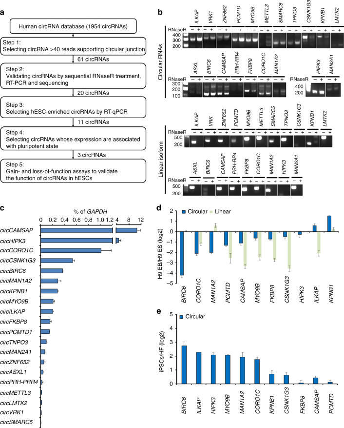
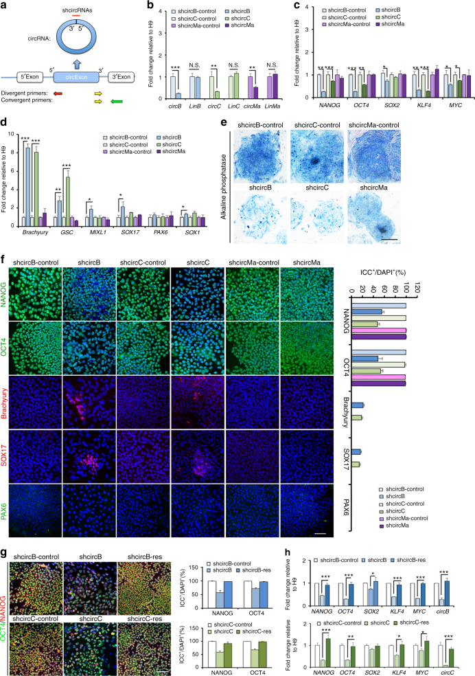
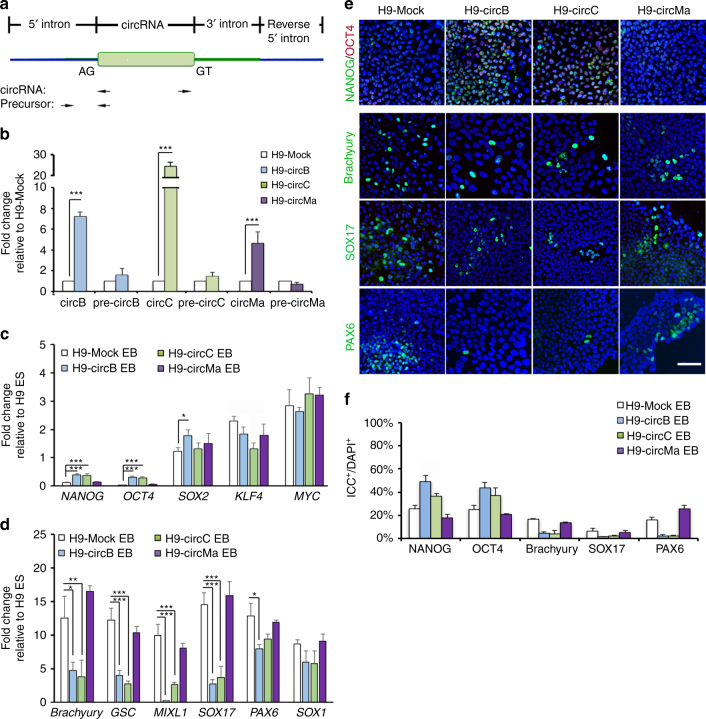
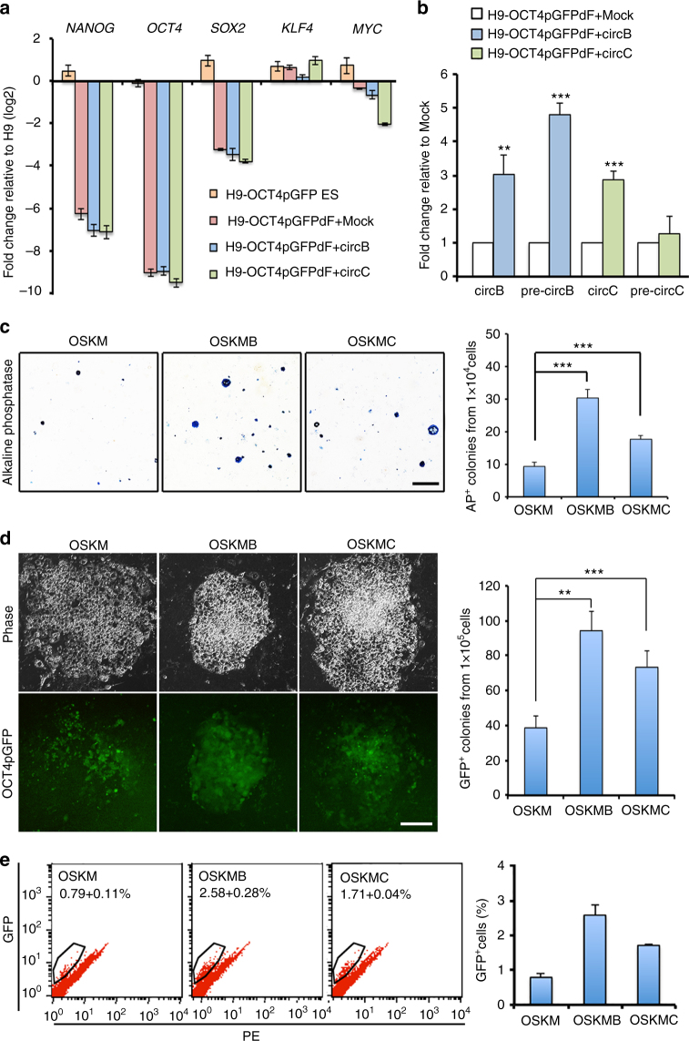
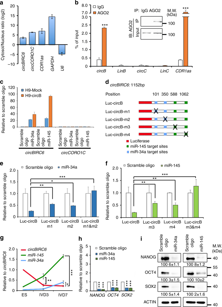
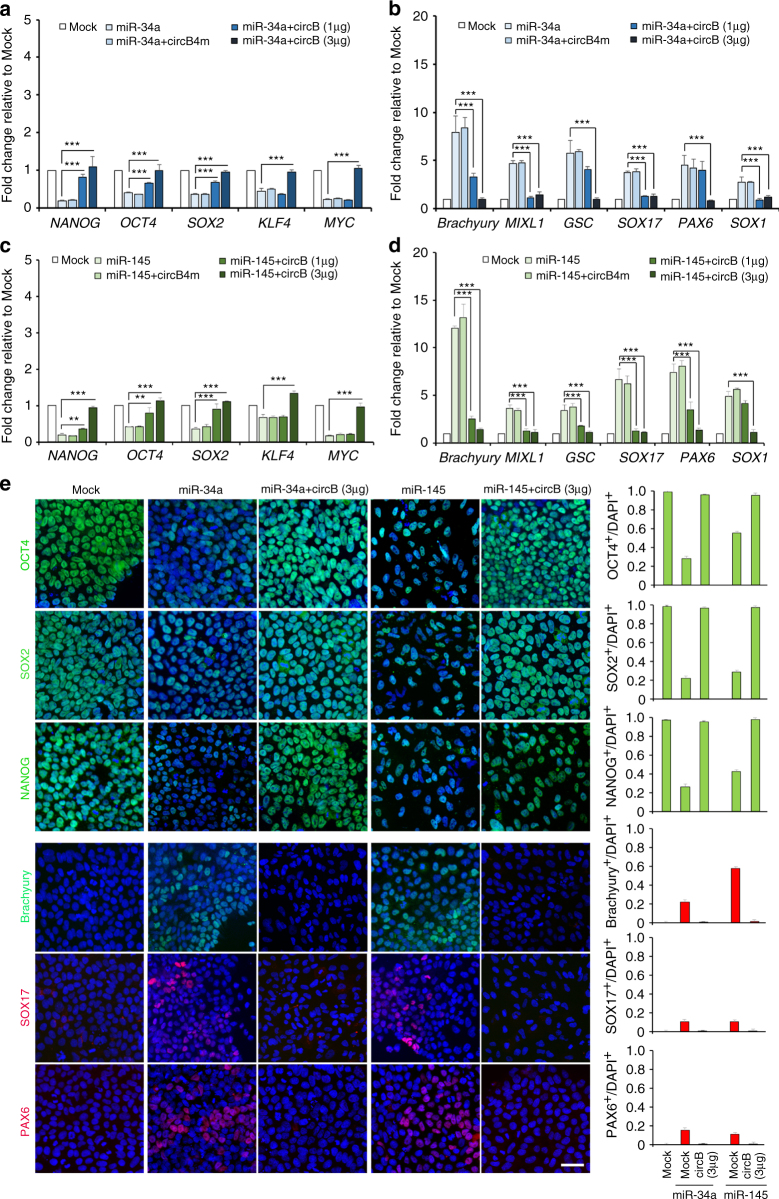
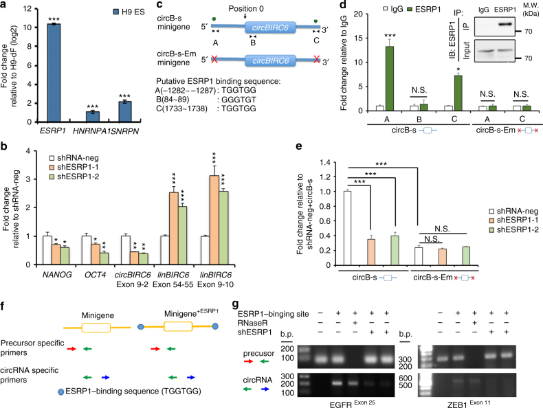
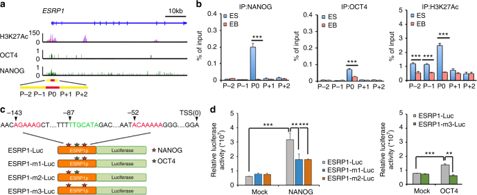
Verification
Claims the paper makes
The claims and statistics of this paper haven’t been checked yet.
Run the check — free, no account needed. It reads the open-access text and checks the sources it cites.
Claims other papers make about this one
Each is checked against this paper’s own text: does it say what the citing paper says it does?
In the citing paper
“In this way, modulation of MRE load with competing endogenous RNAs (ceRNAs), such as with circular RNAs (circRNAs) (96), can act to sequester miRISC from target mRNA, leading to derepression of target mRNA (97).”
From Overview of MicroRNA Biogenesis, Mechanisms of Actions, and Circulation (2018) · cites this paper as [97]
Self-check
Percentages, sums and reported differences re-computed from the paper’s own numbers.
The self-check needs the paper’s full text.
Citations
Cites
An integrated encyclopedia of DNA elements in the human genome
The ENCODE Project Consortium (2012) · Nature · doi:10.1038/nature11247 · Cited by 18,072
Circular RNAs are a large class of animal RNAs with regulatory potency
Memczak, S., Jens, M., Elefsinioti, A., Torti, F., Krueger, J., Rybak, A., Maier, L., Mackowiak, S. D., Gregersen, L. H., Munschauer, M., et al. (2013) · Nature · doi:10.1038/nature11928 · Cited by 7,990
Natural RNA circles function as efficient microRNA sponges
Hansen, T. B., Jensen, T. I., Clausen, B. H., Bramsen, J. B., Finsen, B., Damgaard, C. K., & Kjems, J. (2013) · Nature · doi:10.1038/nature11993 · Cited by 7,985
Circular RNAs are abundant, conserved, and associated with ALU repeats
Jeck, W. R., Sorrentino, J. A., Wang, K., Slevin, M. K., Burd, C. E., Liu, J., Marzluff, W. F., & Sharpless, N. E. (2013) · RNA · doi:10.1261/rna.035667.112 · Cited by 4,388
circRNA biogenesis competes with pre-mRNA splicing
Ashwal-Fluss, R., Meyer, M., Pamudurti, N. R., Ivanov, A., Bartok, O., Hanan, M., Evantal, N., Memczak, S., Rajewsky, N., & Kadener, S. (2014) · Molecular Cell · doi:10.1016/j.molcel.2014.08.019 · Cited by 3,083
The role of site accessibility in microRNA target recognition
Kertesz, M., Iovino, N., Unnerstall, U., Gaul, U., & Segal, E. (2007) · Nature Genetics · doi:10.1038/ng2135 · Cited by 2,460
Circular RNAs in the Mammalian Brain Are Highly Abundant, Conserved, and Dynamically Expressed
Rybak-Wolf, A., Stottmeister, C., Glažar, P., Jens, M., Pino, N., Giusti, S., Hanan, M., Behm, M., Bartok, O., Ashwal-Fluss, R., et al. (2015) · Molecular Cell · doi:10.1016/j.molcel.2015.03.027 · Cited by 2,423
The RNA Binding Protein Quaking Regulates Formation of circRNAs
Conn, S. J., Pillman, K. A., Toubia, J., Conn, V. M., Salmanidis, M., Phillips, C. A., Roslan, S., SCHREIBER, A. W., Gregory, P. A., & Goodall, G. J. (2015) · Cell · doi:10.1016/j.cell.2015.02.014 · Cited by 2,205
Zheng, Q., Bao, C., Guo, W., Li, S., Chen, J., Chen, B., Luo, Y., Lyu, D., Li, Y., Shi, G., et al. (2016) · Nature Communications · doi:10.1038/ncomms11215 · Cited by 2,041
Complementary Sequence-Mediated Exon Circularization
Zhang, X.-O., Wang, H.-B., Zhang, Y., Lu, X., Chen, L.-L., & Yang, L. (2014) · Cell · doi:10.1016/j.cell.2014.09.001 · Cited by 1,999
Cited by
Sorted by citations
Items 101–200 of 346
Lin, X., Chen, W., Wei, F., & Xie, X. (2020) · Molecular Therapy · doi:10.1016/j.ymthe.2020.09.005 · Cited by 44
Li, D., Li, Z., Yang, Y. X., Zeng, X., Li, Y., Du, X., & Zhu, X. (2019) · Environmental Research · doi:10.1016/j.envres.2019.108825 · Cited by 43
Dvir, S., Argoetti, A., Lesnik, C., Roytblat, M., Shriki, K., Amit, M., Hashimshony, T., & Mandel‐Gutfreund, Y. (2021) · Cell Reports · doi:10.1016/j.celrep.2021.109198 · Cited by 39
From circRNAs to fusion circRNAs in hematological malignancies
Babin, L., Andraos, E., Fuchs, S., Pyronnet, S., Brunet, E., & Meggetto, F. (2021) · JCI Insight · doi:10.1172/jci.insight.151513 · Cited by 41
Circular RNA RBPMS inhibits bladder cancer progression via miR-330-3p/RAI2 regulation
Yang, C., Mou, Z., Zhang, Z., Wu, S., Zhou, Q., Chen, Y., Gong, J., Xu, C., Ou, Y., Chen, X., et al. (2021) · Molecular Therapy — Nucleic Acids · doi:10.1016/j.omtn.2021.01.009 · Cited by 40
Zheng, S.-R., Zhang, H.-R., Zhang, Z.-F., Lai, S., Huang, L.-J., Liu, J., Bai, X., Ding, K., & Zhou, J. (2018) · Journal of Cancer · doi:10.7150/jca.24253 · Cited by 40
The roles of circRNAs in cancers: Perspectives from molecular functions
Cao, Y., Sun, J., Chen, Y.-X., Wen, C., & Wei, L. (2020) · Gene · doi:10.1016/j.gene.2020.145182 · Cited by 40
Clinical and functional significance of circular RNAs in cytogenetically normal AML
Papaioannou, D., Volinia, S., Nicolet, D., Świerniak, M., Petri, A., Mrózek, K., Bill, M., Pepe, F., Walker, C. J., Walker, A. E., et al. (2020) · Blood Advances · doi:10.1182/bloodadvances.2019000568 · Cited by 39
Wang, S., Liu, F., Ma, H., Cui, X., Yang, S., & Qin, R. (2020) · Clinical Breast Cancer · doi:10.1016/j.clbc.2020.04.006 · Cited by 39
Yang, P., & Wang, H. H. (2020) · Scientific Reports · doi:10.1038/s41598-020-74452-8 · Cited by 39
Song, P., Zhang, P., Qin, K., Su, F., Gao, K., Liu, X., & Li, Z. (2022) · Talanta · doi:10.1016/j.talanta.2022.123521 · Cited by 37
Shafei, A., ALI, M. A. M., Ghanem, H. G., Shehata, A. I., Abdelgawad, A. A., Handal, H. R., ElSayed, A. S., Ashaal, A. E., Ali, M. M., & El‐Shal, A. S. (2017) · Journal of Cellular Biochemistry · doi:10.1002/jcb.26637 · Cited by 38
Chen, L., Rong, D., Hui, B., He, X., Jiang, W., Xu, Y., Cao, H., Xu, Z., & Tang, W. (2021) · Cell Death Discovery · doi:10.1038/s41420-021-00710-x · Cited by 38
Non-coding RNAs rewire cancer metabolism networks
Lin, X., Wu, Z., Hu, H., Luo, M., & Song, E. (2021) · Seminars in Cancer Biology · doi:10.1016/j.semcancer.2020.12.019 · Cited by 37
CDR1as regulated by hnRNPM maintains stemness of periodontal ligament stem cells via miR‐7/KLF4
Gu, X., Li, X., Jin, Y., Zhang, Z., Li, M., Liu, D., & Wei, F. (2021) · Journal of Cellular and Molecular Medicine · doi:10.1111/jcmm.16541 · Cited by 37
The Role of Circular RNAs in the Physiology and Pathology of the Mammalian Ovary
Zhang, J., Wang, C., Jia, C., Zhang, Y., Qing, X., Zhang, Y., Liu, J., Xu, S., & Pan, Z. (2022) · International Journal of Molecular Sciences · doi:10.3390/ijms232315204 · Cited by 37
Circular RNAs and their participation in stemness of cancer
Lagunas‐Rangel, F. A. (2020) · Medical Oncology · doi:10.1007/s12032-020-01373-x · Cited by 36
Engineering circular RNA regulators to specifically promote circular RNA production
Qi, Y., Han, W., Chen, D., Zhao, J., Bai, L., Huang, F., Dai, Z., Li, G., Chen, C., Zhang, W., et al. (2021) · Theranostics · doi:10.7150/thno.56990 · Cited by 37
The biogenesis, biological functions and modification of Circular RNAs
Liu, S., Guo, X., Shang, Q. J., & Gao, P. (2023) · Experimental and Molecular Pathology · doi:10.1016/j.yexmp.2023.104861 · Cited by 37
Zhang, P., Gao, K., Liang, Y., Su, F., Wang, F., & Li, Z. (2020) · Talanta · doi:10.1016/j.talanta.2020.121021 · Cited by 36
Uhr, K., Sieuwerts, A. M., de Weerd, V., Smid, M., Hammerl, D., Foekens, J. A., & Martens, J. W. M. (2018) · Scientific Reports · doi:10.1038/s41598-018-27987-w · Cited by 34
Chen, F., Guo, L., Di, J., Li, M., Dong, D., & Pei, D. (2021) · Journal of genetics and genomics/Journal of Genetics and Genomics · doi:10.1016/j.jgg.2021.07.017 · Cited by 34
Yang, L., Bin, Z., Hui, S., Li, R., You, B., Wu, P., Han, X., Qian, H., & Xu, W. (2019) · Stem Cells International · doi:10.1155/2019/2316834 · Cited by 31
Circular RNA PRKCI promotes glioma cell progression by inhibiting microRNA-545
Zhang, X., Yang, H., Zhao, L., Li, G., & Duan, Y. (2019) · Cell Death and Disease · doi:10.1038/s41419-019-1863-z · Cited by 33
Ye, Y., Ke, Y., Liu, L., Tong, X., & Yu, J. (2021) · Stem Cells International · doi:10.1155/2021/5177488 · Cited by 33
Han, B., Shaolong, E., Luan, L., Li, N., & Liu, X. (2020) · OncoTargets and Therapy · doi:10.2147/ott.s251436 · Cited by 32
Ma, Y., Xu, Y., Zhang, J., & Zheng, L. (2020) · Reproductive Biology and Endocrinology · doi:10.1186/s12958-020-00653-5 · Cited by 33
Xu, T., Song, X., Wang, Y., Fu, S., & Han, P. (2020) · Frontiers in Cell and Developmental Biology · doi:10.3389/fcell.2020.603516 · Cited by 31
Functions and Potential Applications of Circular RNAs in Cancer Stem Cells
Feng, Z., Meng, S., Zhou, H., Xu, Z., Tang, Y., Li, P., Liu, C., Huang, Y., & Wu, M. (2019) · Frontiers in Oncology · doi:10.3389/fonc.2019.00500 · Cited by 31
Wu, Z., Sun, H., Li, J., & Jin, H. Z. (2019) · Aging · doi:10.18632/aging.102091 · Cited by 31
Profiling and Functional Analysis of Circular RNAs in Porcine Fast and Slow Muscles
Li, B., Yin, D., Li, P., Zhang, Z., Zhang, X., Li, H., Li, R., Hou, L., Liu, H., & Wu, W. (2020) · Frontiers in Cell and Developmental Biology · doi:10.3389/fcell.2020.00322 · Cited by 30
Mou, X., Chenv, J., Zhou, D., Liu, K., Chen, L., Zhou, D., & Hu, Y. (2020) · Molecular Medicine Reports · doi:10.3892/mmr.2020.11486 · Cited by 31
Circular RNAs in stem cells: from basic research to clinical implications
Lu, H., Li, J., Yang, G., Yi, C., Zhang, D., Yu, F., & Ma, Z. (2021) · Bioscience Reports · doi:10.1042/bsr20212510 · Cited by 29
Xu, L., Zhang, Y., Tang, J., Wang, P., Li, L., Yan, X., Zheng, X., Ren, S., Zhang, M., & Xu, M. (2019) · Cancer Epidemiology Biomarkers & Prevention · doi:10.1158/1055-9965.epi-18-0570 · Cited by 28
Non-Coding RNA-Driven Regulation of rRNA Biogenesis
Kaliatsi, E. G., Γιαρίμογλου, Ν., Stathopoulos, C., & Stamatopoulou, V. (2020) · International Journal of Molecular Sciences · doi:10.3390/ijms21249738 · Cited by 28
Direct recognition and sensitive detection of circular RNA with ligation-based PCR
Zhang, P., Guo, N., Gao, K., Su, F., Wang, F., & Li, Z. (2020) · Organic & Biomolecular Chemistry · doi:10.1039/d0ob00625d · Cited by 28
Jakobsen, T., Dahl, M., Dimopoulos, K., Grønbæk, K., Kjems, J., & Kristensen, L. S. (2021) · Cancers · doi:10.3390/cancers13030365 · Cited by 28
Wang, X., Xu, C., Cai, Y., Zou, X., Chao, Y.-Q., Yan, Z., Zou, C., Wu, X., & Tang, L. (2022) · Journal of Allergy and Clinical Immunology · doi:10.1016/j.jaci.2021.10.041 · Cited by 29
Yarmishyn, A. A., Ishola, A. A., Chen, C., Verusingam, N. D., Rengganaten, V., Mustapha, H. A., Chuang, H.-K., Teng, Y., Phung, V., Hsu, P., et al. (2022) · Cancers · doi:10.3390/cancers14040862 · Cited by 28
Exosomal circular RNA: a signature for lung cancer progression
Hussen, B. M., Abdullah, S. R., Faraj, G. S. H., Rasul, M. F., Salihi, A. B., Ghafouri‐Fard, S., Taheri, M. N., & Mokhtari, M. (2022) · Cancer Cell International · doi:10.1186/s12935-022-02793-7 · Cited by 28
Circular RNAs in Embryogenesis and Cell Differentiation With a Focus on Cancer Development
Agostino, S. D., Riccioli, A., Cesaris, P. D., Fontemaggi, G., Blandino, G., Filippini, A., & Fazi, F. (2020) · Frontiers in Cell and Developmental Biology · doi:10.3389/fcell.2020.00389 · Cited by 28
Liu, N., He, J., Fan, D., Gu, Y., Wang, J., Li, H., Zhu, X., Du, Y., Tian, Y., Liu, B., et al. (2022) · Nature Communications · doi:10.1038/s41467-022-32322-z · Cited by 26
CircRNA: A new class of targets for gastric cancer drug resistance therapy
Zheng, Y. F., Li, Z., Wang, Y., Chen, W., Lin, Y., Guo, J., & Ye, G. (2023) · Pathology & Oncology Research · doi:10.3389/pore.2023.1611033 · Cited by 26
Mfossa, A. C. M., Puthenparampil, H. T., Inalegwu, A., Coolkens, A., Baatout, S., Benotmane, M. A., Huylebroeck, D., & Quintens, R. (2019) · Cells · doi:10.3390/cells8080778 · Cited by 24
circ‐ZNF609: A potent circRNA in human cancers
Qian, Y., Yang, L., Li, R., Yang, T., Jia, R.-P., & Ge, Y. (2021) · Journal of Cellular and Molecular Medicine · doi:10.1111/jcmm.16996 · Cited by 24
Emergent Roles of Circular RNAs in Metabolism and Metabolic Disorders
Wu, Y.-L., Li, H.-F., Chen, H.-H., & Lin, H. (2022) · International Journal of Molecular Sciences · doi:10.3390/ijms23031032 · Cited by 25
Self-citationTherapeutic and diagnostic applications of exosomal circRNAs in breast cancer
Gopikrishnan, M., Hephzibah Cathryn, R., Ramanathan, G., Ashour, H. M., Pintus, G., Hammad, M., Kashyap, M. K., Doss, C. G. P., & Zayed, H. (2023) · Functional & Integrative Genomics · doi:10.1007/s10142-023-01083-3 · Cited by 27
Wei, S.-H., Hu, J., Sheng, Z., Zhang, Q., Zhang, J., Zhang, B., Liu, M., & Zhang, C. (2023) · Analytical Chemistry · doi:10.1021/acs.analchem.3c05116 · Cited by 25
Circular RNAs as key regulators in cancer hallmarks: New progress and therapeutic opportunities
Hussen, B. M., Abdullah, S. R., Jaafar, R. M., Rasul, M. F., Aroutiounian, R. M., Harutyunyan, T. A., Liehr, T., Samsami, M., & Taheri, M. N. (2025) · Critical Reviews in Oncology/Hematology · doi:10.1016/j.critrevonc.2024.104612 · Cited by 24
Hong, L., Ma, X., Liu, J., Luo, Y., Lin, J., Shen, Y., & Zhang, L. (2021) · Pulmonary Circulation · doi:10.1177/20458940211000234 · Cited by 24
Decrease of circARID1A retards glioblastoma invasion by modulating miR-370-3p/ TGFBR2 pathway
Li, B., Chen, J., Wu, Y., Luo, H., & Ke, Y. (2022) · International Journal of Biological Sciences · doi:10.7150/ijbs.66673 · Cited by 24
Self-citationXia, W., Mao, Q., Chen, B., Wang, L., Ma, W., Liang, Y., Zhang, T., Dong, G., Xu, L., & Jiang, F. (2019) · Oncogenesis · doi:10.1038/s41389-019-0167-6 · Cited by 24
Hu, H., Tang, J., Wang, H., Guo, X., Tu, C., & Li, Z. (2024) · Cellular & Molecular Biology Letters · doi:10.1186/s11658-024-00662-x · Cited by 23
Wang, Y., Zhang, Y., Su, X., Qiu, Q., Yuan, Y., Weng, C., Zou, S., Tian, Y., Han, W., Liu, P., et al. (2022) · International Journal of Biological Sciences · doi:10.7150/ijbs.69351 · Cited by 23
He, T., Tao, W., Zhang, L., Wang, B.-Y., Li, K., Lü, H., Tang, G., He, Y., & Ling, L. (2022) · Cell Death and Disease · doi:10.1038/s41419-022-04913-7 · Cited by 23
Xue, H., Yu, F., Zhang, X., Liu, L., & Huang, L. (2020) · Ecotoxicology and Environmental Safety · doi:10.1016/j.ecoenv.2020.110455 · Cited by 23
Circular RNAs in stem cell differentiation: a sponge-like role for miRNAs
Zhou, J. P., Qiu, C., Fan, Z., Liu, T., & Liu, T. (2021) · International Journal of Medical Sciences · doi:10.7150/ijms.56457 · Cited by 23
Circular RNAs: history, metabolism, mechanisms of function, and regulatory roles at a glance
Karimi, R., Javandoost, E., Asadmasjedi, N., Atashi, A., Soleimani, A., & Behzadifard, M. (2024) · Annals of Medicine and Surgery · doi:10.1097/ms9.0000000000002761 · Cited by 25
Brozzi, F., & Regazzi, R. (2021) · International Journal of Molecular Sciences · doi:10.3390/ijms22041503 · Cited by 22
A Circular RNA Expressed from the FAT3 Locus Regulates Neural Development
Seeler, S., Andersen, M. S., Sztanka-Toth, T. R., Rybiczka-Tešulov, M., van den Munkhof, M. H., Chang, C.-C., Maimaitili, M., Venø, M. T., Hansen, T. B., Pasterkamp, R. J., et al. (2023) · Molecular Neurobiology · doi:10.1007/s12035-023-03253-7 · Cited by 22
Altered Expression Profile of Circular RNAs in Conjunctival Melanoma
Shang, Q., Li, Y., Wang, H., Ge, S., & Jia, R. (2019) · Epigenomics · doi:10.2217/epi-2019-0029 · Cited by 22
Tip of the Iceberg: Roles of CircRNAs in Cancer Glycolysis
Tan, L., Xian, H.-C., Dai, L., Tang, Y., & Liang, X. (2021) · OncoTargets and Therapy · doi:10.2147/ott.s297140 · Cited by 21
circCYP24A1 promotes Docetaxel resistance in prostate Cancer by Upregulating ALDH1A3
Yin, H., Qin, H., Yang, L., Chen, M., Yang, Y., Zhang, W., Hao, J., Lu, Q., Shi, J., Zhuang, J., et al. (2022) · Biomarker Research · doi:10.1186/s40364-022-00393-1 · Cited by 22
Rahmani-Kukia, N., & Abbasi, A. (2022) · Molecular Therapy — Nucleic Acids · doi:10.1016/j.omtn.2022.08.012 · Cited by 22
Zhang, Y., Yao, H., Li, Y., Yang, L., Zhang, L., Chen, J., Wang, Y., & Li, X. (2021) · Oncology Reports · doi:10.3892/or.2021.8152 · Cited by 21
Zhao, J., Lin, Z., Ying, P., Zhao, Z., Yang, H., Qian, J., Gong, Y. L., Zhou, Y., Dai, Y., Jiao, Y., et al. (2022) · Journal of Crohn s and Colitis · doi:10.1093/ecco-jcc/jjac154 · Cited by 20
Bi, T., Lu, Q., Pan, X., Dong, F., Hu, Y., Xu, Z., Xiu, P., Liu, Z., & Li, J. (2023) · Cell Cycle · doi:10.1080/15384101.2023.2249302 · Cited by 20
Rengganaten, V., Huang, C.-J., Wang, M., Chien, Y., Tsai, P., Lan, Y., Ong, H. T., Chiou, S., & Choo, K. (2023) · BMC Cancer · doi:10.1186/s12885-023-11571-1 · Cited by 20
Hsa_circ_0015278 Regulates FLT3-ITD AML Progression via Ferroptosis-Related Genes
Jiang, J., Feng, J. Q., Song, X., Yang, Q., Zhao, H., Zhao, R., He, X., Tian, Y., Wang, L., & Liu, Y. (2022) · Cancers · doi:10.3390/cancers15010071 · Cited by 20
Ruan, X., Liu, Y., Wang, P., Liu, L., Ma, T., Xue, Y., Dong, W., Zhao, Y., Tiange, E., Lin, H., et al. (2023) · Cell Death and Disease · doi:10.1038/s41419-023-06269-y · Cited by 19
Circular RNAs: Diversity of Functions and a Regulatory Nova in Oral Medicine: A Pilot Review
Wang, H., Cheng, F., Wang, M., Yang, S., & Wei, F. (2019) · Cell Transplantation · doi:10.1177/0963689719837917 · Cited by 19
Liu, J., Zhu, H., Fu, L., & Xu, T. (2021) · Frontiers in Genetics · doi:10.3389/fgene.2021.653051 · Cited by 19
Divisato, G., Piscitelli, S., Elia, M. A., Cascone, E., & Parisi, S. (2021) · Biomolecules · doi:10.3390/biom11081074 · Cited by 19
Cai, Y., Zhao, X., Chen, D., Zhang, F., Chen, Q., Shao, C.-C., Ouyang, Y.-X., Feng, J. L., Cui, L., Chen, M., et al. (2021) · Molecular Therapy — Nucleic Acids · doi:10.1016/j.omtn.2021.09.013 · Cited by 19
The function and clinical implication of circular RNAs in lung cancer
Ren, W., Yuan, Y., Peng, J., Mutti, L., & Jiang, X. (2022) · Frontiers in Oncology · doi:10.3389/fonc.2022.862602 · Cited by 18
Xie, F., Zhao, Y. F., Wang, S., Ma, J., Wang, X., & Qian, L. (2018) · Journal of Cellular Biochemistry · doi:10.1002/jcb.27614 · Cited by 18
Biological functions of circRNAs and their progress in livestock and poultry
Huang, Y., Wang, Y., Zhang, C., & Sun, X. (2020) · Reproduction in Domestic Animals · doi:10.1111/rda.13816 · Cited by 18
Circr, a Computational Tool to Identify miRNA:circRNA Associations
Dori, M., Caroli, J., & Forcato, M. (2022) · Frontiers in Bioinformatics · doi:10.3389/fbinf.2022.852834 · Cited by 17
Zhang, R., Pan, Y., Zou, C.-X., An, Q., Cheng, J., Li, P.-J., Zheng, Z., Pan, Y., Feng, W., Yang, S., et al. (2022) · BMC Genomics · doi:10.1186/s12864-022-08518-4 · Cited by 17
ESRP1-Induced CD44 v3 Is Important for Controlling Pluripotency in Human Pluripotent Stem Cells
Kim, Y., Kim, H., Lee, J., Choi, J.-K., Han, E., Jeong, J. E., & Cho, Y. S. (2018) · Stem Cells · doi:10.1002/stem.2864 · Cited by 17
The Secret Garden of Neuronal circRNAs
Gasparini, S., Licursi, V., Presutti, C., & Mannironi, C. (2020) · Cells · doi:10.3390/cells9081815 · Cited by 15
Chen, X., Liu, C., Ji, L., Wang, N., Liu, Y., Wang, M., & Ruan, L. (2021) · Biochemical and Biophysical Research Communications · doi:10.1016/j.bbrc.2021.04.028 · Cited by 17
CircITCH: A Circular RNA With Eminent Roles in the Carcinogenesis
Ghafouri‐Fard, S., Khoshbakht, T., Taheri, M. N., & Jamali, E. (2021) · Frontiers in Oncology · doi:10.3389/fonc.2021.774979 · Cited by 17
Signal Amplification-Based Biosensors and Application in RNA Tumor Markers
Li, H., Zhang, Z., Gan, L., Fan, D., Sun, X., Qian, Z., Liu, X., & Huang, Y. (2023) · Sensors · doi:10.3390/s23094237 · Cited by 16
Post-transcriptional dynamics and RNA homeostasis in autophagy and cancer
Kolapalli, S. P., Nielsen, T. M., & Frankel, L. B. (2023) · Cell Death and Differentiation · doi:10.1038/s41418-023-01201-5 · Cited by 15
The role of circHmbox1(3,4) in ferroptosis-mediated cognitive impairments induced by manganese
Wei, S., Ma, X., Liang, G., He, J., Wang, J., Chen, H., Lu, W., Qin, H., & Zou, Y. (2024) · Journal of Hazardous Materials · doi:10.1016/j.jhazmat.2024.135212 · Cited by 16
Tang, L., Ji, Y., Ni, C., Xu, Z., Shen, Y., Lu, H., Zhang, C., & Yang, S. (2025) · Advanced Science · doi:10.1002/advs.202411869 · Cited by 16
Emerging roles of circular RNAs in stem cells
Wang, M., Wu, J., Wu, P., & Li, Y. (2022) · Genes & Diseases · doi:10.1016/j.gendis.2022.05.015 · Cited by 15
Chen, K., Liu, Z., Lu, J., Yang, S., Hu, X., & Liang, G. (2024) · Cardiovascular Drugs and Therapy · doi:10.1007/s10557-024-07557-1 · Cited by 16
Pu, J., Yan, X., & Zhang, H. (2024) · Journal of Advanced Research · doi:10.1016/j.jare.2024.11.032 · Cited by 15
Chen, D., Wu, W., Yi, L., Feng, Y., Chang, C., Chen, S., Gao, J., Chen, G., & Zhen, G. (2021) · Frontiers in Molecular Biosciences · doi:10.3389/fmolb.2021.703307 · Cited by 15
Functions and clinical significance of circular RNAs in acute myeloid leukemia
Zhou, M., Gao, X., Zheng, X., & Jing, L. (2022) · Frontiers in Pharmacology · doi:10.3389/fphar.2022.1010579 · Cited by 14
CircNDST1 Regulates Bovine Myoblasts Proliferation and Differentiation via the miR-411a/Smad4 Axis
Yang, Z., Song, C., Jiang, R., Huang, Y., Lan, X., Lei, C., Qi, X., Zhang, C., Huang, B., & Chen, H. (2022) · Journal of Agricultural and Food Chemistry · doi:10.1021/acs.jafc.1c08167 · Cited by 14
Sun, X., Kang, Y. Q., Li, M., Li, Y., & Song, J. (2022) · Biochimica et Biophysica Acta (BBA) - Gene Regulatory Mechanisms · doi:10.1016/j.bbagrm.2022.194888 · Cited by 15
Krishnan, R., Murari, L. S. S., Akshaya, R. L., Gomathi, K., Saranya, I., Das, U. R., Satishkumar, S., & Selvamurugan, N. (2022) · International Journal of Biological Macromolecules · doi:10.1016/j.ijbiomac.2022.11.176 · Cited by 13
Noncoding RNAs: Potential players in the self‐renewal of mammalian spermatogonial stem cells
Bie, B., Wang, Y., Li, L., Fang, H., Liu, L., & Sun, J. (2018) · Molecular Reproduction and Development · doi:10.1002/mrd.23041 · Cited by 13
A circular RNA map for human induced pluripotent stem cells of foetal origin
Barilani, M., Cherubini, A., Peli, V., Polveraccio, F., Bollati, V., Guffanti, F., GOBBO, A. D., Lavazza, C., Giovanelli, S., Elvassore, N., et al. (2020) · EBioMedicine · doi:10.1016/j.ebiom.2020.102848 · Cited by 13
Wei, T., Sui, H., Su, Y., Cheng, W., Liu, Y., He, Z., Ji, Q., & Xu, C.-L. (2020) · Molecular Medicine Reports · doi:10.3892/mmr.2020.11607 · Cited by 13
Role of circular RNAs in lung cancer
Babayev, M., & Silveyra, P. (2024) · Frontiers in Genetics · doi:10.3389/fgene.2024.1346119 · Cited by 13
Imaging and quantification of human and viral circular RNAs
Jaijyan, D. K., Yang, S., Ramasamy, S., Gu, A., Zeng, M., Subbian, S., Tyagi, S., & Zhu, H. (2024) · Nucleic Acids Research · doi:10.1093/nar/gkae583 · Cited by 12
Metadata
- Authors
- Yu, C.-Y., Li, T.-C., Wu, Y.-Y., Yeh, C.-H., Chiang, W., Chuang, C.-Y., & Kuo, H.-C.
- Affiliations
- Institute of Cellular and Organismic Biology, Academia Sinica (TW)
- Genomics Research Center, Academia Sinica (TW)
- National Taiwan University (TW)
- Corresponding author
- Kuo, Hung-Chih
- Venue
- Nature Communications
- Published
- 2017-10-27
- Volume
- 8
- Issue
- 1
- Pages
- 1149-1149
- ISSN
- 2041-1723 (ISSN-L)
- PMC
- PMC5658440
- OpenAlex
- W2766512489
- Cited by
- 336
- References
- 43
- Citations by year
- 2026: 10 · 2025: 20 · 2024: 26 · 2023: 23 · 2022: 63 · 2021: 72 · 2020: 50 · 2019: 43 · 2018: 26 · 2017: 2
- FWCI
- 13.74
- Type
- journal article
- Language
- en
- Open access
- gold
- doi.org
- Locations
- Nature Communications (publishedVersion, cc-by)
- PubMed Central (publishedVersion, cc-by)
- PubMed (publishedVersion)
- RePEc: Research Papers in Economics (submittedVersion)
- DOAJ (DOAJ: Directory of Open Access Journals) (submittedVersion, cc-by-sa)
- Funders
- Academia Sinica (AS-104-TP-B09, 105-0210-01-13-01, AS‐103‐TP‐B10)
- Ministry of Science and Technology, Taiwan (MOST103-2321-B001-065)
- National Health Research Institutes (NHRI-EX104-10320SI, EX104)
- Topics
- Life Sciences › Biochemistry, Genetics and Molecular Biology › Molecular Biology › Circular RNAs in diseases
- Life Sciences › Biochemistry, Genetics and Molecular Biology › Molecular Biology › RNA Research and Splicing
- Life Sciences › Biochemistry, Genetics and Molecular Biology › Molecular Biology › Pluripotent Stem Cells Research
- Keywords
- circular RNA, human embryonic stem cells, miR-34a, miR-145, microRNA sponge, ESRP1, splicing factors, Oct4, Nanog, cell differentiation, circRNA biogenesis
- MeSH terms
- Nanog Homeobox Protein, RNA, Circular, Animals, Cell Differentiation, Exoribonucleases, Humans, Mice, Inbred ICR, Microfilament Proteins, RNA, Gene Expression Profiling, MicroRNAs, Pluripotent Stem Cells, Octamer Transcription Factor-3, Inhibitor of Apoptosis Proteins, Mice, Embryonic Stem Cells, Argonaute Proteins
About these checks
Registry, retraction, Cites and statistics results are computed, not judged, with one exception: a printed reference that no index lists is read once by a model before it counts as a citation error. Claims are extracted by a model and kept only when their quote appears verbatim in the text; each cited source is then checked, with a model’s help, for whether it says what the claim relies on. Those verdicts can be wrong — read the claim’s quote and the cited source before relying on one.
Statistics and arithmetic are recomputed, not judged.
Confidence bands are shown only for fields calibrated on our annotated test set. None is calibrated yet, so no band is shown.
What counts as a claim ›How we measure confidence ›How we check this ›
Are you an author, or do you see a problem with this page? Request a review
