

The DOI matches its Crossref record; 70 of the 70 references it cites were found in a registry or an index.
Competitive repopulation of an empty microglial niche yields functionally distinct subsets of microglia-like cells
Open accessAbstract
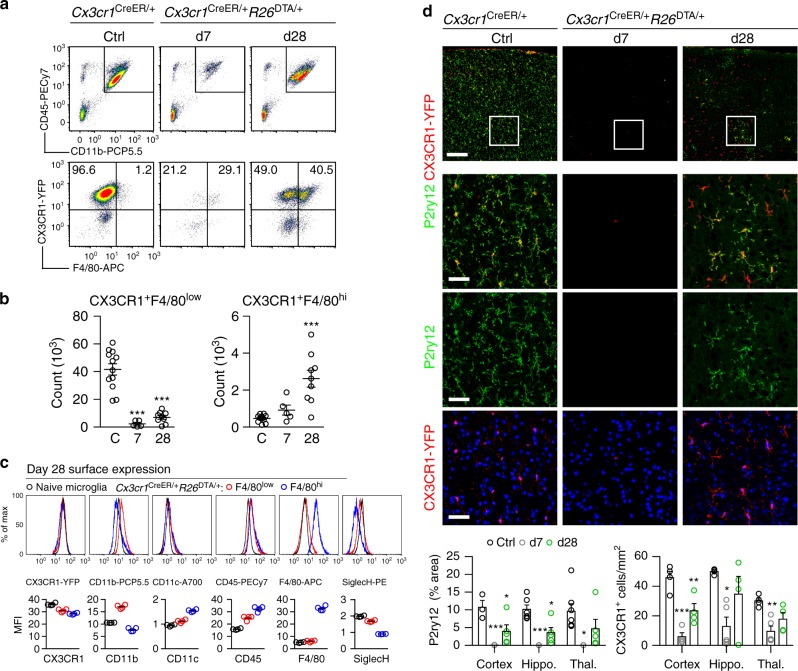
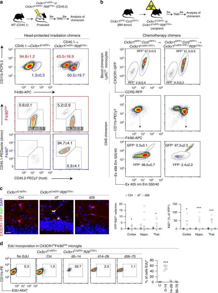
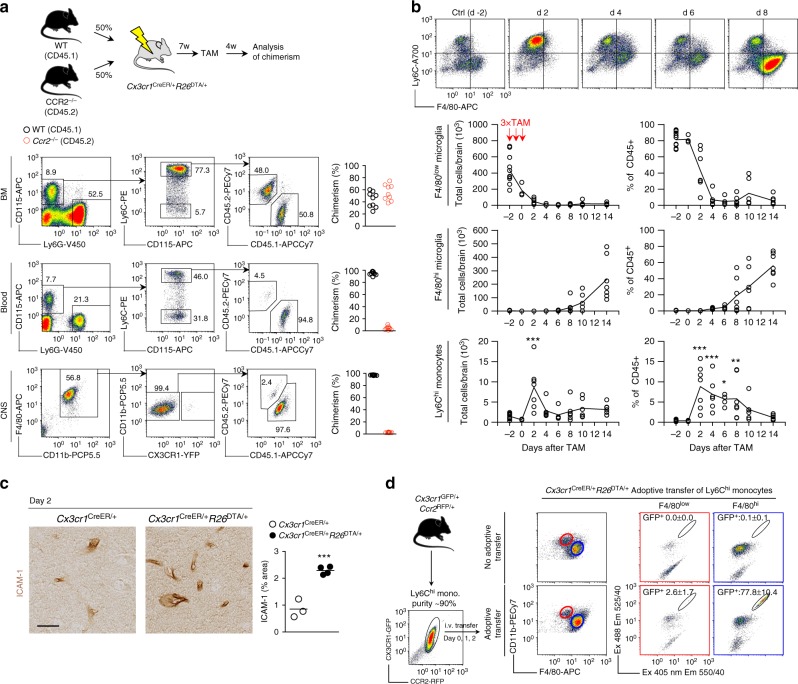
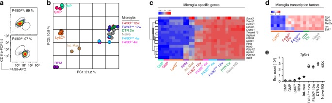
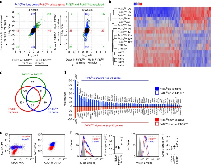
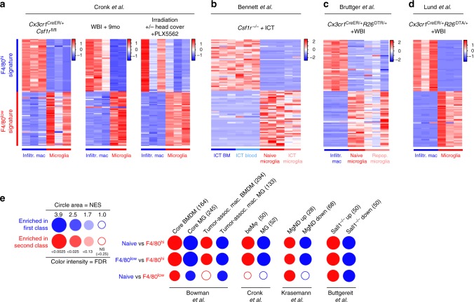
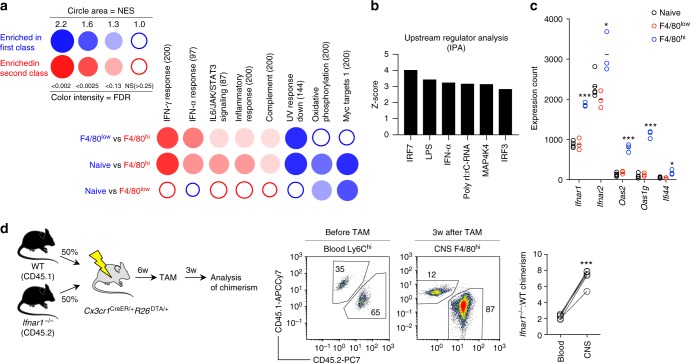
Circulating monocytes can compete for virtually any tissue macrophage niche and become long-lived replacements that are phenotypically indistinguishable from their embryonic counterparts. As the factors regulating this process are incompletely understood, we studied niche competition in the brain by depleting microglia with >95% efficiency using Cx3cr1 CreER/+ R26 DTA/+ mice and monitored long-term repopulation. Here we show that the microglial niche is repopulated within weeks by a combination of local proliferation of CX3CR1 + F4/80 low Clec12a – microglia and infiltration of CX3CR1 + F4/80 hi Clec12a + macrophages that arise directly from Ly6C hi monocytes. This colonization is independent of blood brain barrier breakdown, paralleled by vascular activation, and regulated by type I interferon. Ly6C hi monocytes upregulate microglia gene expression and adopt microglia DNA methylation signatures, but retain a distinct gene signature from proliferating microglia, displaying altered surface marker expression, phagocytic capacity and cytokine production. Our results demonstrate that monocytes are imprinted by the CNS microenvironment but remain transcriptionally, epigenetically and functionally distinct.
Overview
The claims and statistics of this paper haven’t been checked yet.
Run the check — free, no account needed. It reads the open-access text and checks the sources it cites.
Integrity signals
Registry
Cites
Statistics
Figures
Verification
Claims the paper makes
The claims and statistics of this paper haven’t been checked yet.
Run the check — free, no account needed. It reads the open-access text and checks the sources it cites.
Claims other papers make about this one
Each is checked against this paper’s own text: does it say what the citing paper says it does?
The cited source doesn’t support the claim as written.
In the citing paper
“Moreover, recent studies have reported that during aging or neurodegeneration, bone marrow-derived blood monocytes can infiltrate the brain through a disrupted blood-brain barrier (BBB) and differentiate into microglia-like cells, exhibiting transcriptional and functional similarity to brain-resident microglia13–15.”
From Somatic cancer variants enriched in Alzheimer’s disease microglia-like cells drive inflammatory and proliferative states (2026) · cites this paper as [13]
Checked by a reasoning model — may be wrong.
Experimental — accuracy not yet measuredThe cited source doesn’t support the claim as written.
In the citing paper
“Interestingly, MoMg and to a lesser extend HSC-M F displayed expression of Sall1 , as compared to leptomeningeal M F ( Fig. 4f ), suggesting that also engrafted cells in BM chimeras can acquire this proposed 'microglia signature' marker 14,18,19 .”
From Clonal hematopoiesis-associated motoric deficits caused by monocyte-derived microglia accumulating in aging mice (2025) · cites this paper as [19]
Checked by a reasoning model — may be wrong.
Experimental — accuracy not yet measuredThe cited source says what the claim attributes to it.
In the citing paper
“Marrow-derived cells may have an ameliorative effect on neurodegenerative phenotypes in mice33,34, possibly due to their superior phagocytic capacity35.”
From Clonal hematopoiesis is associated with protection from Alzheimer’s disease (2023) · cites this paper as [35]
The cited source says what the claim attributes to it.
In the citing paper
“This includes, for instance, lack of expression of the transcriptional repressor Spalt Like Transcription Factor 1 (Sall1), a proposed hallmark of microglia identity 14,18,19 .”
From Clonal hematopoiesis-associated motoric deficits caused by monocyte-derived microglia accumulating in aging mice (2025) · cites this paper as [19]
Self-check
Percentages, sums and reported differences re-computed from the paper’s own numbers.
The self-check needs the paper’s full text.
Citations
Cites
1 retracted
Microglia-dependent synapse loss in type I interferon-mediated lupus
Bialas, A. R., Presumey, J., Das, A., van der Poel, C. E., Lapchak, P. H., Mesin, L., Victora, G., Tsokos, G. C., Mawrin, C., Herbst, R., et al. (2017) · Nature · doi:10.1038/nature22821 · Cited by 197
RetractedThe Molecular Signatures Database Hallmark Gene Set Collection
Liberzon, A., Birger, C., Thorvaldsdóttir, H., Ghandi, M., Mesirov, J. P., & Tamayo, P. (2015) · Cell Systems · doi:10.1016/j.cels.2015.12.004 · Cited by 13,807
Fate Mapping Analysis Reveals That Adult Microglia Derive from Primitive Macrophages
Ginhoux, F., Greter, M., Leboeuf, M., Nandi, S., See, P., Gokhan, S., Mehler, M. F., Conway, S. J., Ng, L. G., Stanley, E. R., et al. (2010) · Science · doi:10.1126/science.1194637 · Cited by 5,175
Fate Mapping Reveals Origins and Dynamics of Monocytes and Tissue Macrophages under Homeostasis
Yona, S., Kim, K.-W., Wolf, Y., Mildner, A., Varol, D., Breker, M., Strauss-Ayali, D., Viukov, S., Guilliams, M., Misharin, A., et al. (2013) · Immunity · doi:10.1016/j.immuni.2012.12.001 · Cited by 3,245
Krasemann, S., Madore, C., Cialic, R., Baufeld, C., Calcagno, N., El Fatimy, R., Beckers, L., O’Loughlin, E., Xu, Y., Fanek, Z., et al. (2017) · Immunity · doi:10.1016/j.immuni.2017.08.008 · Cited by 2,979
Self-citationMicroglia promote learning-dependent synapse formation through brain-derived neurotrophic factor
Parkhurst, C. N., Yang, G., Ninan, I., Savas, J. N., Yates, J. R., Lafaille, J. J., Hempstead, B. L., Littman, D. R., & Gan, W.-B. (2013) · Cell · doi:10.1016/j.cell.2013.11.030 · Cited by 2,599
A Lineage of Myeloid Cells Independent of Myb and Hematopoietic Stem Cells
Schulz, C., Perdiguero, E. G., Chorro, L., Szabo-Rogers, H., Cagnard, N., Kierdorf, K., Prinz, M., Wu, B., Jacobsen, S. E. W., Pollard, J. W., et al. (2012) · Science · doi:10.1126/science.1219179 · Cited by 2,578
Identification of a unique TGF-β–dependent molecular and functional signature in microglia
Butovsky, O., Jedrychowski, M. P., Moore, C. S., Cialic, R., Lanser, A. J., Gabriely, G., Koeglsperger, T., Dake, B., Wu, P. M., Doykan, C. E., et al. (2014) · Nature Neuroscience · doi:10.1038/nn.3599 · Cited by 2,543
Self-citationTissue-resident macrophages originate from yolk-sac-derived erythro-myeloid progenitors
Gomez Perdiguero, E., Klapproth, K., Schulz, C., Busch, K., Azzoni, E., Crozet, L., Garner, H., Trouillet, C., de Bruijn, M. F., Geissmann, F., et al. (2015) · Nature · doi:10.1038/nature13989 · Cited by 2,339
Hashimoto, D., Chow, A., Noizat, C., Teo, P., Beasley, M. B., Leboeuf, M., Becker, C. D., See, P., Price, J., Lucas, D., et al. (2013) · Immunity · doi:10.1016/j.immuni.2013.04.004 · Cited by 2,311
Cited by
Sorted by citations
Items 201–249 of 249
Xavier, A. M., Belhocine, S., & Gosselin, D. (2019) · WIREs Systems Biology and Medicine · doi:10.1002/wsbm.1449 · Cited by 1
Microglial Replacement and COURIER or SPIT for Neuronal Gene EditingPreprint
Renteln, M. (2024) · Preprints.org · doi:10.20944/preprints202404.1620.v3
Activation of Microglia and Macrophages in Neurodegenerative Diseases
Salamero‐Boix, A., Schulz, M., & Sevenich, L. (2021) · Neuromethods · doi:10.1007/978-1-0716-1712-0_2
VEGF-C expression in TAMs rewires the metastatic fate of breast cancer cellsPreprint
Banerjee, K., Kerzel, T., Bekkhus, T., Ferreira, S. D. S., Wallmann, T., Wallerius, M., Landwehr, L., Agardy, D. A., Schauer, N., Malmerfeldt, A., et al. (2022) · bioRxiv (Cold Spring Harbor Laboratory) · doi:10.1101/2022.01.26.468593
Wada, H., Otsuka, R., Germeraad, W. T. V., Murata, T., Kondo, T., & Seino, K. (2022) · Research Square · doi:10.21203/rs.3.rs-2236823/v1
Timmerman, R. (2022) · Utrecht University Repository (Utrecht University) · doi:10.33540/1603
Zhou, K., Zisiadis, G. A., Havermans, M., Dominguez, C. A., Ohshima, M., Rodrigues, C. F. D., & Blomgren, K. (2023) · Research Square · doi:10.21203/rs.3.rs-2681591/v1
Unleashing the Potential of Natural Killer Cells in Immunotherapy for Glioblastoma and Brain Tumors
Farhangnia, P., Khorramdelazad, H., Ghiaee, P., Fard, S. R., & Delbandi, A. (2023) · Interdisciplinary cancer research · doi:10.1007/16833_2023_186
Etebar, F. (2023) · Queensland University of Technology · doi:10.5204/thesis.eprints.244774
Ma, W., Zhao, L., Xu, B., et al. (2024) · eLife · doi:10.7554/elife.90695.2.sa1
Ma, W., Zhao, L., Xu, B., et al. (2024) · eLife · doi:10.7554/elife.90695.2.sa2
Ma, W., Zhao, L., Xu, B., Fariss, R. N., Redmond, T. M., Zou, J., Wong, W. T., & Li, W. (2024) · eLife · doi:10.7554/elife.90695.2.sa0
Revision to the origins of the mononuclear phagocyte
Cuda, C. M., & Perlman, H. (2024) · Elsevier eBooks · doi:10.1016/b978-0-443-23947-2.00047-3
Pido-Lopez, J., Shaban, E., Moula, S. E., Chritchely, B., Whittaker, T. E., Svensson, S., Virmouni, S. A., Kalef-Ezra, E., Carr, L., Hassell, J. K., et al. (2024) · SSRN Electronic Journal · doi:10.2139/ssrn.5026639
Microglia Adopt Temporally Specific States after Irradiation, Correlating with Neuronal AsynchronyPreprint
Romero, A. L., Preka, E., Pizzirusso, G., Arroyo‐García, L. E., Zisiadis, G. A., Oliva‐Vilarnau, N., Ruchiy, Y., Seitz, T., Zhou, K., Isla, A. G., et al. (2025) · bioRxiv (Cold Spring Harbor Laboratory) · doi:10.1101/2025.02.15.638339
A restarted life: Resetting microglia innate immune memory
Cheng, F., Yan, B., Chen, F., & Lei, P. (2025) · Neuroscience & Biobehavioral Reviews · doi:10.1016/j.neubiorev.2025.106206
Ontogeny and function of microglia and nerve-associated macrophages
Mayer, M., Taveras, E. F., & Mass, E. (2025) · Advances in immunology · doi:10.1016/bs.ai.2025.04.002
CSF1R inhibition during cranial radiotherapy reshapes the immune environment and glial dynamicsPreprint
Leter, Y. M., Heijningen, C. L. V., Tolboom, Z. J., de Gooijer, M. C., Compter, A., van Tellingen, O., Schagen, S. B., & Kuil, L. E. (2025) · Research Square · doi:10.1101/2025.10.11.681366
Additional file 5 of CSF1R antagonism results in increased supraspinal infiltration in EAE
Wang, M., Caryotakis, S. E., Smith, G., Nguyen, A. V., Pleasure, D., & Soulika, A. M. (2024) · Figshare · doi:10.6084/m9.figshare.26709314
Additional file 1 of CSF1R antagonism results in increased supraspinal infiltration in EAE
Wang, M., Caryotakis, S. E., Smith, G., Nguyen, A. V., Pleasure, D., & Soulika, A. M. (2024) · Figshare · doi:10.6084/m9.figshare.25658576
Additional file 3 of CSF1R antagonism results in increased supraspinal infiltration in EAE
Wang, M., Caryotakis, S. E., Smith, G., Nguyen, A. V., Pleasure, D., & Soulika, A. M. (2024) · Figshare · doi:10.6084/m9.figshare.26709308
Hohsfield, L. A., Najafi, A. R., Ghorbanian, Y., Soni, N., Hingco, E. E., Kim, S. J., Jue, A. D., Swarup, V., Inlay, M. A., & Green, K. N. (2020) · Figshare · doi:10.6084/m9.figshare.12981238
Additional file 7 of CSF1R antagonism results in increased supraspinal infiltration in EAE
Wang, M., Caryotakis, S. E., Smith, G., Nguyen, A. V., Pleasure, D., & Soulika, A. M. (2024) · Figshare · doi:10.6084/m9.figshare.26709320
Muñoz‐Castro, C., Mejias‐Ortega, M., Sánchez‐Mejías, E., Navarro, V., Trujillo‐Estrada, L., Jiménez, S., León, J. A. G., Fernandez-Valenzuela, J. J., Sanchez-Mico, M. V., Romero‐Molina, C., et al. (2024) · Figshare · doi:10.6084/m9.figshare.26571967
Additional file 8 of CSF1R antagonism results in increased supraspinal infiltration in EAE
Wang, M., Caryotakis, S. E., Smith, G., Nguyen, A. V., Pleasure, D., & Soulika, A. M. (2024) · Figshare · doi:10.6084/m9.figshare.26709323
Additional file 4 of CSF1R antagonism results in increased supraspinal infiltration in EAE
Wang, M., Caryotakis, S. E., Smith, G., Nguyen, A. V., Pleasure, D., & Soulika, A. M. (2024) · Figshare · doi:10.6084/m9.figshare.26709311
Additional file 2 of CSF1R antagonism results in increased supraspinal infiltration in EAE
Wang, M., Caryotakis, S. E., Smith, G., Nguyen, A. V., Pleasure, D., & Soulika, A. M. (2024) · Figshare · doi:10.6084/m9.figshare.25658669
Additional file 6 of CSF1R antagonism results in increased supraspinal infiltration in EAE
Wang, M., Caryotakis, S. E., Smith, G., Nguyen, A. V., Pleasure, D., & Soulika, A. M. (2024) · Figshare · doi:10.6084/m9.figshare.26709317
Church, K. A., Rodriguez, D., Vanegas, D., Gutiérrez, I. L., Cardona, S. M., Madrigal, J. L. M., Kaur, T., & Cardona, A. E. (2023) · Figshare · doi:10.6084/m9.figshare.22611893
Nagata, K., NISHIMURA-YANO, Y., Hosoki, A., Kudo, K., Daino, K., Nishimura, M., & Imaoka, T. (2026) · Journal of Radiation Research · doi:10.1093/jrr/rrag007
Chataigner, M. (2021) · theses.fr (ABES) · doi:10.70675/a4317690zc4c8z4ba6za738zd457c6bb9b74
Guérinot, C. (2022) · theses.fr (ABES) · doi:10.70675/db4b8c16zc80ez4024z89a8z6f7ce20ca860
Blood endothelial cells control the homeostasis of non-classical monocytes
Thierry, G. R. (2021) · theses.fr (ABES) · doi:10.70675/67143778z4fd3z4a9bza25bz923d37c8e077
Mechanisms controlling microglial colonization of the forebrain
Bridlance, C. (2023) · theses.fr (ABES) · doi:10.70675/5879aa36z8649z48f9zb359zf349ce3cb429
Impact of Listeria monocytogenes on microglia and cerebral homeostasis
Levallois, S. (2023) · theses.fr (ABES) · doi:10.70675/cf058269z5911z4582z94fezcb6761f74ddd
Promoting donor microglial replacement through augmented conditioning or radiation sensitivity
Lund, T. C., Durose, W., Schindhelm, E. A., Constans, M., Nolan, E. E., Gupta, A. O., Braaten, K., & Orchard, P. J. (2026) · Molecular Therapy · doi:10.1016/j.ymthe.2026.04.012
Pido-Lopez, J., Moula, S. E., Shaban, E., Stamatiou, K., Critchley, B. J., Whittaker, T. E., Svensson, S., Anjomani‐Virmouni, S., Kalef-Ezra, E., Carr, L., et al. (2026) · Cell Reports Medicine · doi:10.1016/j.xcrm.2026.102803
Díaz, D., Alonso, E. M. B., Pérez-Revuelta, L., Zapata-Acevedo, J. F., Torossian, F., Anginot, A., Alonso, J. R., Bousse‐Kerdilès, M. L., & Weruaga, E. (2026) · Journal of Tissue Engineering · doi:10.1177/20417314261442787
Hardwired and rewired: Tissue macrophages in homeostasis and cancer
Mohei, H., Deren, D. A. V., & Haldar, M. (2026) · Journal of Leukocyte Biology · doi:10.1093/jleuko/qiag046
The brain’s sweet spot: why microglia can run on fructose metabolism
Eshac, Y., Cabang, J., & Miska, J. M. (2026) · Frontiers in Immunology · doi:10.3389/fimmu.2026.1864729
Deng, S., Qiu, R., Wu, J., & Jiang, H. (2026) · Journal of Neuroinflammation · doi:10.1186/s12974-026-03996-9
Nolt, G. L., MacLean, S. M., Golden, L. R., Thorpe, S. P., Stephens, I. O., Funnell, J. L., Brock, C. R., Lucido, C. C., Smith, L., Hernandez, G., et al. (2027) · Neurobiology of Aging · doi:10.1016/j.neurobiolaging.2026.08.005
Aggravated brain injury after neonatal hypoxic ischemia in microglia-depleted mice.
TSUJI, S., 村上, 節., Tsuji, S., MARTINO, E. D., Mukai, T., Tsuji, S., MURAKAMI, T., Harris, R. A., Blomgren, K., & Ådén, U. (2020) · Institutional Repositories DataBase (IRDB)
Self-citationHove, H. V., Bastos, J., Kancheva, D., Vara‐Pérez, M., Dorion, M., Westermann, F., Ulutekin, C., Shakeri, H., Ali, L., Lievens, A. L., et al. (2026) · Immunity · doi:10.1016/j.immuni.2026.09.002
The microglial lineage: from molecular states to context-dependent functions
Li, C., Wu, Z., Xiang, X., & Li, H. (2026) · Cell Insight · doi:10.1016/j.cellin.2026.100378
Ghosh, M., & Pearse, D. D. (2026) · Cells · doi:10.3390/cells15201851
Wlodarek, L., & Li, R. (2026) · bioRxiv (Cold Spring Harbor Laboratory) · doi:10.64898/2026.10.15.759429
Human brain cellular xenografts in neuroimmunology and beyond
Fattorelli, N., Martínez‐Muriana, A., Perry, V. H., & Mancuso, R. (2026) · Immunity · doi:10.1016/j.immuni.2026.09.024
Metadata
- Authors
- Lund, H., Pieber, M., Parsa, R., Han, J., Grommisch, D., Ewing, E., Kular, L., Needhamsen, M., Espinosa, A., Nilsson, E., Överby, A. K., Butovsky, O., Jagodic, M., Zhang, X.-M., & Harris, R. A.
- Affiliations
- Karolinska University Hospital (SE)
- Umeå University (SE)
- Brigham and Women's Hospital (US)
- Corresponding author
- Harris, Robert A.
- ORCID
- Venue
- Nature Communications
- Published
- 2018-11-19
- Volume
- 9
- Issue
- 1
- Pages
- 4845-4845
- ISSN
- 2041-1723 (ISSN-L)
- PMC
- PMC6242869
- OpenAlex
- W2901905540
- Cited by
- 249
- References
- 70
- Citations by year
- 2026: 23 · 2025: 36 · 2024: 39 · 2023: 33 · 2022: 36 · 2021: 26 · 2020: 39 · 2019: 17
- FWCI
- 9.20
- Type
- journal article
- Language
- en
- Open access
- gold
- doi.org
- Locations
- Nature Communications (publishedVersion, cc-by)
- Europe PMC (PubMed Central) (publishedVersion, other-oa)
- PubMed Central (publishedVersion, cc-by)
- PubMed (publishedVersion)
- KTH Publication Database DiVA (KTH Royal Institute of Technology) (submittedVersion, other-oa)
- RePEc: Research Papers in Economics (submittedVersion)
- DOAJ (DOAJ: Directory of Open Access Journals) (submittedVersion, cc-by-sa)
- DOAJ (DOAJ: Directory of Open Access Journals) (submittedVersion)
- Funders
- Hjärnfonden
- Karolinska Institutet
- Vetenskapsrådet
- Alzheimerfonden
- Science for Life Laboratory
- Servier
- National Institute of Neurological Disorders and Stroke (R01NS088137)
- Topics
- Life Sciences › Neuroscience › Neurology › Neuroinflammation and Neurodegeneration Mechanisms
- Life Sciences › Neuroscience › Developmental Neuroscience › Neurogenesis and neuroplasticity mechanisms
- Life Sciences › Neuroscience › Neurology › Barrier Structure and Function Studies
- Keywords
- microglia, niche competition, CX3CR1, CLEC12A, Ly6Chi monocytes, microglia-like cells, type I interferon, DNA methylation, epigenetic imprinting, phagocytosis, microglial proliferation, brain macrophages, vascular activation, blood-brain barrier
- MeSH terms
- CX3C Chemokine Receptor 1, Animals, Antigens, Differentiation, Antigens, Ly, Bacterial Proteins, Bone Marrow Cells, Brain, Gene Expression Regulation, Interferon Type I, Luminescent Proteins, Macrophages, Mice, Transgenic, Monocytes, Phagocytosis, Receptors, Mitogen, Whole-Body Irradiation, Signal Transduction, Microglia, Genes, Reporter, Transplantation Chimera, Cell Lineage, DNA Methylation, Adoptive Transfer, Lectins, C-Type, Cell Proliferation, Green Fluorescent Proteins, Mice
About these checks
Registry, retraction, Cites and statistics results are computed, not judged, with one exception: a printed reference that no index lists is read once by a model before it counts as a citation error. Claims are extracted by a model and kept only when their quote appears verbatim in the text; each cited source is then checked, with a model’s help, for whether it says what the claim relies on. Those verdicts can be wrong — read the claim’s quote and the cited source before relying on one.
Statistics and arithmetic are recomputed, not judged.
Confidence bands are shown only for fields calibrated on our annotated test set. None is calibrated yet, so no band is shown.
What counts as a claim ›How we measure confidence ›How we check this ›
Are you an author, or do you see a problem with this page? Request a review
