

The DOI matches its Crossref record; 66 of the 66 references it cites were found in a registry or an index.
Microglia are an essential component of the neuroprotective scar that forms after spinal cord injury
Abstract
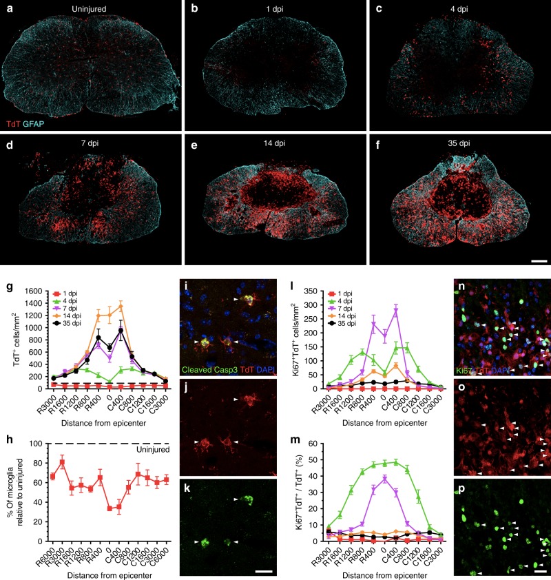
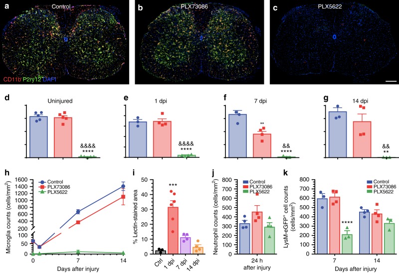
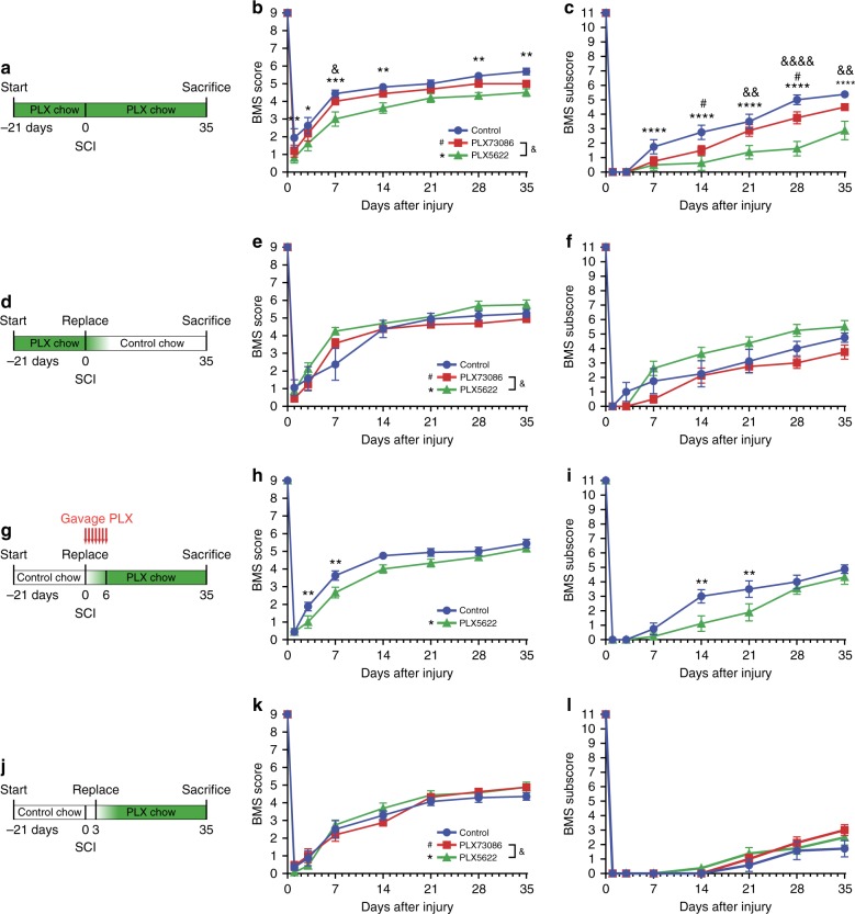
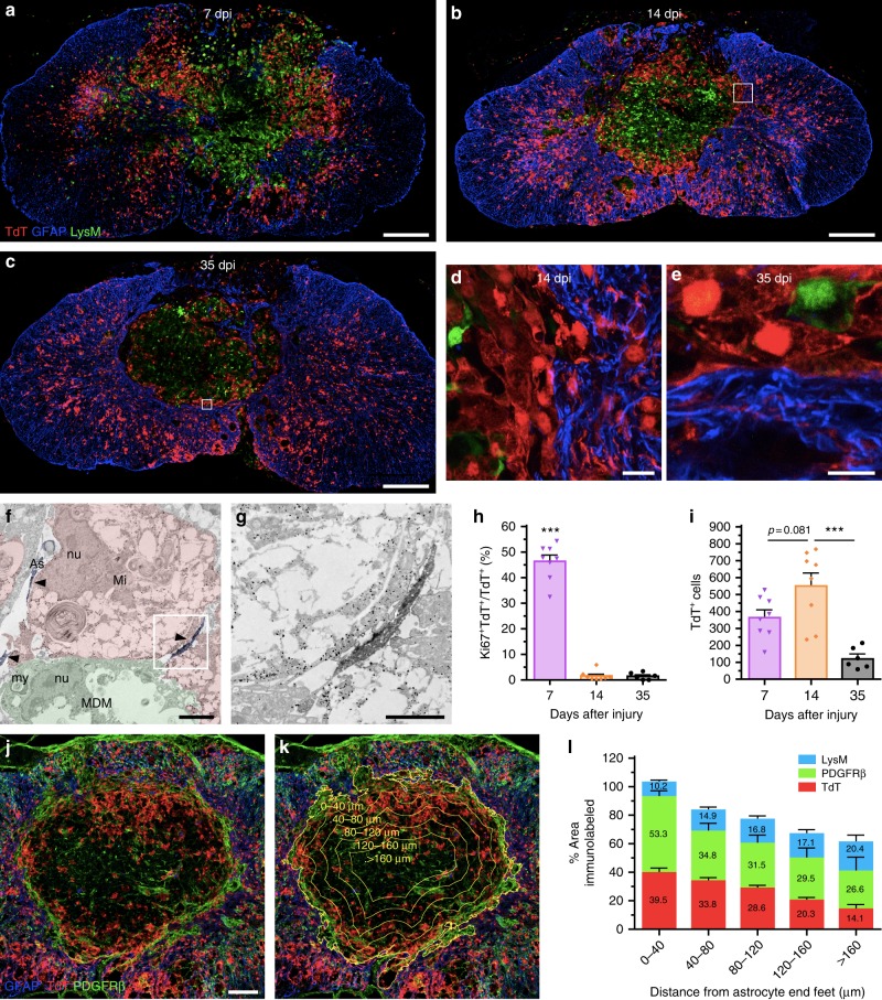
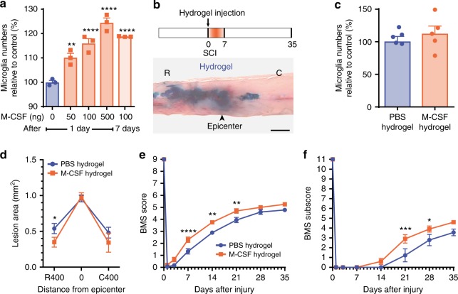
The role of microglia in spinal cord injury (SCI) remains poorly understood and is often confused with the response of macrophages. Here, we use specific transgenic mouse lines and depleting agents to understand the response of microglia after SCI. We find that microglia are highly dynamic and proliferate extensively during the first two weeks, accumulating around the lesion. There, activated microglia position themselves at the interface between infiltrating leukocytes and astrocytes, which proliferate and form a scar in response to microglia-derived factors, such as IGF-1. Depletion of microglia after SCI causes disruption of glial scar formation, enhances parenchymal immune infiltrates, reduces neuronal and oligodendrocyte survival, and impairs locomotor recovery. Conversely, increased microglial proliferation, induced by local M-CSF delivery, reduces lesion size and enhances functional recovery. Altogether, our results identify microglia as a key cellular component of the scar that develops after SCI to protect neural tissue.
Overview
The claims and statistics of this paper haven’t been checked yet.
Run the check — free, no account needed. It reads the open-access text and checks the sources it cites.
Integrity signals
Registry
Cites
Statistics
Figures
Verification
Claims the paper makes
The claims and statistics of this paper haven’t been checked yet.
Run the check — free, no account needed. It reads the open-access text and checks the sources it cites.
Claims other papers make about this one
Each is checked against this paper’s own text: does it say what the citing paper says it does?
In the citing paper
“Given that peripheral monocytes have been shown to participate in CNS disease via both infiltration and repopulation of neuroglia following microglia depletion (4–10), it is important to determine if CSF1R inhibition causes functional changes in peripheral immune cells that can become part of the CNS (3, 4, 10–14).”
From CSF1R inhibition by a small-molecule inhibitor is not microglia specific; affecting hematopoiesis and the function of macrophages (2020) · cites this paper as [4]
In the citing paper
“Previous studies have suggested that microglia depletion by CSF1R inhibition can either promote or exacerbate neurodegeneration (4, 10).”
From CSF1R inhibition by a small-molecule inhibitor is not microglia specific; affecting hematopoiesis and the function of macrophages (2020) · cites this paper as [4]
Self-check
Percentages, sums and reported differences re-computed from the paper’s own numbers.
The self-check needs the paper’s full text.
Citations
Cites
Neurotoxic reactive astrocytes are induced by activated microglia
Liddelow, S. A., Guttenplan, K. A., Clarke, L. E., Bennett, F. C., Bohlen, C. J., Schirmer, L., Bennett, M. L., Münch, A. E., Chung, W.-S., Peterson, T. C., et al. (2017) · Nature · doi:10.1038/nature21029 · Cited by 7,971
Fate Mapping Analysis Reveals That Adult Microglia Derive from Primitive Macrophages
Ginhoux, F., Greter, M., Leboeuf, M., Nandi, S., See, P., Gokhan, S., Mehler, M. F., Conway, S. J., Ng, L. G., Stanley, E. R., et al. (2010) · Science · doi:10.1126/science.1194637 · Cited by 5,175
Fate Mapping Reveals Origins and Dynamics of Monocytes and Tissue Macrophages under Homeostasis
Yona, S., Kim, K.-W., Wolf, Y., Mildner, A., Varol, D., Breker, M., Strauss-Ayali, D., Viukov, S., Guilliams, M., Misharin, A., et al. (2013) · Immunity · doi:10.1016/j.immuni.2012.12.001 · Cited by 3,245
Cell types in the mouse cortex and hippocampus revealed by single-cell RNA-seq
Zeisel, A., Muñoz-Manchado, A. B., Codeluppi, S., Lönnerberg, P., La Manno, G., Juréus, A., Marques, S., Munguba, H., He, L., Betsholtz, C., et al. (2015) · Science · doi:10.1126/science.aaa1934 · Cited by 3,124
Bracken, M. B., Shepard, M. J., Collins, W. F., Holford, T. R., Young, W., Baskin, D. S., Eisenberg, H. M., Flamm, E., Leo-Summers, L., Maroon, J., et al. (1990) · New England Journal of Medicine · doi:10.1056/nejm199005173222001 · Cited by 2,715
Microglia promote learning-dependent synapse formation through brain-derived neurotrophic factor
Parkhurst, C. N., Yang, G., Ninan, I., Savas, J. N., Yates, J. R., Lafaille, J. J., Hempstead, B. L., Littman, D. R., & Gan, W.-B. (2013) · Cell · doi:10.1016/j.cell.2013.11.030 · Cited by 2,599
A Lineage of Myeloid Cells Independent of Myb and Hematopoietic Stem Cells
Schulz, C., Perdiguero, E. G., Chorro, L., Szabo-Rogers, H., Cagnard, N., Kierdorf, K., Prinz, M., Wu, B., Jacobsen, S. E. W., Pollard, J. W., et al. (2012) · Science · doi:10.1126/science.1219179 · Cited by 2,578
Tissue-resident macrophages originate from yolk-sac-derived erythro-myeloid progenitors
Gomez Perdiguero, E., Klapproth, K., Schulz, C., Busch, K., Azzoni, E., Crozet, L., Garner, H., Trouillet, C., de Bruijn, M. F., Geissmann, F., et al. (2015) · Nature · doi:10.1038/nature13989 · Cited by 2,339
Kigerl, K. A., Gensel, J. C., Ankeny, D. P., Alexander, J. K., Donnelly, D. J., & Popovich, P. G. (2009) · The Journal of Neuroscience · doi:10.1523/jneurosci.3257-09.2009 · Cited by 2,130
Elmore, M. R. P., Najafi, A. R., Koike, M. A., Dagher, N. N., Spangenberg, E. E., Rice, R. A., Kitazawa, M., Matusow, B., Nguyen, H., West, B. L., et al. (2014) · Neuron · doi:10.1016/j.neuron.2014.02.040 · Cited by 1,968
Cited by
Repopulating Microglia Promote Brain Repair in an IL-6-Dependent Manner
Willis, E. F., MacDonald, K. P. A., Nguyen, Q. H., Garrido, A. L., Gillespie, E. R., Harley, S. B. R., Bartlett, P. F., Schroder, W. A., Yates, A. G., Anthony, D. C., et al. (2020) · Cell · doi:10.1016/j.cell.2020.02.013 · Cited by 520
Astrocytes in human central nervous system diseases: a frontier for new therapies
Verkhratsky, A., Butt, A., Li, B., Illes, P., Zorec, R., Semyanov, A., Tang, Y., & Sofroniew, M. V. (2023) · Signal Transduction and Targeted Therapy · doi:10.1038/s41392-023-01628-9 · Cited by 428
Lei, F., Cui, N., Zhou, C., Chodosh, J., Vavvas, D. G., & Paschalis, E. I. (2020) · Proceedings of the National Academy of Sciences · doi:10.1073/pnas.1922788117 · Cited by 322
Zhou, X., Wahane, S., Friedl, M.-S., Kluge, M., Friedel, C. C., Avrampou, K., Zachariou, V., Guo, L., Zhang, B., He, X., et al. (2020) · Nature Neuroscience · doi:10.1038/s41593-020-0597-7 · Cited by 275
Shaping Neuronal Fate: Functional Heterogeneity of Direct Microglia-Neuron Interactions
Cserép, C., Pósfai, B., & Dénes, Á. (2021) · Neuron · doi:10.1016/j.neuron.2020.11.007 · Cited by 256
Plemel, J. R., Stratton, J. A., Michaels, N. J., Rawji, K. S., Zhang, E., Sinha, S., Baaklini, C. S., Dong, Y., Ho, M., Thorburn, K., et al. (2020) · Science Advances · doi:10.1126/sciadv.aay6324 · Cited by 208
Microglia regulation of central nervous system myelin health and regeneration
Kent, S. A., & Miron, V. E. (2024) · Nature Reviews Immunology · doi:10.1038/s41577-023-00907-4 · Cited by 183
Microglia as hackers of the matrix: sculpting synapses and the extracellular space
Crapser, J. D., Arreola, M. A., Tsourmas, K. I., & Green, K. N. (2021) · Cellular & Molecular Immunology · doi:10.1038/s41423-021-00751-3 · Cited by 178
Gut microbiota–driven brain Aβ amyloidosis in mice requires microglia
Dodiya, H. B., Lutz, H. L., Weigle, I. Q., Patel, P., Michalkiewicz, J., Roman-Santiago, C. J., Zhang, C. M., Liang, Y., Srinath, A., Zhang, X., et al. (2022) · Journal of Experimental Medicine · doi:10.1084/jem.20200895 · Cited by 120
White matter microglia heterogeneity in the CNS
Amor, S., McNamara, N. B., Gerrits, E., Marzin, M. C., Kooistra, S. M., Miron, V. E., & Nutma, E. (2022) · Acta Neuropathologica · doi:10.1007/s00401-021-02389-x · Cited by 89
Xia, Y., Zhu, J., Yang, R., Wang, H., Li, Y., & Fu, C. (2023) · Frontiers in Immunology · doi:10.3389/fimmu.2023.1141601 · Cited by 87
Kisucká, A., Bimbová, K., Bačová, M., Gálik, J., & Lukáčová, N. (2021) · Cells · doi:10.3390/cells10081943 · Cited by 84
OʼShea, T. M., Wollenberg, A. L., Kim, J. H., Ao, Y., Deming, T. J., & Sofroniew, M. V. (2020) · Nature Communications · doi:10.1038/s41467-020-19906-3 · Cited by 70
The role of neural stem cells in regulating glial scar formation and repair
Nicaise, A. M., D’Angelo, A., Ionescu, R.-B., Krzak, G., Willis, C. M., & Pluchino, S. (2022) · Cell and Tissue Research · doi:10.1007/s00441-021-03554-0 · Cited by 65
Glial functions in the blood-brain communication at the circumventricular organs
Miyata, S. (2022) · Frontiers in Neuroscience · doi:10.3389/fnins.2022.991779 · Cited by 66
Johnson, N. R., Yuan, P., Castillo, E., Lopez, T. P., Yue, W., Bond, A., Rivera, B. M., Sullivan, M. C., Hirouchi, M., Giles, K., et al. (2023) · Nature Communications · doi:10.1038/s41467-022-35753-w · Cited by 46
Single-cell and spatial RNA sequencing identify perturbators of microglial functions with aging
Dong, Y., Jain, R. W., Lozinski, B. M., D’Mello, C., Visser, F., Ghorbani, S., Zandee, S., Brown, D. I., Prat, A., Xue, M., et al. (2022) · Nature Aging · doi:10.1038/s43587-022-00205-z · Cited by 32
Karg, M. M., Moorefield, M., Hoffmann, E., Philipose, H., Krasniqi, D., Hoppe, C., Shu, D. Y., Shirahama, S., Ksander, B. R., & Saint-Geniez, M. (2023) · Immunity & Ageing · doi:10.1186/s12979-023-00358-4 · Cited by 27
Reid, K. M., Kitchener, E. J. A., Butler, C. A., Cockram, T. O. J., & Brown, G. C. (2022) · Frontiers in Immunology · doi:10.3389/fimmu.2022.859686 · Cited by 23
Metadata
- Authors
- Bellver-Landete, V., Bretheau, F., Mailhot, B., Vallières, N., Lessard, M., Janelle, M.-E., Vernoux, N., Tremblay, M.-È., Fuehrmann, T., Shoichet, M. S., & Lacroix, S.
- Venue
- Nature Communications
- Published
- 2019-01-31
- Volume
- 10
- Issue
- 1
- ISSN
- 2041-1723
- PMC
- PMC6355913
- Cited by
- 594
- References
- 66
- Language
- en
- Licence
- creativecommons.org/licenses/by/4.0 (tdm)
- creativecommons.org/licenses/by/4.0 (vor)
About these checks
Registry, retraction, Cites and statistics results are computed, not judged, with one exception: a printed reference that no index lists is read once by a model before it counts as a citation error. Claims are extracted by a model and kept only when their quote appears verbatim in the text; each cited source is then checked, with a model’s help, for whether it says what the claim relies on. Those verdicts can be wrong — read the claim’s quote and the cited source before relying on one.
Statistics and arithmetic are recomputed, not judged.
Confidence bands are shown only for fields calibrated on our annotated test set. None is calibrated yet, so no band is shown.
What counts as a claim ›How we measure confidence ›How we check this ›
Are you an author, or do you see a problem with this page? Request a review
Data sources
Bibliographic data: Semantic Scholar, OpenAlex, Crossref.
Contains information from the Semantic Scholar Academic Graph Datasets, made available under the ODC Attribution License (ODC-BY 1.0).
Semantic Scholar’s data is described in: Kinney, R., et al. (2023). The Semantic Scholar Open Data Platform. arXiv:2301.10140.
