

The DOI matches its Crossref record; 57 of the 57 references it cites were found in a registry or an index.
Large-scale discovery of protein interactions at residue resolution using co-evolution calculated from genomic sequences
Open accessAbstract
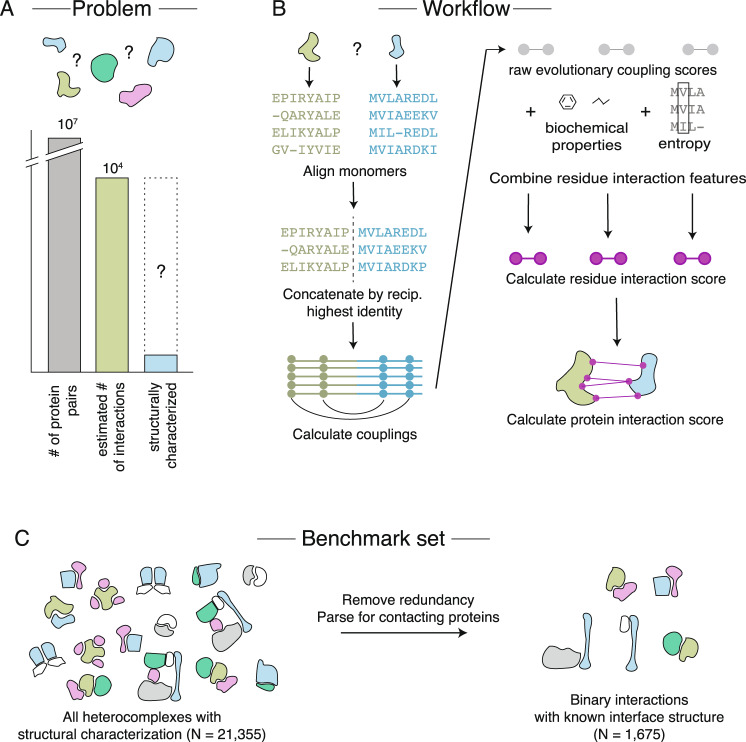
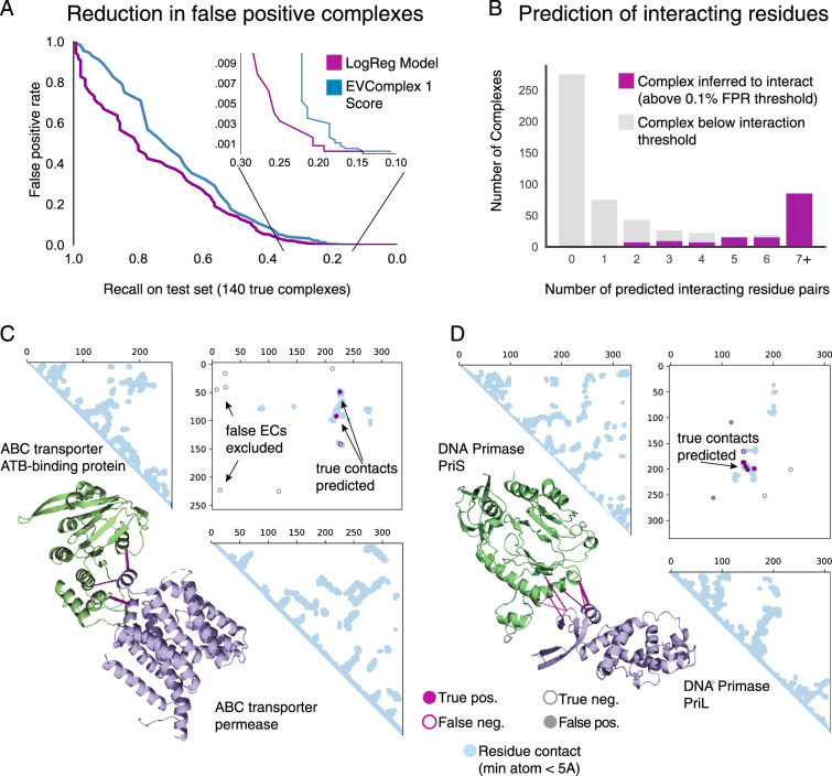
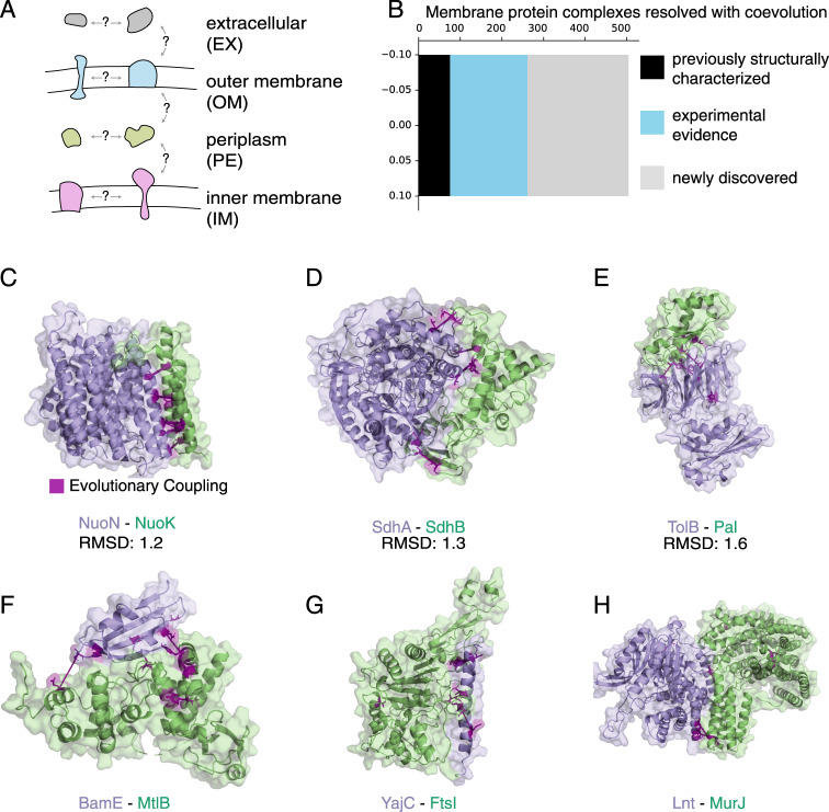
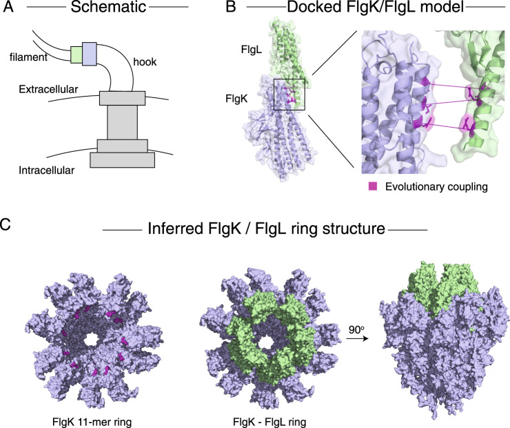
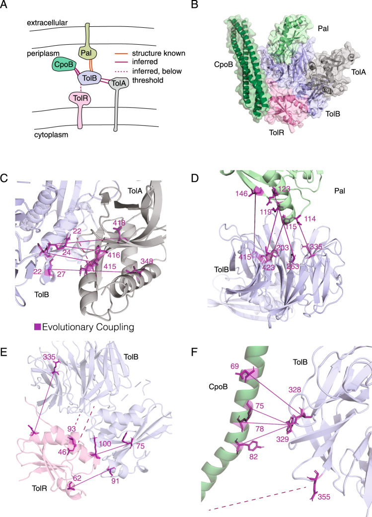
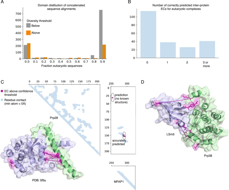
Increasing numbers of protein interactions have been identified in high-throughput experiments, but only a small proportion have solved structures. Recently, sequence coevolution-based approaches have led to a breakthrough in predicting monomer protein structures and protein interaction interfaces. Here, we address the challenges of large-scale interaction prediction at residue resolution with a fast alignment concatenation method and a probabilistic score for the interaction of residues. Importantly, this method (EVcomplex2) is able to assess the likelihood of a protein interaction, as we show here applied to large-scale experimental datasets where the pairwise interactions are unknown. We predict 504 interactions de novo in the E. coli membrane proteome, including 243 that are newly discovered. While EVcomplex2 does not require available structures, coevolving residue pairs can be used to produce structural models of protein interactions, as done here for membrane complexes including the Flagellar Hook-Filament Junction and the Tol/Pal complex.
Overview
The claims and statistics of this paper haven’t been checked yet.
Run the check — free, no account needed. It reads the open-access text and checks the sources it cites.
Integrity signals
Registry
Cites
Statistics
Figures
Verification
Claims the paper makes
The claims and statistics of this paper haven’t been checked yet.
Run the check — free, no account needed. It reads the open-access text and checks the sources it cites.
Claims other papers make about this one
Each is checked against this paper’s own text: does it say what the citing paper says it does?
In the citing paper
“Because residues at protein-protein interfaces are expected to coevolve, the likelihood that any two proteins interact can be assessed by identifying and aligning the ortholog sequences of the two proteins in many different species, joining them to create paired multiple sequence alignments (pMSA), and then determining the extent to which changes in the sequences of orthologs for the first protein”
From Computed structures of core eukaryotic protein complexes (2021) · cites this paper as [10]
Self-check
Percentages, sums and reported differences re-computed from the paper’s own numbers.
The self-check needs the paper’s full text.
Citations
Cites
Scikit-learn: Machine Learning in Python
Pedregosa, F., Varoquaux, G., Gramfort, A., Michel, V., Thirion, B., Grisel, O., Blondel, M., Müller, A., Nothman, J., Louppe, G., et al. (2012) · arXiv · doi:10.48550/arxiv.1201.0490 · Cited by 71,016
Kabsch, W., & Sander, C. (1983) · Biopolymers · doi:10.1002/bip.360221211 · Cited by 15,320
STRING v10: protein–protein interaction networks, integrated over the tree of life
Szklarczyk, D., Franceschini, A., Wyder, S., Forslund, K., Heller, D., Huerta-Cepas, J., Simonovic, M., Roth, A., Santos, A., Tsafou, K. P., et al. (2015) · Nucleic Acids Research · doi:10.1093/nar/gku1003 · Cited by 10,436
Inference of Macromolecular Assemblies from Crystalline State
Krissinel, E., & Henrick, K. (2007) · Journal of Molecular Biology · doi:10.1016/j.jmb.2007.05.022 · Cited by 10,007
UniProt: the Universal Protein knowledgebase
Apweiler, R. (2003) · Nucleic Acids Research · doi:10.1093/nar/gkh131 · Cited by 7,593
Accelerated Profile HMM Searches
Eddy, S. R. (2011) · PLoS Computational Biology · doi:10.1371/journal.pcbi.1002195 · Cited by 6,975
The Pfam protein families database in 2019
El-Gebali, S., Mistry, J., Bateman, A., Eddy, S. R., Luciani, A., Potter, S. C., Qureshi, M., Richardson, L. J., Salazar, G. A., Smart, A., et al. (2019) · Nucleic Acids Research · doi:10.1093/nar/gky995 · Cited by 4,602
Systematic identification of protein complexes in Saccharomyces cerevisiae by mass spectrometry
Ho, Y., Gruhler, A., Heilbut, A. M., Bader, G. D., Moore, L., Adams, S., Millar, A., Taylor, P. P., Bennett, K. L., Boutilier, K., et al. (2002) · Nature · doi:10.1038/415180a · Cited by 3,573
HADDOCK: A Protein−Protein Docking Approach Based on Biochemical or Biophysical Information
Dominguez, C., Boelens, R., & Bonvin, A. M. J. J. (2003) · Journal of the American Chemical Society · doi:10.1021/ja026939x · Cited by 3,187
Towards a proteome-scale map of the human protein–protein interaction network
Rual, J.-F. M., Venkatesan, K., Hao, T., Hirozane-Kishikawa, T., Dricot, A., Li, N., Berriz, G. F., Gibbons, F. D., Dreze, M., Ayivi-Guedehoussou, N., et al. (2005) · Nature · doi:10.1038/nature04209 · Cited by 2,904
Research works printed but not linked: 1
- Marcotte, C. & Marcotte, E. Predicting functional linkages from gene fusion. Appl. Bioinf. 1, 1–8 (2002).
Cited by
Sorted by citations
Protein complex prediction with AlphaFold-Multimer
Evans, R., O’Neill, M., Pritzel, A., Antropova, N., Senior, A., Green, T., Žídek, A., Bates, R., Blackwell, S., Yim, J., et al. (2021) · doi:10.1101/2021.10.04.463034 · Cited by 3,808
Improved prediction of protein-protein interactions using AlphaFold2
Bryant, P., Pozzati, G., & Elofsson, A. (2021) · doi:10.1101/2021.09.15.460468 · Cited by 1,022
AlphaFold2 and its applications in the fields of biology and medicine
Yang, Z., Zeng, X., Zhao, Y. C., & Chen, R. (2023) · Signal Transduction and Targeted Therapy · doi:10.1038/s41392-023-01381-z · Cited by 633
Computed structures of core eukaryotic protein complexes
Humphreys, I. R., Pei, J., Baek, M., Krishnakumar, A., Anishchenko, I., Ovchinnikov, S., Zhang, J., Ness, T. J., Banjade, S., Bagde, S. R., et al. (2021) · Science · doi:10.1126/science.abm4805 · Cited by 542
Protein design and variant prediction using autoregressive generative models
Shin, J.-E., Riesselman, A. J., Kollasch, A. W., McMahon, C., Simon, E., Sander, C., Manglik, A., Kruse, A. C., & Marks, D. S. (2021) · Nature Communications · doi:10.1038/s41467-021-22732-w · Cited by 320
Self-citation
Metadata
- Authors
- Green, A. G., Elhabashy, H., Brock, K. P., Maddamsetti, R., Kohlbacher, O., & Marks, D. S.
- Affiliations
- Harvard University (US)
- Max Planck Institute for Developmental Biology (DE)
- Universitätsklinikum Tübingen (DE)
- Broad Institute (US)
- Corresponding author
- Kohlbacher, Oliver
- Marks, Debora S.
- ORCID
- Venue
- Nature Communications
- Published
- 2021-03-02
- Volume
- 12
- Issue
- 1
- Pages
- 1396-1396
- ISSN
- 2041-1723 (ISSN-L)
- PMC
- PMC7925567
- OpenAlex
- W3133793061
- Cited by
- 114
- References
- 57
- Citations by year
- 2026: 14 · 2025: 17 · 2024: 24 · 2023: 25 · 2022: 21 · 2021: 13
- FWCI
- 6.51
- Type
- journal article
- Language
- en
- Open access
- gold
- doi.org
- Locations
- Nature Communications (publishedVersion, cc-by)
- PubMed Central (publishedVersion, cc-by)
- PubMed (publishedVersion)
- RePEc: Research Papers in Economics (submittedVersion)
- DOAJ (DOAJ: Directory of Open Access Journals) (submittedVersion, cc-by-sa)
- Funders
- National Science Foundation (DGE1144152)
- U.S. Department of Health & Human Services | National Institutes of Health (T15LM007092)
- Topics
- Life Sciences › Biochemistry, Genetics and Molecular Biology › Molecular Biology › Protein Structure and Dynamics
- Life Sciences › Biochemistry, Genetics and Molecular Biology › Molecular Biology › Machine Learning in Bioinformatics
- Life Sciences › Biochemistry, Genetics and Molecular Biology › Molecular Biology › Genomics and Phylogenetic Studies
- Keywords
- protein-protein interaction, membrane protein complexes
- MeSH terms
- Amino Acids, Bacterial Proteins, Base Sequence, Escherichia coli, Eukaryotic Cells, Membrane Proteins, Protein Binding, Genome, Bacterial, Evolution, Molecular, Proteome, Protein Interaction Mapping, Molecular Docking Simulation
About these checks
Registry, retraction, Cites and statistics results are computed, not judged, with one exception: a printed reference that no index lists is read once by a model before it counts as a citation error. Claims are extracted by a model and kept only when their quote appears verbatim in the text; each cited source is then checked, with a model’s help, for whether it says what the claim relies on. Those verdicts can be wrong — read the claim’s quote and the cited source before relying on one.
Statistics and arithmetic are recomputed, not judged.
Confidence bands are shown only for fields calibrated on our annotated test set. None is calibrated yet, so no band is shown.
What counts as a claim ›How we measure confidence ›How we check this ›
Are you an author, or do you see a problem with this page? Request a review
